OBJECTIVE:Endoscopic third ventriculostomy with choroid plexus cauterization (ETV/CPC) has decreased rates of shunt dependence in infants with hydrocephalus. The ETV Success Score (ETVSS) is the standard for predicting the 6-month success rate for ETV in children based on age, hydrocephalus etiology, and shunt history. However, the ETVSS does not account for the impact of CPC or preoperative ventricular volume. It also relies on the independence of each contributing variable and does not predict success beyond 6 months. In this study, the authors used a machine learning approach to create a tool specifically for ETV/CPC to predict the likelihood of success at 1 year. METHODS:The records of 206 pediatric patients younger than 2 years of age who received ETV/CPC as a primary, definitive treatment for hydrocephalus at a single institution between 2009 and 2021 were reviewed for patient demographics, presenting characteristics, and medical history. Data on corrected age at surgery, frontal occipital horn ratio (FOHR), hydrocephalus etiology, and whether there was prior CSF diversion were used in developing logistic regression, XGBoost, random forest, and gradient boosting algorithms to predict the percentage likelihood of ETV/CPC failure within postoperative year 1. RESULTS:Unlike the ETVSS, etiology of hydrocephalus did not substantially influence any model's predictions and was removed. A logistic regression model produced the best area under the receiver operating characteristic curve (AUROC) and was used as the final model. Three features selected for the final model (corrected age, prior CSF diversion, and FOHR) proved relevant. The logistic regression predictor had an AUROC of 0.85 (compared to 0.66 for the ETVSS) and an outcome classification accuracy of 76%. The model correctly classified its predictions for success slightly more accurately than its predictions for failure (sensitivity 78%, specificity 75%). CONCLUSIONS:This model can reliably predict the likelihood of ETV/CPC failure at 1 year. Its outperformance of the ETVSS is likely due to the impact of CPC, the novel use of preoperative ventricle size as a predictive parameter, and accounting for variable interdependence. Further validation in additional patient populations is needed.
Delineating the epileptogenic zone (EZ) is essential for achieving seizure freedom in drug-resistant epilepsy (DRE). Conventionally, seizure onset derived from ictal intracranial EEG (iEEG) approximates the EZ, but acquiring ictal data can be challenging. Interictal iEEG abnormalities offer abundant, non-seizure-dependent markers of the epileptogenic tissue; however, these biomarkers offer limited specificity. Here, we propose a machine learning framework that integrates interictal spike and ripple features to predict the epileptogenic contacts targeted for surgical removal and the patient’s surgical outcome. We retrospectively analyzed iEEG data from 62 children with DRE [34 with good outcome (Engel I)], automatically detected spikes and ripples, and computed temporal, spectral, and spatial features for each channel. Using combinations of these features and the resected contacts as targets, we trained Random Forest classifiers using only good outcome patients to estimate epileptogenic contacts. Spike-based and combined spike and ripple features outperformed individual biomarkers in predicting the epileptogenic contacts with an area under the receiver operating characteristic curve of 0.89 and 74% spatial overlap with resection. Although most individual features and classifiers predicted outcome, the combined feature model performed best (i.e., sensitivity 88%, specificity 68%, and accuracy 79%). Our findings demonstrate that integrating multimodal interictal features improves the identification of epileptogenic contacts providing valuable prognostic insights for epilepsy surgery.
OBJECTIVE:It is unclear whether shunt survival differs for infants in whom prior endoscopic third ventriculostomy with choroid plexus cauterization (ETV/CPC) failed compared with those treated initially by shunt placement. Results among four previous studies spanning a range of age groups have suggested either no effect or a protective effect of prior endoscopic treatment on shunt survival. The aim of this study was to determine whether prior ETV/CPC positively impacts shunt survival among infants with hydrocephalus regardless of etiology. METHODS:The authors conducted a historical cohort review of all patients under 12 months corrected age who underwent ventriculoperitoneal shunt (VPS) placement either with or without prior ETV/CPC at their institution between December 2008 and December 2023. Statistical analyses included Cox regression modeling, Kaplan-Meier survival curves, and log-rank testing to evaluate the time to shunt failure. RESULTS:The study included 122 infants with and 46 infants without prior ETV/CPC who underwent VPS placement. There were no statistically significant differences between groups regarding sex, race, gestational age at birth, or corrected age at initial treatment for hydrocephalus. The distribution of etiologies and initial frontal-occipital horn ratios (FOHRs) were similar. The median pre-shunt FOHR was greater in the ETV/CPC group. The patients in the primary shunt placement group were all candidates for ETV/CPC based on retrospective review. Seventy-one infants (58%) with prior ETV/CPC remained free of shunt revision compared with 17 infants (37%) with primary shunt placement (OR 2.4, 95% CI 1.1-5.1; p = 0.01). The estimated 2-year shunt survival probability was 75% with and 50% without prior ETV/CPC (log-rank p = 0.001). The estimated 5-year survival was 60% with and 45% without prior ETV/CPC (p = 0.045). CONCLUSIONS:Infants who underwent ETV/CPC prior to VPS placement had better shunt survival than those who did not. ETV/CPC might reduce the risk of subsequent shunt failure, indicating the potential benefit of the procedure even when it fails.
Aromatic ʟ-amino acid decarboxylase (AADC) deficiency is a rare pediatric neurotransmitter disorder that typically necessitates lifelong care, and that carries a risk of childhood mortality. Eladocagene exuparvovec gene therapy is designed to restore AADC production. Study GT-002 (NCT04903288) is a phase 2, multicenter, open-label trial assessing the pharmacodynamics, safety, and efficacy of eladocagene exuparvovec administered to the putamen bilaterally in pediatric patients with AADC deficiency using a magnetic resonance (MR)-compatible cannula. Patients received eladocagene exuparvovec at 1.8 × 10¹¹ vector genomes via the SmartFlow MR-compatible cannula in a single operative session. Endpoints include the change from baseline in cerebrospinal fluid homovanillic acid levels, motor milestone achievement, and safety. Here we report results from 48 weeks of follow-up. Mean (SD) cerebrospinal fluid homovanillic acid levels increased from baseline (22.5 [32.3] nmol/L; n = 13) to week 48 (55.3 [45.6] nmol/L; change from baseline: 28.3 [13.7] nmol/L; p = 0.0003; n = 9), indicating de novo dopamine production. At baseline (n = 13), all patients showed severe motor developmental delay; at week 48 (n = 12), nine achieved full head control, four could sit unassisted, two could stand with support, and two could walk independently to a toy. Overall, 260 treatment-emergent adverse events were reported in 13 patients; 259 were deemed unrelated and one likely unrelated to the MR-compatible cannula. No treatment-emergent adverse events led to study withdrawal and no deaths occurred. This study provides further evidence of the favorable pharmacodynamic, efficacy, and safety profile of eladocagene exuparvovec in children with AADC deficiency; intraputaminal administration using an MR-compatible cannula was well tolerated. Study GT-002 (NCT04903288) provides further evidence of the favourable pharmacodynamic, efficacy and safety profile of eladocagene exuparvovec gene therapy in children with AADC deficiency over 48 weeks and demonstrates that intraputaminal administration using an MR-compatible cannula was well tolerated, allowing for real-time MRI confirmation of cannula placement and infusate coverage, and for accurate dosing to the putamen.
ABSTRACT Background and Objectives Deep sequencing of brain tissue in the research setting has established that mosaic variants are a major cause of malformations of cortical development (MCDs) and epilepsy. However, genetic testing in the clinical setting primarily detects germline variants using clinically accessible samples. We aimed to determine the diagnostic yield and clinical utility of deep sequencing in the clinical setting to identify pathogenic mosaic variants for this population. Methods We performed a retrospective cohort analysis of individuals at Boston Children’s Hospital with MCDs with or without epilepsy who received clinical deep sequencing between September 2017 and February 2026. Demographic, clinical, and genetic testing data were abstracted from the medical record. For individuals without systemic features, we classified brain tissue as an affected tissue sample. For individuals with systemic features, we classified brain or relevant non-brain tissue as affected. The primary outcome was the diagnostic yield of clinical deep sequencing performed using affected vs unaffected tissue samples. The secondary outcome was the clinical utility of genetic diagnoses. Results Our cohort included 37 individuals (19/37 (51%) female, 18/37 (49%) male) with MCDs, of whom 35/37 (95%) had epilepsy (25 with brain tissue samples available from epilepsy surgery) and 8/37 (22%) had systemic features. Most (35/37 (95%)) had dysplasia phenotypes on MRI and 12/27 (44%) with pathology available had Focal Cortical Dysplasia Type I or II. The diagnostic yield was 53% (17/32; 16 mosaic and 1 germline variant) when clinical deep sequencing was performed using an affected tissue sample vs 0% (0/6) using an unaffected tissue sample (p=0.016). Of the diagnosed cases, 13/17 (76%) had testing performed on brain tissue (1 with systemic features) and 4/17 (24%) on non-brain tissue (3 buccal and 1 duodenal tissue, all with systemic features). All but one diagnosis involved the mTOR pathway. All diagnoses had clinical utility. Discussion Clinical deep sequencing, when performed using an affected tissue sample, has high diagnostic yield and clinical utility for individuals with MCDs, especially dysplasia phenotypes, and epilepsy. Our findings support implementation of clinical deep sequencing for this population, especially as the genetic diagnoses have implications for emerging precision therapies.
Inferring object identity from incomplete information is a ubiquitous challenge for the visual system. Here, we study the neural mechanisms underlying processing of minimally recognizable configurations (MIRCs) and their subparts, which are unrecognizable (sub-MIRCs). MIRCs and sub-MIRCs are very similar at the pixel level, yet they lead to a dramatic gap in recognition performance. To evaluate how the brain processes such images, we invasively record human neurophysiological responses. Correct identification of MIRCs is associated with a dynamic interplay of feedback and feedforward mechanisms between frontal and temporal areas. Interpretation of sub-MIRC images improves dramatically after exposure to the corresponding full objects. This rapid and unsupervised learning is accompanied by changes in neural responses in the temporal cortex. These results are at odds with purely feedforward models of object recognition and suggest a role for the frontal lobe in providing top-down signals related to object identity in difficult visual tasks.
OBJECTIVE:This single-center retrospective study examined the response to initial standard therapy in children with infantile epileptic spasms syndrome (IESS) associated with surgically-remediable lesion and evaluated the risk factors for drug resistance. We assessed whether the failure of the first standard therapy for surgically-remediable IESS predicted eventual drug resistance. METHODS:New-onset IESS with surgically-remediable lesions was included. Regression analysis was performed to identify risk factors for drug resistance. Kaplan-Meier survival analysis stratified by the response to first standard therapy was conducted to explore if earlier recognition of drug-resistant epilepsy (DRE) was possible. RESULTS:We identified 61 patients (57% female) with IESS and surgically-remediable lesion (median follow-up of 52 months). First standard treatment started at a median of 15 days after IESS onset resulted in favorable initial response in 31%. Response rate to second standard therapy among those who failed first treatment was 53%, with an overall response rate to sequential standard therapy of 63.8%. Relapses (epileptic spasms/focal seizures) were frequent (59%). At last follow-up, 41% (n = 25) remained drug responsive. The cumulative proportion of survival free of drug resistance was 57% at 2 months, dropping to 38% at 36 months after IESS diagnosis. The odds for DRE increased with extensive magnetic resonance imaging (MRI) abnormality (odds ratio [OR] = 38.5) and congenital-structural abnormality (OR = 23.3) and decreased with older age at IESS onset (OR = 0.68). Kaplan-Meier survival curve differed significantly between responders and non-responders to first standard therapy (p = .02). In the drug-resistant group (n = 36), 34 underwent surgery with Engel class I outcome in 76.5%. SIGNIFICANCE:Although two-thirds of surgically-remediable IESS exhibited an initial response to medical therapy, relapses were frequent. The majority progressed to DRE during follow-up, particularly those with younger age at IESS onset, congenital-structural etiology, or extensive MRI abnormalities. Patients at risk for DRE can be recognized early by the lack of response to first standard therapy (OR 4.15). Our findings can help reduce delays for surgical referral in patients with surgically-remediable IESS.
BACKGROUND AND OBJECTIVES:Deep brain stimulation (DBS) is considered off-label and investigational in pediatric populations with some exceptions. There are limited data on the relative rates of complications after DBS across different indications and targets in children. This study aimed to evaluate the safety of DBS surgery for children with movement disorders (MDs; dystonia, chorea, or tic disorders), drug-resistant epilepsy (DRE), or neurodevelopmental disorders, namely, self-injurious behavior (SIB). METHODS:Data were collected both prospectively and retrospectively from children implanted with DBS through the North American multicenter Child and Youth CompreHensIve Longitudinal Database for Deep Brain Stimulation and included demographic, clinical, operative, and postoperative variables. Complications included infection, noninfectious surgical site findings (dehiscence or seroma), hardware-related issues (disconnection or impedance change), intracranial injury, or other complications. The primary outcome was major complications, defined as any adverse event causing permanent neurologic injury or requiring surgical intervention. The secondary outcome was minor complications, defined as nonmajor complications. Generalized linear models were used to assess for any significant associations with complications. RESULTS:A total of 130 children and youth (mean age 12.2 ± 4.2; range 3-18) years and weighing 12.5-126.6 kg underwent DBS. The most common indication was MD (77, 59.2%), followed by DRE (47, 36.2%) and SIB (6, 4.6%). Major complications occurred in 11.5%, with a greater likelihood in MD (n = 12, 15.6%) compared with DRE (n = 2, 4.3%; odds ratio [OR] 3.55, 95% CI 2.66-4.73, p < 0.001) and significantly associated with lower weight at surgery (p < 0.001) and urgent intervention (p = 0.028). These included infection (6.2%), hardware malfunction (3.1%), and wound dehiscence (0.8%). Minor complications were also higher with MD compared with DRE (OR 1.83, 95% CI 1.16-2.89, p = 0.010) occurring in 22 participants (16.9%; 14 MD, 7 DRE, 1 SIB), including infection (6.2%), high impedance (1.5%), unrelated hydrocephalus (0.8%), perioperative worsening of symptoms (3.8%), incidental tract hemorrhage (2.3%), and noninfectious peri-electrode cystic changes (0.8%). DISCUSSION:DBS-associated complications were low across multiple pediatric indications and targets, with MD associated with higher risk of major complications. Limitations include a focus on surgical postoperative complications and not stimulation-related adverse outcomes. These findings demonstrate the safety profile of DBS in children in a large cohort.
Spikes are the most established interictal epilepsy biomarkers. Yet, they suffer from low specificity since they are partially concordant with the epileptogenic zone and are often found in non-epileptogenic areas. High-frequency oscillations, classified as ripples and fast ripples, are considered more specific biomarkers compared with spikes. Ripples occur more often than fast ripples but are believed to be less specific, since they are more frequently generated by physiological mechanisms. Here, we examine the temporal relationship between spikes, ripples and fast ripples, and assess the ability of these biomarkers (and their combinations) to delineate the epileptogenic zone and predict outcome. We hypothesize that spikes on ripples (temporal co-occurrence of spikes and ripples) can identify the epileptogenic zone and predict outcome better than spikes or ripples. We analysed intracranial EEG data from 40 children with drug-resistant epilepsy. Spikes, ripples and fast ripples were classified based on their temporal occurrence. Their rates were compared with resection by performing a receiver operating characteristic analysis. The resection ratio, quantifying the extent of each biomarker's removal, was computed, and correlated with patients' outcome. Spikes on ripples were seen in all patients; fast ripples were seen in 43% of patients. In good outcome patients, fast ripple and spike on ripple rates were higher inside resection (P = 0.027; P = 0.003, respectively). Fast ripples and spikes on ripples resection ratio predicted outcome (P < 0.05). For fast ripples, outcome was predicted in 82% of patients; this proportion was higher than the one for spikes (48%, P = 0.015) and ripples (40%, P = 0.003), and spikes on ripples (53%, P = 0.034). Fast ripples were the most accurate (82%) to predict outcome; spikes on ripples were the most precise (positive predictive value = 90%). Spike rate and spikes on ripples performance to predict the epileptogenic zone were correlated (r = 0.36, P = 0.035). For patients with frequent spikes, spikes on ripples accuracy to predict outcome reached 70%. Fast ripples are the best biomarker, but they can be seen in only half of patients with drug-resistant epilepsy. Spikes on ripples are a good alternative with more universal applicability since they can be seen in all patients while their resection predicts good outcome; their performance is improved in patients with frequent spikes. Overall, in the absence of fast ripples, spike on ripple areas should be targeted during surgery.
Somatic mosaic variants contribute to focal epilepsy, with variants often present only in brain tissue and not in blood or other samples typically assayed for genetic testing. Thus, genetic analysis for mosaic variants in focal epilepsy has been limited to patients with drug-resistant epilepsy who undergo surgical resection and have resected brain tissue samples available. Stereo-EEG (sEEG) has become part of the evaluation for many patients with focal drug-resistant epilepsy, and sEEG electrodes provide a potential source of small amounts of brain-derived DNA. We aimed to identify, validate, and assess the distribution of deleterious mosaic variants in epilepsy-associated genes in DNA extracted from trace brain tissue on individual sEEG electrodes. We enrolled a prospective cohort of 10 paediatric patients with drug-resistant epilepsy who had sEEG electrodes implanted for invasive monitoring. We extracted unamplified DNA and in parallel performed whole-genome amplification from trace brain tissue on each sEEG electrode. We also extracted DNA from resected brain tissue and blood/saliva samples where available. We performed deep sequencing (panel and exome) and analysis for candidate germline and mosaic variants. We validated candidate mosaic variants and assessed the variant allele fraction in amplified and unamplified electrode-derived DNA and across electrodes. We extracted unamplified DNA and performed whole-genome amplification from >150 individual electrodes from 10 individuals. Immunohistochemistry confirmed the presence of neurons in the brain tissue on electrodes. Deep sequencing and analysis demonstrated similar depth of coverage between amplified and unamplified DNA samples but significantly more potential mosaic variants in amplified samples. We validated four deleterious mosaic variants in epilepsy-associated genes in electrode-derived DNA in three patients who underwent laser ablation and did not have resected brain tissue samples available. Three of the four variants were detected in both amplified and unamplified electrode-derived DNA, with higher variant allele fraction observed in DNA from electrodes in closest proximity to the electrical seizure focus in one case. We demonstrate that mosaic variants can be identified and validated from DNA extracted from trace brain tissue on individual sEEG electrodes in patients with drug-resistant focal epilepsy, from both unamplified and amplified electrode-derived DNA. Our findings support a relationship between the extent of regional genetic abnormality and electrophysiology and suggest that with further optimization, this minimally invasive diagnostic approach holds promise for advancing precision medicine for patients with drug-resistant epilepsy as part of the surgical evaluation.
Pediatric cerebral palsy patients carry frequent medical comorbidities and disproportionately consume hospital resources after neurosurgical procedures. We implemented an institutional pre-operative gastrointestinal (GI) optimization protocol to improve outcomes and decrease resource utilization. All 323 intrathecal baclofen surgeries from 2000 to 2023 were categorized relative to protocol implementation on July 1, 2017. Outcomes and resource utilization were compared. The protocol change resulted in significantly fewer hospital readmissions (p = 0.001) for constipation, eliminating them and GI-related emergency visits. There was a reduction of 27 hospital days for constipation-related readmission (median 1 day per patient, IQR 1, 2 days). No differences were reported between the experimental groups including demographics or GI comorbidities. Post-operative complications within 30 days were comparable between groups regarding urinary tract infections, surgical-site infections, and spinal fluid leak. There were no differences in post-operative length of stay between groups. A GI optimization protocol can eliminate a frequent source of hospital readmissions and GI-related emergency department visits after baclofen pump surgery, even accounting for baseline GI comorbidities. Preventing readmissions and emergency visits translates to lower hospital resource utilization and improves quality of care. Future efforts are warranted to improve outcomes and care efficiency for our most complex and resource-intensive patients.
Surgical success for patients with focal drug resistant epilepsy (DRE) relies on accurate localization of the epileptogenic zone (EZ). Currently, no exam delineates this zone unambiguously. Instead, the EZ is approximated by the area where seizures begin, which is identified manually through a tedious process that is prone to errors and biases. More importantly, resection of this area does not always predict good surgical outcome. Here, we propose an artificially intelligent, patient-specific framework that automatically identifies the EZ requiring little to no input from clinicians, without having to wait for a seizure to occur. The framework transforms interictal intracranial electroencephalography data into spatiotemporal representations of brain activity discriminating the interictal epileptogenic network from background activity. The epileptogenic network delineates the EZ with high precision and predicts surgical outcome. Our framework eliminates the need for manual data inspection, reduces prolonged monitoring, and enhances surgical planning for DRE patients.
OBJECTIVE Intrathecal baclofen (ITB) pumps are commonly used in pediatric patients with cerebral palsy (CP) and medically refractory spasticity. However, catheter malfunction and associated risk factors are not well understood. The aim of this study was to examine potential risk factors for spinal catheter malfunction and characterize postoperative follow-up to understand the clinical consequences. METHODS Patients who received ITB pump replacement or revision at Boston Children's Hospital between 2010 and 2023 were retrospectively reviewed. The spinal catheter revision cohort (SCRC) included patients whose spinal catheter was occluded requiring lumbar catheter revision. The second cohort included abdominal pump replacements only (APRC). Between-group comparisons and multivariable regression identified factors associated with catheter revision and postoperative outcomes. RESULTS Forty-one (33.6%) patients underwent spinal catheter revision and were compared with 81 patients (66.4%) who underwent abdominal pump replacement only. Younger age at surgery and an elevated preoperative lower-extremity modified Ashworth scale grade were associated with spinal catheter revision (p < 0.05). Catheter model type, tip location, and history of spinal fusion were not associated with obstruction. Postoperatively, SCRC patients experienced a higher rate of infection (17.1%) relative to APRC patients (0%) within 30 days from their ITB pump replacement procedure (p < 0.05) and greater likelihood of subsequent ITB system removal compared with the APRC (24.4% vs 7.4%, p < 0.05). Although not differing preoperatively, SCRC patients had lower postoperative ITB doses when compared with the APRC group (median dose 143 vs 350 g/day, p < 0.05) at hospital discharge and remained statistically different at the 6-month and 1-year follow-ups (p < 0.05). There were no postoperative differences in baclofen overdose, withdrawal, or median number of hospital readmissions within 30 days. Overall, 31.7% of spinal catheter revisions were unanticipated by the clinical team at time of surgery. CONCLUSIONS Younger age at surgery and increased preoperative lower-extremity tone may be risk factors for catheter obstruction, resulting in a higher rate of postoperative infection and subsequent ITB pump removal compared with pump replacement alone. Spinal catheter occlusion can complicate revision or replacement procedures, especially when unanticipated. Routine clinical assessment may be inadequate for diagnosing insidious catheter malfunction. Catheter occlusion deserves further study, and routine assessment of catheter patency may be warranted to prevent suboptimal tone therapy.
AbstractBackgroundAromatic ʟ‐amino acid decarboxylase (AADC) deficiency is a rare, severe neurological disorder caused by pathogenic variants in the dopa decarboxylase (DDC) gene, resulting in a combined deficiency of monoamine neurotransmitters. Clinically, patients present with a range of dysfunctions that impact motor, autonomic, and cognitive development. The constellation of symptoms of AADC deficiency varies among patients, and clinical presentation falls across a wide spectrum. However, most patients with AADC deficiency experience significant impairments when compared with children with normal development, irrespective of genotype, phenotype, or disease severity. Further, AADC deficiency is associated with increased mortality.MethodsIn response to the recent approval of a disease‐modifying gene therapy for AADC deficiency, this review presents considerations for the selection of patients for treatment.ConclusionSuggested clinical criteria to determine whether a patient is a candidate for gene therapy are: (1) genetically and biochemically confirmed AADC deficiency; (2) lack of achievement of gross motor milestones and/or persistence of clinically significant movement disorders; (3) persistent neurocognitive or systemic symptoms secondary to AADC deficiency despite standard medical therapy; and (4) informed parental/guardian decision and consent to treatment.
OBJECTIVE:To deconstruct the epileptogenic networks of patients with drug-resistant epilepsy (DRE) using source functional connectivity (FC) analysis; unveil the FC biomarkers of the epileptogenic zone (EZ); and develop machine learning (ML) models to estimate the EZ using brief interictal electroencephalography (EEG) data.METHODS:We analyzed scalp EEG from 50 patients with DRE who had surgery. We reconstructed the activity (electrical source imaging [ESI]) of virtual sensors (VSs) across the whole cortex and computed FC separately for epileptiform and non-epileptiform EEG epochs (with or without spikes). In patients with good outcome (Engel 1a), four cortical regions were defined: EZ (resection) and three non-epileptogenic zones (NEZs) in the same and opposite hemispheres. Region-specific FC features in six frequency bands and three spatial ranges (long, short, inner) were compared between regions (Wilcoxon sign-rank). We developed ML classifiers to identify the VSs in the EZ using VS-specific FC features. Cross-validation was performed using good outcome data. Performance was compared with poor outcomes and interictal spike localization.RESULTS:FC differed between EZ and NEZs (p < .05) during non-epileptiform and epileptiform epochs, showing higher FC in the EZ than its homotopic contralateral NEZ. During epileptiform epochs, the NEZ in the epileptogenic hemisphere showed higher FC than its contralateral NEZ. In good outcome patients, the ML classifiers reached 75% accuracy to the resection (91% sensitivity; 74% specificity; distance from EZ: 38 mm) using epileptiform epochs (gamma and beta frequency bands) and 62% accuracy using broadband non-epileptiform epochs, both outperforming spike localization (accuracy = 47%; p < .05; distance from EZ: 57 mm). Lower performance was seen in poor outcomes.SIGNIFICANCE:We present an FC approach to extract EZ biomarkers from brief EEG data. Increased FC in various frequencies characterized the EZ during epileptiform and non-epileptiform epochs. FC-based ML models identified the resection better in good than poor outcome patients, demonstrating their potential for presurgical use in pediatric DRE.
BACKGROUND Focal epilepsy caused by a posterior fossa lesion is a rare phenomenon. In these cases, seizure onset typically occurs during the first few months of life, with episodes of epileptic hemifacial spasms and abnormal eye movements. Patients often present with drug-resistant epilepsy and often require resection for the best chance of seizure freedom. OBSERVATIONS The authors present the case of a 19-month-old male with intractable epileptic hemifacial spasms and a dorsally exophytic right brainstem and middle cerebellar peduncle hamartoma, following 2 prior subtotal resections. The authors recommended a third suboccipital craniotomy with intraoperative electrocorticography, which revealed interictal spiking from an intralesional depth electrode. Near-total resection led to durable seizure freedom. LESSONS Although posterior fossa lesions are rarely associated with epileptiform activity, this case demonstrates that pediatric patients with epileptic hemifacial spasms associated with a posterior fossa lesion may respond favorably to resection. Furthermore, this case demonstrates that intralesional electrocorticography can detect epileptic activity in posterior fossa lesions, which may predict postoperative seizure outcomes. https://thejns.org/doi/10.3171/CASE2452
Purpose: Stereoelectroencephalography (SEEG) is widely performed on individuals with medically refractory epilepsy for whom invasive seizure localization is desired. Despite increasing adoption in many centers across the world, no standardized electrode naming convention exists, generating confusion among both clinical and research teams. Methods: We have developed a novel nomenclature, named the Standardized Electrode Nomenclature for SEEG Applications system. Concise, unique, informative, and unambiguous labels provide information about entry point, deep targets, and relationships between electrodes. Inter-rater agreement was evaluated by comparing original electrode names from 10 randomly sampled cases (including 136 electrodes) with those prospectively assigned by four additional blinded raters. Results: The Standardized Electrode Nomenclature for SEEG Application system was prospectively implemented in 40 consecutive patients undergoing SEEG monitoring at our institution, creating unique electrode names in all cases, and facilitating implantation design, SEEG recording and mapping interpretation, and treatment planning among neurosurgeons, neurologists, and neurophysiologists. The inter-rater percent agreement for electrode names among two neurosurgeons, two epilepsy neurologists, and one neurosurgical fellow was 97.5%. Conclusions: This standardized naming convention, Standardized Electrode Nomenclature for SEEG Application, provides a simple, concise, reproducible, and informative method for specifying the target(s) and relative position of each SEEG electrode in each patient, allowing for successful sharing of information in both the clinical and research settings. General adoption of this nomenclature could pave the way for improved communication and collaboration between institutions.
AbstractObjectiveInterictal biomarkers are critical for identifying the epileptogenic focus. However, spikes and ripples lack specificity while fast ripples lack sensitivity. These biomarkers propagate from more epileptogenic onset to areas of spread. The pathophysiological mechanism of these propagations is elusive. Here, we examine zones where spikes and high frequency oscillations co‐occur (SHFO), the spatiotemporal propagations of spikes, ripples, and fast ripples, and evaluate the spike–ripple onset overlap (SRO) as an epilepsy biomarker.MethodsWe retrospectively analyzed intracranial EEG data from 41 patients with drug‐resistant epilepsy. We mapped propagations of spikes, ripples, and fast ripples, and identified their onset and spread zones, as well as SHFO and SRO. We then estimated the SRO prognostic value in predicting surgical outcome and compared it to onset and spread zones of spike, ripple, and fast ripple propagations, and SHFO.ResultsWe detected spikes and ripples in all patients and fast ripples in 12 patients (29%). We observed spike and ripple propagations in 40 (98%) patients. Spike and ripple onsets overlapped in 35 (85%) patients. In good outcome patients, SRO showed higher specificity and precision (p < 0.05) in predicting resection compared to onset and zones of spikes, ripples, and SHFO. Only SRO resection predicted outcome (p = 0.01) with positive and negative predictive values of 82% and 57%, respectively.InterpretationSRO is a specific and precise biomarker of the epileptogenic zone whose removal predicts outcome. SRO is present in most patients with drug‐resistant epilepsy. Such a biomarker may reduce prolonged intracranial monitoring and improve outcome.
 Joseph Madsen合作论文数Department of Neurosurgery, Boston Children's Hospital;Harvard Medical School34
 Phillip L. Pearl合作论文数School of Medicine and Health Sciences, George Washington University21
 Jeffrey Bolton合作论文数20
 John Ragheb合作论文数14
 Andres Lozano合作论文数Department of Surgery, Temerty Faculty of Medicine, University of Toronto12
 Francesco T. Mangano合作论文数Division of Pediatric Neurosurgery, Cincinnati Children's Hospital Medical Center;Department of Neurosurgery, University of Cincinnati;College of Medicine, University of Cincinnati10
 Tamilia Eleonora合作论文数Center for Integrated Research, Università Campus Bio-Medico di Roma10
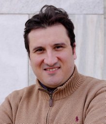 Christos Papadelis合作论文数Lab for Human Brain Dynamics;Brain Science Institue;Lab for Human Brain Dynamics, Brain Science Institue10
 Brent R O'Neill合作论文数10Mark Iantosca合作论文数9Tae Sung Park合作论文数9
 Cormac O. Maher合作论文数Department of Adult Neurosurgery, Stanford University9
 Nathan R Selden合作论文数Dept Neurol Surg, Oregon Hlth & Sci Univ9
 Chevis Shannon合作论文数American Society for Reproductive Medicine9
 Stephanie Greene合作论文数9
 Richard G. Ellenbogen合作论文数Department of Radiology, University of Washington;Department of Neurological Surgery, School of Medicine, University of Washington;Department of Global Health, University of Washington9Durham Susan R合作论文数9
 J Gordon Mc Comb合作论文数Keck School of Medicine, University of Southern California9
 Strahle Jennifer M合作论文数Department of Neurosurgery, University of Michigan9
 P. David Adelson合作论文数Department of Emergency Medicine, College of Medicine - Tucson, University of Arizona;Children's Neuroscience Institute9
 Chern Joshua J合作论文数9Matthew D. Smyth合作论文数Epilepsy Research Laboratory, University of California at San Francisco9John C. Wellons合作论文数Pediatric Neurosurgery Children’s Hospital9Arnold H. Menezes合作论文数9
 Gerald Arthur Grant合作论文数Department of Neurosurgery, School of Medicine, Stanford University;Department of Neurosurgery, Duke University9
 Sanjay P. Prabhu合作论文数Department of Radiology, Children’s Hospital Boston and Harvard Medical School9Daniel J. Guillaume合作论文数9
 Robert F Keating合作论文数Department of Neurosurgery, Children’s National Medical Center9
 Douglas L. Brockmeyer合作论文数Division of Pediatric Neurosurgery, Primary Children's Hospital, University of Utah9
 Tord D Alden合作论文数St. Louis Children's Hospital, Washington University School of Medicine9
 Gregory W. Albert合作论文数College of Medicine, University of Arkansas for Medical Sciences9
 Richard C. E. Anderson合作论文数9Jeffrey R. Leonard合作论文数9Daniel E. Couture合作论文数8
 Manish N Shah合作论文数8Melissa Tsuboyama合作论文数8Raheel Ahmed合作论文数8
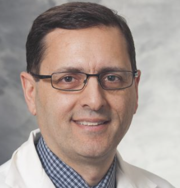 Bermans Iskandar合作论文数Department of Neurological Surgery, School of Medicine and Public Health, University of Wisconsin-Madison8
 Jeffrey P. Greenfield合作论文数Neurological Surgery, Weill Cornell Medicine, Cornell University8
 Hankinson Todd C合作论文数School of Medicine, University of Colorado Anschutz Medical Campus8
 Johnston James M合作论文数8Daniels David J合作论文数8Gross Naina L合作论文数8
 David D Limbrick合作论文数8
 Philipp R Aldana合作论文数Department of Neurosurgery, University of Florida College of Medicine, Jacksonville, FL8
 Ramin Eskandari合作论文数Department of Neurosurgery, University of Utah8
 Gerald Francis Tuite合作论文数Tampa General Hospital8
 Michael Scott Perry合作论文数8David F. Bauer合作论文数8




