Prospective, multi-institutional surgical data collection in pediatric neuro-oncology remains limited despite substantial variation in operative and perioperative management across institutions. To address this, we are developing the NeuroPoint Alliance (NPA) Quality Outcomes Database (QOD) Pediatric Tumor Surgery Registry. Here, we used a modified Delphi process to define a core outcome set for the registry. A modified Delphi study was conducted among pediatric neurosurgeons serving as site principal investigators for the proposed registry. Candidate data elements were rated on a 9-point Likert scale. Consensus for inclusion was predefined as ≥70
Objective: Unicoronal craniosynostosis is the premature fusion of one coronal suture resulting in supra-orbital, midface and skull base asymmetry. While spring-assisted cranioplasty is known to be effective for sagittal craniosynostosis, there is little reported regarding its use in unicoronal synostosis. The purpose of this study is to report a new technique and experience with spring-assisted hinged cranioplasty for unicoronal synostosis. Methods: Demographic data, complications, and hospital course for 5 consecutive patients that underwent spring-assisted cranioplasty for nonsyndromic unicoronal synostosis since 2022 at our institution were reviewed. The surgical technique, which utilizes a fronto-orbital hinged cranioplasty with removal of the fused suture and placement of springs, is described. Results: Five patients were included, with an average age at time of cranioplasty of 6.4 months. There were 3 complications, 1 superficial infection treated with oral antibiotics and 2 patients requiring return to OR for removal of vicryl suture found to be inhibiting spring expansion. Conclusion: We describe a surgical technique utilizing a hinged cranioplasty design for spring-assisted correction of unicoronal synostosis. The theorized mechanism of correction employs dynamic remodeling, which provides some correction for skull base and midface deformities with reduced reliance on caregiver compliance compared to helmet therapy or distraction.
BACKGROUND AND OBJECTIVES: Magnetic resonance-guided laser interstitial thermal therapy (MRgLITT) is an emerging minimally invasive alternative for epileptogenic cerebral cavernous malformations (CCMs). Available evidence is limited to small, single-center series. This multicenter study aimed to evaluate the efficacy and safety of MRgLITT for epileptogenic CCMs (eCCMs) in heterogeneous real-world practice and identify factors that may guide patient selection and technique. METHODS: In this multicenter retrospective cohort study with no overlap to previous series, 23 adult patients with eCCMs underwent MRgLITT at 6 institutions between 2015 and 2023. Seizure outcomes were analyzed for 19 patients with ≥ 1-year follow-up. Patient characteristics and adverse events were reported for all patients. RESULTS: Of 23 patients, 60.9% (14/23) were men, and median age was 50.5 (range 18.0-70.4 years). The median duration from epilepsy diagnosis to treatment was 10 years (range 1-42 years). Lesions were targeted in the left hemisphere in 60.9% (14/23) and to the temporal lobe in 65.2% (15/23). Engel class I seizure freedom was achieved in 57.8% (11/19) at 1 year, all of whom remained seizure free at last follow-up (median 2.2 years, range 1.0-8.6 years). Engel class I-II (favorable) outcomes were achieved in 68.4% (13/19) at 1 year and 73.7% (14/19) at last follow-up. Shorter duration of epilepsy before treatment was associated with seizure freedom (median 8.4 vs 25.5 years, P = .026). Fully concordant presurgical workup was associated with favorable outcome (odds ratio not estimable, 95% CI [0.81, ∞], P = .045) at last follow-up. Perioperative hemorrhage was detected in 3 patients (13.0%), all asymptomatic, with no subsequent hemorrhage during follow-up. Three patients (13.0%) experienced transient neurological deficits. One (4.3%) had a persistent deficit at last follow-up. CONCLUSION: MRgLITT is a promising minimally invasive option for carefully selected patients with eCCMs. Shorter epilepsy duration and concordant presurgical evaluation may predict favorable outcome.
BACKGROUND:In children with Chiari type I malformation and syringomyelia, neurosurgical posterior fossa decompression (PFD) provides clinical improvement, but whether duraplasty (incising the dura and placing a dural graft) improves outcomes is unclear. METHODS:We conducted a multicenter, cluster-randomized, controlled trial of PFD with duraplasty (PFD-D) as compared with PFD alone. Persons 21 years of age or younger with cerebellar tonsillar ectopia of at least 5 mm and a maximum syrinx diameter of 3.0 to 9.9 mm were enrolled at 38 centers. Centers were cluster-randomized: all the participants within each center underwent the same intervention. The primary outcome was surgical complications within 6 months. Secondary outcomes were clinical improvement, syrinx reduction, and repeat decompression at 10 to 24 months and the change in overall health-related quality of life at 6 to 24 months. RESULTS:A total of 162 participants were included in the trial, of whom 78 were assigned to undergo PFD-D and 84 to undergo PFD alone. The percentage of participants with complications within 6 months was 14% with PFD-D and 6% with PFD (adjusted odds ratio, 2.59; 95% confidence interval [CI], 0.86 to 7.84; P = 0.11). At 24 months, the percentage of participants with clinical improvement was 58% with PFD-D and 46% with PFD; the mean (±SD) syrinx reduction was 3.08±2.33 mm and 1.22±1.79 mm, respectively; and the percentage of participants with repeat decompression was 3% and 14%. Changes in health-related quality of life were similar in the two groups. CONCLUSIONS:The percentage of participants with surgical complications did not differ significantly between those who underwent PFD-D and and those who underwent PFD alone. Larger trials are needed to determine the relative benefits and risks of these two procedures. (Funded by the Patient-Centered Outcomes Research Institute and others; ClinicalTrials.gov number, NCT02669836.).
BACKGROUND:Blunt cerebrovascular injury (BCVI), defined as an injury occurring to the carotid and/or vertebral arteries, occurs in ~1% of pediatric blunt trauma patients and is associated with morbidity and mortality. Our objective was to evaluate the sensitivity and specificity of the McGovern score, a pediatric-specific screening tool for BCVI, and describe the effect of its implementation on the use of additional imaging for BCVI and the detection of BCVI. METHODS:This was a retrospective cohort study of patients below 16 years old presenting with blunt trauma to the Pediatric Emergency Department of a tertiary care level 1 pediatric trauma center pre- (July 1, 2020, to November 30, 2021) and post- (December 1, 2021, to December 31, 2022) implementation of McGovern scoring into the clinical decision algorithm for blunt trauma. Patient characteristics, diagnostic studies used [including computed tomography angiography (CTA) or magnetic resonance angiography (MRA) of the neck vessels], and outcomes (BCVI, stroke, mortality), were obtained from the medical record and compared pre-McGovern versus post-McGovern score implementation. RESULTS:A total of 1189 patients were included in the study; 664 p reimplementation of the McGovern scoring and 525 postimplementation. Median age was 6 years (IQR 2 to 11), and 668 (56%) were trauma activations (leveled traumas), with no significant differences in patient characteristics between the 2 cohorts. Imaging for BCVI was performed in 13 (2.0%) patients in the preimplementation group and 27 (5.0%) patients in the postimplementation group ( P =0.003). BCVI was detected in 12/1189 patients overall (1.0%); 2 in the preimplementation group (0.3%), and 10 (1.9%) in the postimplementation group ( P =0.007). In the postimplementation group, the sensitivity of the McGovern score was 90% while the specificity was 96.7%. CONCLUSIONS:The implementation of the McGovern score into the pediatric trauma decision algorithm was associated with the detection of an increased number of BCVIs compared to the preimplementation group, with good sensitivity and specificity, but a significant increase in the use of imaging.
The hippocampus is crucial for forming new episodic memories. While its role in encoding spatial and temporal information (where and when) is well understood, how it encodes objects (what) remains unclear due to the high dimensionality of object space. Rather than encoding each object separately, the hippocampus may encode object categories to reduce complexity. Here, an experimental-modeling approach to investigate how the hippocampus encodes visual memory categories in humans is developed. Spikes are recorded from hippocampal CA3 and CA1 neurons in 24 epilepsy patients performing a delayed match-to-sample task involving five image categories. An interpretable memory decoding model is employed to decode memory categories from hippocampal spiking activity and identify the spatio-temporal characteristics of hippocampal encoding. Using this model, the optimal temporal resolutions for decoding each visual memory category per neuron are estimated. Results indicate that visual memory categories can be decoded from hippocampal spike patterns, supporting the presence of category-specific coding. Hippocampal neuron ensembles encode memory categories in a distributed manner, akin to a population code, while individual neurons use a temporal code. Additionally, CA3 and CA1 neurons exhibit similar and redundant memory category information, likely due to strong and diffuse feedforward synaptic connections from CA3 to CA1 regions.
Magnetic resonance imaging-guided laser interstitial thermal therapy (LITT) has emerged as a minimally invasive alternative to open surgical resection for drug-resistant epilepsy, with increasing adoption in advanced epilepsy centers over the past decade. This review provides a comprehensive overview of LITT applications and outcomes in treating various epilepsy etiologies. Stereotactic laser amygdalohippocampectomy (SLAH) via LITT for temporal lobe epilepsy achieves Engel Class 1 outcomes in 58
OBJECTIVE Patients with a history of surgery for single-suture craniosynostosis (SSC) as an infant often wish to participate in sports later in childhood. However, there are no established guidelines from neurosurgeons and craniofacial surgeons to guide parents in which sports their child should or should not participate. Therefore, this study aimed to evaluate the attitudes and practice patterns of experienced neurosurgeons and craniofacial surgeons regarding the counseling of caregivers of these patients about sports participation. METHODS A survey was administered to neurosurgeons and craniofacial plastic surgeons of the Synostosis Research Group (SynRG), a group of 9 North American institutions, to identify attitudes toward sports participation in patients with past SSC surgery. Survey responses were collected anonymously in REDCap. Questions regarding specific sports participation recommendations for patients who underwent surgery as an infant for SSC with ideal healing and for those who required a delayed cranioplasty were answered. Questions pertained to patients with nonsyndromic SSC without associated Chiari malformation, syrinx, or other intracranial/intraspinal anomalies. RESULTS Overall, 20 surgeons were invited to participate in the survey, with 18 (90%) (9 neurosurgeons and 9 craniofacial plastic surgeons) fully completing it. Only 1 (5.6%) surgeon counseled against any sports participation for patients with ideal healing. If cranioplasty was required, 39%-50% of surgeons counseled against some participation (most commonly restricting football/rugby, boxing, ice hockey, lacrosse, and wrestling), depending on the extent of the cranioplasty. Overall, more plastic surgeons (56%-67%) counseled against sports participation (including lower-contact sports such as baseball/softball, basketball, gymnastics, and soccer) than neurosurgeons (22%-33%) in patients who required cranioplasty. CONCLUSIONS SynRG surgeons generally did not counsel against sports participation (including contact sports) for children with a history of SSC surgery as an infant who had ideal healing. In patients requiring cranioplasty, 39%-50% of surgeons recommended against high-contact sports participation.
The hippocampus is crucial for forming new episodic memories. While the encoding of spatial and temporal information (where and when) in the hippocampus is well understood, the encoding of objects (what) remains less clear due to the high dimensions of object space. Rather than encoding each individual object separately, the hippocampus may instead encode categories of objects to reduce this dimensionality. In this study, we developed and applied a combined experimental-modeling approach to investigate how the hippocampus encodes visual memory categories in humans. We recorded spikes from hippocampal CA3 and CA1 neurons in 24 epilepsy patients performing a visual delayed match-to-sample (DMS) task involving five image categories. An ensemble multi-temporal-resolution classification model was employed to decode these visual memory categories from the hippocampal spiking activity with moderate numbers of trials. This model enables the identification of the spatio-temporal characteristics of hippocampal encoding through its interpretable representations. Using this model, we estimated the optimal temporal resolutions for decoding each visual memory category for each neuron in the ensemble. Results indicate that visual memory categories can be decoded from hippocampal spike patterns despite the short data length, supporting the presence of category-specific coding in the human hippocampus. We found that hippocampal neuron ensembles encode visual memory categories in a distributed manner, akin to a population code, while individual neurons use a temporal code. Additionally, CA3 and CA1 neurons exhibit similar and redundant information regarding visual memory categories, likely due to the strong and diffuse feedforward synaptic connections from the CA3 region to the CA1 region.
Background: Individuals with genetic or nutritional rickets frequently suffer from craniosynostosis. While cranial vault remodeling is most often performed, other approaches, such as distraction osteogenesis and endoscopic suturectomy, are infrequently reported. Here, we discuss the status of the current literature and describe 2 patients with rachitic craniosynostosis, one of whom had previously undergone endoscopic suturectomy, who both successfully underwent distraction osteogenesis. Case description: Two male patients with rickets-associated craniosynostosis, 1 genetic and 1 nutritional, underwent posterior-vault distraction osteogenesis (PVDO). The patient with genetic rickets previously underwent endoscopic-assisted cranial suturectomy of the sagittal suture with barrel-staving at an outside hospital and subsequently re-fused all cranial sutures 19 months postoperatively necessitating reintervention. Complete distraction was confirmed by X-ray and both patients recovered without complication and without re-synostosis at 18 month follow-up. The patient with nutritional rickets required subsequent fronto-facial advancement because of poor midface growth. Conclusion: PVDO effectively treats rachitic craniosynostosis associated with both genetic and nutritional etiologies. Long-term follow up is warranted to monitor for skull-base involvement and associated midface retrusion in these patients.
Traumatic brain injury (TBI) is a leading cause of mortality year after year in the United States and abroad. The impact of life years lost secondary to TBI remains a growing problem with significant health care expenditure year after year. The core tenets of TBI management have evolved over the years but many remain similar to those developed decades ago. Technological advances continue to pave the way for new monitoring techniques in a quest to improve patient outcomes. Continued efforts toward quality research trials are necessary to answer ongoing questions key to improving TBI care. This will require the combined efforts of specialists across many disciplines as well as cooperation of the many patients and families they have treated.
OBJECTIVE As many as 5% of normocephalic children may have a prematurely fused sagittal suture, yet the clinical significance and best course of management of this finding remain unclear. Providers in the Synostosis Research Group were surveyed to create a multicenter consensus on an optimal treatment and monitoring algorithm for this condition. METHODS A four-round modified Delphi method was utilized. The first two rounds consisted of anonymous surveys distributed to 10 neurosurgeons and 9 plastic surgeons with expertise in craniosynostosis across 9 institutions, and presented 3 patients (aged 3 years, 2 years, and 2 months) with incidentally discovered fused sagittal sutures, normal cephalic indices, and no parietal dysmorphology. Surgeons were queried about their preferred term for this entity and how best to manage these patients. Results were synthesized to create a treatment algorithm. The third and fourth feedback rounds consisted of open discussion of the algorithm until no further concerns arose. RESULTS Most surgeons preferred the term "premature fusion of the sagittal suture" (93%). At the conclusion of the final round, all surgeons agreed to not operate on the 3- and 2-year-old patients unless symptoms of intracranial hypertension or papilledema were present. In contrast, 50% preferred to operate on the 2-month-old. However, all agreed to utilize shared decision-making, taking into account any concerns about future head shape and neurodevelopment. Panelists agreed that patients over 18 months of age without signs or symptoms suggesting elevated intracranial pressure (ICP) should not undergo surgical treatment. CONCLUSIONS Through the Delphi method, a consensus regarding management of premature fusion of the sagittal suture was obtained from a panel of North American craniofacial surgeons. Without signs or symptoms of ICP elevation, surgery is not recommended in patients over 18 months of age. However, for children younger than 18 months, surgery should be discussed with caregivers using a shared decision-making process.
ObjectiveHere, we demonstrate the first successful use of static neural stimulation patterns for specific information content. These static patterns were derived by a model that was applied to a subject’s own hippocampal spatiotemporal neural codes for memory.ApproachWe constructed a new model of processes by which the hippocampus encodes specific memory items via spatiotemporal firing of neural ensembles that underlie the successful encoding of targeted content into short-term memory. A memory decoding model (MDM) of hippocampal CA3 and CA1 neural firing was computed which derives a stimulation pattern for CA1 and CA3 neurons to be applied during the encoding (sample) phase of a delayed match-to-sample (DMS) human short-term memory task.Main resultsMDM electrical stimulation delivered to the CA1 and CA3 locations in the hippocampus during the sample phase of DMS trials facilitated memory of images from the DMS task during a delayed recognition (DR) task that also included control images that were not from the DMS task. Across all subjects, the stimulated trials exhibited significant changes in performance in 22.4% of patient and category combinations. Changes in performance were a combination of both increased memory performance and decreased memory performance, with increases in performance occurring at almost 2 to 1 relative to decreases in performance. Across patients with impaired memory that received bilateral stimulation, significant changes in over 37.9% of patient and category combinations was seen with the changes in memory performance show a ratio of increased to decreased performance of over 4 to 1. Modification of memory performance was dependent on whether memory function was intact or impaired, and if stimulation was applied bilaterally or unilaterally, with nearly all increase in performance seen in subjects with impaired memory receiving bilateral stimulation.SignificanceThese results demonstrate that memory encoding in patients with impaired memory function can be facilitated for specific memory content, which offers a stimulation method for a future implantable neural prosthetic to improve human memory.
Background: Spring-assisted surgery (SAS) and cranial vault remodeling (CVR) are widely used surgical techniques to correct sagittal craniosynostosis (SC). The authors evaluated changes in regional morphology of patients with SC who had undergone SAS or CVR, using the frontal bossing index (FBI), occipital bulleting index, vertex narrowing index (VNI), and scaphocephalic severity index (SCI) to capture differences in anterior protrusion, posterior protrusion, width restriction, and global dysmorphology, respectively. Methods: Indices were measured on computed tomography and 3-dimensional photographs (n = 788) of 257 patients with SC from 2001 through 2022 who underwent SAS (n = 177) or CVR (n = 80). Short-term and long-term outcomes were evaluated. Results: Mean age at time of surgery was older in the CVR cohort (CVR, 22.55 ± 16.00 years; SAS, 4.56 ± 2.24 years) (P < 0.05). Before surgery, the SAS cohort had more severe regional dysmorphology in FBI, VNI, and SCI (P < 0.05) relative to the CVR cohort. Frontal bossing consistently improved over time in both cohorts. Head width improved as soon as 0 to 6 months postoperatively (P < 0.05), but it consistently regressed after the initial improvement in both cohorts. Global head shape initially improved in the CVR cohort but regressed over time. In contrast, patients in the SAS group maintained improvements in SCI over time, with significantly better percentage change in every age interval (P < 0.05). Conclusions: The FBI, occipital bulleting index, VNI, and SCI are useful tools for monitoring head shape and growth. SAS and CVR achieve similar morphologic outcomes, despite more severe preoperative morphology in the SAS group. Trending index changes with growth between the 2 cohorts suggest improved longevity of correction in the SAS group. CLINICAL QUESTION/LEVEL OF EVIDENCE: Therapeutic, III.
Spinal cord injury affects the ability of the brain to send and receive signals from the body systems that control sensory, motor, and autonomic function. It remains a significant cause of morbidity and mortality and represents a tremendous financial cost to society. Motor vehicles and falls represent leading causes of spinal cord injuries. The injuries can be complete or incomplete. A careful neurological examination and radiographic assessment can help determine the location of the spinal cord injury. Treatment is dependent on the appropriate diagnosis and involves stabilization of the patient and prevention of secondary injury to the spinal cord.
OBJECTIVE:Congenital anomalies of the atlanto-occipital articulation may be present in patients with Chiari malformation type I (CM-I). However, it is unclear how these anomalies affect the biomechanical stability of the craniovertebral junction (CVJ) and whether they are associated with an increased incidence of occipitocervical fusion (OCF) following posterior fossa decompression (PFD). The objective of this study was to determine the prevalence of condylar hypoplasia and atlas anomalies in children with CM-I and syringomyelia. The authors also investigated the predictive contribution of these anomalies to the occurrence of OCF following PFD (PFD+OCF). METHODS:The authors analyzed the prevalence of condylar hypoplasia and atlas arch anomalies for patients in the Park-Reeves Syringomyelia Research Consortium database who underwent PFD+OCF. Condylar hypoplasia was defined by an atlanto-occipital joint axis angle (AOJAA) ≥ 130°. Atlas assimilation and arch anomalies were identified on presurgical radiographic imaging. This PFD+OCF cohort was compared with a control cohort of patients who underwent PFD alone. The control group was matched to the PFD+OCF cohort according to age, sex, and duration of symptoms at a 2:1 ratio. RESULTS:Clinical features and radiographic atlanto-occipital joint parameters were compared between 19 patients in the PFD+OCF cohort and 38 patients in the PFD-only cohort. Demographic data were not significantly different between cohorts (p > 0.05). The mean AOJAA was significantly higher in the PFD+OCF group than in the PFD group (144° ± 12° vs 127° ± 6°, p < 0.0001). In the PFD+OCF group, atlas assimilation and atlas arch anomalies were identified in 10 (53%) and 5 (26%) patients, respectively. These anomalies were absent (n = 0) in the PFD group (p < 0.001). Multivariate regression analysis identified the following 3 CVJ radiographic variables that were predictive of OCF occurrence after PFD: AOJAA ≥ 130° (p = 0.01), clivoaxial angle < 125° (p = 0.02), and occipital condyle-C2 sagittal vertical alignment (C-C2SVA) ≥ 5 mm (p = 0.01). A predictive model based on these 3 factors accurately predicted OCF following PFD (C-statistic 0.95). CONCLUSIONS:The authors' results indicate that the occipital condyle-atlas joint complex might affect the biomechanical integrity of the CVJ in children with CM-I and syringomyelia. They describe the role of the AOJAA metric as an independent predictive factor for occurrence of OCF following PFD. Preoperative identification of these skeletal abnormalities may be used to guide surgical planning and treatment of patients with complex CM-I and coexistent osseous pathology.
Traumatic brain injury is a disruption in the normal function of the brain due to force that results in alteration of the normal brain function. TBI can be mild, moderate, or severe and can even result in death depending on the extent of damage to the brain. It is a major health concern with an estimated $76.5 billion in direct and indirect costs annually. The primary injury cannot be reversed, so prevention is the best treatment, but early diagnosis and management can help prevent further injury.
Introduction Blunt cerebrovascular injury (BCVI) is an increasingly detected pattern in trauma with significant morbidity, putting patients at risk for subsequent stoke. Complex screening protocols exist to determine who should undergo CT angiography of the neck (CTAN) to evaluate for BCVI. Once identified, stroke incidence may be reduced with appropriate treatment across grades. We hypothesize that an expanded and simplified method for identifying patients with clinical suspicion for BCVI based upon injury above the clavicle (ATC) will illustrate a previously undiagnosed cohort of patients. Methods A single-institution retrospective review of adult (age ≥18 years) blunt trauma patients with BCVI from January 1, 2010 to December 31, 2019 was conducted at a tertiary academic medical center. Patients undergoing CTAN were divided into 2 groups based upon qualification by either the expanded Denver criteria or clinical evidence of any injury ATC. Results A total of 219 patients were diagnosed with BCVI (25 566 blunt trauma admissions, .9% incidence). Seventeen patients (8%) who did not satisfy expanded Denver were diagnosed with BCVI by ATC, most commonly undergoing CTAN due to facial trauma (n = 8). There were no differences in distribution of carotid artery injuries (CAI) and vertebral artery injuries (VAI) in the expanded Denver criteria group compared to the ATC group. Conclusions CTAN for blunt trauma with any injury ATC is an easy-to-use screening tool and may be seamlessly included with initial whole-body imaging.
BACKGROUND:Magnetic resonance-guided laser interstitial thermal therapy (MRgLITT) is a minimally invasive alternative to surgical resection for drug-resistant mesial temporal lobe epilepsy (mTLE). Reported rates of seizure freedom are variable and long-term durability is largely unproven. Anterior temporal lobectomy (ATL) remains an option for patients with MRgLITT treatment failure. However, the safety and efficacy of this staged strategy is unknown. METHODS:This multicentre, retrospective cohort study included 268 patients consecutively treated with mesial temporal MRgLITT at 11 centres between 2012 and 2018. Seizure outcomes and complications of MRgLITT and any subsequent surgery are reported. Predictive value of preoperative variables for seizure outcome was assessed. RESULTS:Engel I seizure freedom was achieved in 55.8% (149/267) at 1 year, 52.5% (126/240) at 2 years and 49.3% (132/268) at the last follow-up ≥1 year (median 47 months). Engel I or II outcomes were achieved in 74.2% (198/267) at 1 year, 75.0% (180/240) at 2 years and 66.0% (177/268) at the last follow-up. Preoperative focal to bilateral tonic-clonic seizures were independently associated with seizure recurrence. Among patients with seizure recurrence, 14/21 (66.7%) became seizure-free after subsequent ATL and 5/10 (50%) after repeat MRgLITT at last follow-up≥1 year. CONCLUSIONS:MRgLITT is a viable treatment with durable outcomes for patients with drug-resistant mTLE evaluated at a comprehensive epilepsy centre. Although seizure freedom rates were lower than reported with ATL, this series represents the early experience of each centre and a heterogeneous cohort. ATL remains a safe and effective treatment for well-selected patients who fail MRgLITT.
 Matthew D. Smyth合作论文数Epilepsy Research Laboratory, University of California at San Francisco12
 Strahle Jennifer M合作论文数Department of Neurosurgery, University of Michigan11Lisa R. David合作论文数10Gautam Popli合作论文数9
 Francesco T. Mangano合作论文数Division of Pediatric Neurosurgery, Cincinnati Children's Hospital Medical Center;Department of Neurosurgery, University of Cincinnati;College of Medicine, University of Cincinnati9
 Chevis Shannon合作论文数American Society for Reproductive Medicine9
 Johnston James M合作论文数9
 David D Limbrick合作论文数9Mark Iantosca合作论文数8Tae Sung Park合作论文数8
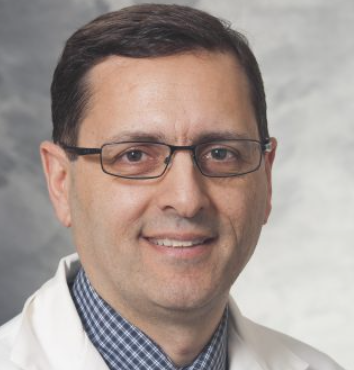 Bermans Iskandar合作论文数Department of Neurological Surgery, School of Medicine and Public Health, University of Wisconsin-Madison8
 Eric S. Jackson合作论文数Department of Communicative Sciences and Disorders, Steinhardt School of Culture, Education, and Human Development, New York University8
 Cormac O. Maher合作论文数Department of Adult Neurosurgery, Stanford University8
 Nathan R Selden合作论文数Dept Neurol Surg, Oregon Hlth & Sci Univ8
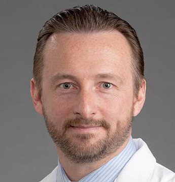 Adrian W. Laxton合作论文数Atrium Health Wake Forest Baptist8
 Stephanie Greene合作论文数8
 Richard G. Ellenbogen合作论文数Department of Radiology, University of Washington;Department of Neurological Surgery, School of Medicine, University of Washington;Department of Global Health, University of Washington8Durham Susan R合作论文数8
 J Gordon Mc Comb合作论文数Keck School of Medicine, University of Southern California8
 P. David Adelson合作论文数Department of Emergency Medicine, College of Medicine - Tucson, University of Arizona;Children's Neuroscience Institute8
 Chern Joshua J合作论文数8John C. Wellons合作论文数Pediatric Neurosurgery Children’s Hospital8Arnold H. Menezes合作论文数8
 Gerald Arthur Grant合作论文数Department of Neurosurgery, School of Medicine, Stanford University;Department of Neurosurgery, Duke University8
 Scellig Stone合作论文数University of Toronto, University Health Network8Daniel J. Guillaume合作论文数8
 William E. Whitehead合作论文数Division of Gastroenterology and Hepatology, Department of Medicine, School of Medicine, University of North Carolina at Chapel Hill;Center for Functional Gastrointestinal Disorders, University of North Carolina at Chapel Hill8
 Robert F Keating合作论文数Department of Neurosurgery, Children’s National Medical Center8
 Douglas L. Brockmeyer合作论文数Division of Pediatric Neurosurgery, Primary Children's Hospital, University of Utah8
 Tord D Alden合作论文数St. Louis Children's Hospital, Washington University School of Medicine8
 Gregory W. Albert合作论文数College of Medicine, University of Arkansas for Medical Sciences8
 Richard C. E. Anderson合作论文数8John Ragheb合作论文数8Jeffrey R. Leonard合作论文数8
 Brent R O'Neill合作论文数8
 Manish N Shah合作论文数7Greg Olavarria合作论文数7
 Jeffrey P. Greenfield合作论文数Neurological Surgery, Weill Cornell Medicine, Cornell University7
 Gregory G. Heuer合作论文数Department of Neurosurgery, Perelman School of Medicine, University of Pennsylvania7
 Hankinson Todd C合作论文数School of Medicine, University of Colorado Anschutz Medical Campus7Daniels David J合作论文数7Gross Naina L合作论文数7Scott D. Wait合作论文数7
 Philipp R Aldana合作论文数Department of Neurosurgery, University of Florida College of Medicine, Jacksonville, FL7
 Ramin Eskandari合作论文数Department of Neurosurgery, University of Utah7
 Gerald Francis Tuite合作论文数Tampa General Hospital7David F. Bauer合作论文数7
 James C. Torner合作论文数Department of Epidemiology, The University of Iowa7Michael S Muhlbauer合作论文数6




