The use of artificial intelligence to process diffraction images is challenged by the need to assemble large and precisely designed training data sets. To address this, a codebase called Resonet was developed for synthesizing diffraction data and training residual neural networks on these data. Here, two per-pattern capabilities of Resonet are demonstrated: (i) interpretation of crystal resolution and (ii) identification of overlapping lattices. Resonet was tested across a compilation of diffraction images from synchrotron experiments and X-ray free-electron laser experiments. Crucially, these models readily execute on graphics processing units and can thus significantly outperform conventional algorithms. While Resonet is currently utilized to provide real-time feedback for macromolecular crystallography users at the Stanford Synchrotron Radiation Lightsource, its simple Python-based interface makes it easy to embed in other processing frameworks. This work highlights the utility of physics-based simulation for training deep neural networks and lays the groundwork for the development of additional models to enhance diffraction collection and analysis.
Protein crystallographers value rapid feedback during beamtime for guiding experiments. Commonly, a human visually scans diffraction images for signs of crystal quality. Recently, we have developed a framework for replacing this human interpreter: we have built models whose inputs are diffraction images, and whose outputs are parameters of interest. Notably, these parameters include crystal resolution/B - factor, and the presence of multi -lattice scatteri ng, determined on a per-shot basis. Most importantly, we train the models using simulated diffraction images, providing us full control over the space of learnable parameters. The simulations, however, are sufficiently realistic such that the trained models p erform well when tested on real user data. These diffraction AI models, which we collectively call XRAI, can easily be incorporated at different user facilities, due to their simplicity, requiring a single image array as input. During the talk, we will discuss the development of these models, from their underlying architectures to the simulation software used to create their training data. We will also explore how XRAI is incorporated in the Structural and Molecular Biology bea mlines at the Stanford Sync hrotron, and how other beamline groups might tune the models to their specific needs.
Hydrogenases display a wide range of catalytic rates and biases in reversible hydrogen gas oxidation catalysis. The interactions of the iron-sulfur-containing catalytic site with the local protein environment are thought to contribute to differences in catalytic reactivity, but this has not been demonstrated. The microbe Clostridium pasteurianum produces three [FeFe]-hydrogenases that differ in catalytic bias by exerting a disproportionate rate acceleration in one direction or the other that spans a remarkable 6 orders of magnitude. The combination of high-resolution structural work, biochemical analyses, and computational modeling indicates that protein secondary interactions directly influence the relative stabilization/destabilization of different oxidation states of the active site metal cluster. This selective stabilization or destabilization of oxidation states can preferentially promote hydrogen oxidation or proton reduction and represents a simple yet elegant model by which a protein catalytic site can confer catalytic bias.
The crystal structure of the trans-acyltransferase (AT) from the disorazole polyketide synthase (PKS) was determined at room temperature to a resolution of 2.5 Å using a new method for the direct delivery of the sample into an X-ray free-electron laser. A novel sample extractor efficiently delivered limited quantities of microcrystals directly from the native crystallization solution into the X-ray beam at room temperature. The AT structure revealed important catalytic features of this core PKS enzyme, including the occurrence of conformational changes around the active site. The implications of these conformational changes for polyketide synthase reaction dynamics are discussed.
The Stanford Automated Mounter System, a system for mounting and dismounting cryo-cooled crystals, has been upgraded to increase the throughput of samples on the macromolecular crystallography beamlines at the Stanford Synchrotron Radiation Lightsource. This upgrade speeds up robot maneuvers, reduces the heating/drying cycles, pre-fetches samples and adds an air-knife to remove frost from the gripper arms. Sample pin exchange during automated crystal quality screening now takes about 25 s, five times faster than before this upgrade.
Significance A major problem in determining the crystal structures of metalloenzymes is that the reducing power of X-rays often changes the oxidation state of the metal center, thereby complicating important mechanistic conclusions on enzyme function. This reduction is especially problematic in studying Fe(IV)=O intermediates, which are powerful oxidants used by many metalloenzymes. This problem can be circumvented using the Stanford Linear Coherent Light Source (LCLS), which generates intense X-ray pulses on the femtosecond time scale and enables structure determinations with no reduction of metal centers. Here, we report the crystal structure of the Fe(IV)=O peroxidase intermediate called compound I using data obtained from the LCLS. We also present kinetic and computational results that, together with crystal structures, provide important mechanistic insights.
Higher throughput methods to mount and collect data from multiple small and radiation-sensitive crystals are important to support challenging structural investigations using microfocus synchrotron beamlines. Furthermore, efficient sample-delivery methods are essential to carry out productive femtosecond crystallography experiments at X-ray free-electron laser (XFEL) sources such as the Linac Coherent Light Source (LCLS). To address these needs, a high-density sample grid useful as a scaffold for both crystal growth and diffraction data collection has been developed and utilized for efficient goniometer-based sample delivery at synchrotron and XFEL sources. A single grid contains 75 mounting ports and fits inside an SSRL cassette or uni-puck storage container. The use of grids with an SSRL cassette expands the cassette capacity up to 7200 samples. Grids may also be covered with a polymer film or sleeve for efficient room-temperature data collection from multiple samples. New automated routines have been incorporated into the Blu-Ice/DCSS experimental control system to support grids, including semi-automated grid alignment, fully automated positioning of grid ports, rastering and automated data collection. Specialized tools have been developed to support crystallization experiments on grids, including a universal adaptor, which allows grids to be filled by commercial liquid-handling robots, as well as incubation chambers, which support vapor-diffusion and lipidic cubic phase crystallization experiments. Experiments in which crystals were loaded into grids or grown on grids using liquid-handling robots and incubation chambers are described. Crystals were screened at LCLS-XPP and SSRL BL12-2 at room temperature and cryogenic temperatures.
Determining the interconverting conformations of dynamic proteins in atomic detail is a major challenge for structural biology. Conformational heterogeneity in the active site of the dynamic enzyme cyclophilin A (CypA) has been previously linked to its catalytic function, but the extent to which the different conformations of these residues are correlated is unclear. Here we compare the conformational ensembles of CypA by multitemperature synchrotron crystallography and fixed-target X-ray free-electron laser (XFEL) crystallography. The diffraction-before-destruction nature of XFEL experiments provides a radiation-damage-free view of the functionally important alternative conformations of CypA, confirming earlier synchrotron-based results. We monitored the temperature dependences of these alternative conformations with eight synchrotron datasets spanning 100-310 K. Multiconformer models show that many alternative conformations in CypA are populated only at 240 K and above, yet others remain populated or become populated at 180 K and below. These results point to a complex evolution of conformational heterogeneity between 180-–240 K that involves both thermal deactivation and solvent-driven arrest of protein motions in the crystal. The lack of a single shared conformational response to temperature within the dynamic active-site network provides evidence for a conformation shuffling model, in which exchange between rotamer states of a large aromatic ring in the middle of the network shifts the conformational ensemble for the other residues in the network. Together, our multitemperature analyses and XFEL data motivate a new generation of temperature- and time-resolved experiments to structurally characterize the dynamic underpinnings of protein function.
Significance The extremely short and bright X-ray pulses produced by X-ray free-electron lasers unlock new opportunities in crystallography-based structural biology research. Efficient methods to deliver crystalline material are necessary due to damage or destruction of the crystal by the X-ray pulse. Crystals for the first experiments were 5 µm or smaller in size, delivered by a liquid injector. We describe a highly automated goniometer-based approach, compatible with crystals of larger and varied sizes, and accessible at cryogenic or ambient temperatures. These methods, coupled with improvements in data-processing algorithms, have resulted in high-resolution structures, unadulterated by the effects of radiation exposure, from only 100 to 1,000 diffraction images.
AutoDrug is software based upon the scientific workflow paradigm that integrates the Stanford Synchrotron Radiation Lightsource macromolecular crystallography beamlines and third-party processing software to automate the crystallography steps of the fragment-based drug-discovery process. AutoDrug screens a cassette of fragment-soaked crystals, selects crystals for data collection based on screening results and user-specified criteria and determines optimal data-collection strategies. It then collects and processes diffraction data, performs molecular replacement using provided models and detects electron density that is likely to arise from bound fragments. All processes are fully automated, i.e. are performed without user interaction or supervision. Samples can be screened in groups corresponding to particular proteins, crystal forms and/or soaking conditions. A single AutoDrug run is only limited by the capacity of the sample-storage dewar at the beamline: currently 288 samples. AutoDrug was developed in conjunction with RestFlow, a new scientific workflow-automation framework. RestFlow simplifies the design of AutoDrug by managing the flow of data and the organization of results and by orchestrating the execution of computational pipeline steps. It also simplifies the execution and interaction of third-party programs and the beamline-control system. Modeling AutoDrug as a scientific workflow enables multiple variants that meet the requirements of different user groups to be developed and supported. A workflow tailored to mimic the crystallography stages comprising the drug-discovery pipeline of CoCrystal Discovery Inc. has been deployed and successfully demonstrated. This workflow was run once on the same 96 samples that the group had examined manually and the workflow cycled successfully through all of the samples, collected data from the same samples that were selected manually and located the same peaks of unmodeled density in the resulting difference Fourier maps.
Structure-property investigation of crystalline amino acids is an important challenge since interactions between individual molecular fragments or even structural domains in the structure can simulate interactions in more complicated biological systems such as proteins and peptides.Besides, crystalline amino acids are applied as drugs, as piezoelectric and nonlinear optical materials.Therefore understanding a crystal structure response to variation in temperature and pressure is significant in such applications.Cysteine is a remarkable amino acid because its side-chain residue contains a sulfhydryl group involved in formation of additional labile hydrogen bonds (S-H…S or S-H…O).The presence of these very weak bonds in the structure allows cysteine to take a peculiar place between hydrophobic (no contribution of side-chains to H-bonds) and hydrophilic amino acids (with that contribution).In the present contribution we discuss an evolution of chiral and racemic cysteine crystal structures on cooling and on increasing pressure followed by X-ray crystallography and Raman spectroscopy.We also compare their behavior with that of cysteine crystalline salts and derivatives.The study was supported by the Projects of RAS (21.44, 5.6.4),SB RAS (Projects 13 & 109), grants from RFBR (09-03-00451, 10-03-00252), a BRHE grant from the CRDF (RUX0-008-NO-06) and FASI (RF) Contracts No GK P2529 & 16.740.11.0166.
Complete automation of the macromolecular crystallography experiment has been achieved at SSRL through the combination of robust mechanized experimental hardware and a flexible control system with an intuitive user interface. These highly reliable systems have enabled crystallography experiments to be carried out from the researchers' home institutions and other remote locations while retaining complete control over even the most challenging systems. A breakthrough component of the system, the Stanford Auto-Mounter (SAM), has enabled the efficient mounting of cryocooled samples without human intervention. Taking advantage of this automation, researchers have successfully screened more than 200 000 samples to select the crystals with the best diffraction quality for data collection as well as to determine optimal crystallization and cryocooling conditions. These systems, which have been deployed on all SSRL macromolecular crystallography beamlines and several beamlines worldwide, are used by more than 80 research groups in remote locations, establishing a new paradigm for macromolecular crystallography experimentation.
C486consisting of predominantly -Ti (P6 3 /mmc) [1].The Ti-6Al-4V rod was machined using both conventional and high-pressure jet-assisted methods.The depth profile of residual stress was measured using xray diffraction.It was found that the compress residual stress is higher and the deeper under which the compress residual stress exists, for sample cut by high-pressure jet-assisted than for sample cut by conventional method [2].Using transmission electron microscopy the cross-section of the surface layer was found to consist of a thin outer layer with nano-sized crystals ( 10 nm) and the substrate of large grains with very high density of dislocations.Electron diffraction reveals that the nano-sized outer layer is highly textured.Furthermore, the study shows that the nano-sized layer has twice the thickness for the high-pressure jetassisted cut sample ( 1,000 nm) than for the conventionally cut sample ( 500 nm).This shows that high-pressure jet-assisted cutting resulted in a thick and highly modified outer layer and provides an explanation for the large and deep compressed residual stress after high-pressure jet-assisted cutting of Ti-6Al-4V.
Since June 2005, the macromolecular crystallography users of the Stanford Synchrotron Radiation Laboratory (SSRL) have had the option to conduct diffraction experiments from their home institutions and other remote locations by means of advanced software tools that enable network-based control of highly automated beam lines. Remote experimenters have access to the same tools as local users, and have the capability to mount, center, and screen crystalline samples, and to collect, analyze, and backup diffraction data. Automated sample mounting is accomplished with the Stanford Auto-Mounting System (SAM) [1–3 Cohen, A. E., McPhillips, S. E., Song, J., Miller, M. D. and for the SSRL SMB and JCSG Groups. 2005. Automation of High-Throughput Protein Crystal Screening at SSRL. Synch. Rad. News, 18: 28–35. Van den Bedem, H., Miller, M. D., Wolf, G. and for the SSRL SMB and JCSG Groups. 2003. Towards Automated Data Collection at the Stanford Synchrotron Radiation Laboratory. Synch. Rad. News, 16: 15 Cohen, A. E., Ellis, P. J., Miller, M. D., Deacon, A. M. and Phizackerley, R. P. 2002. An Automated System to Mount Cryo-Cooled Protein Crystals on a Synchrotron Beamline, Using Compact Sample Cassettes and a Small-Scale Robot. J. Appl. Cryst., 35: 720–726. ], beamline and experimental control is carried out using Blu-Ice/DCS [4 McPhillips, T. M., McPhillips, S. E., Chiu, H. -J., Cohen, A. E., Deacon, A. M., Ellis, P. J., Garman, E., González, A., Sauter, N. K., Phizackerley, R. P., Soltis, S. M and Kuhn, P. 2002. Blu-Ice and the Distributed Control System: Software for Data Acquisition and Instrument Control at Macromolecular Crystallography Beamlines. J. Synchrotron Rad., 9: 401–406. [Crossref], [PubMed], [Web of Science ®] , [Google Scholar]], and additional remote monitoring of the experiment and data backup is supported with several web-based applications [5 Eriksson, T., Chiu, H.-J., Sharp, K., McPhillips, T., McPhillips, S., Sauter, N., Soltis, M. and Kuhn, P. 2002. Collaboratory for Macromolecular Crystallography at SSRL. Acta Cryst., A58(Supplement): C73 [Google Scholar]]. The highly graphical applications and computational resources at SSRL are accessed through a client/server application that uses minimal resources on the client side and has a typical response close to that obtained at the beamline.
Macromolecular crystallography has proven to be the most effective method of determining the structures of biological macromolecules.Each year thousands of data collections that form the basis for such structure determinations are carried out at the ESRF.To cope with this high demand, it is essential to maximise beamline efficiency and to simplify beamline control procedures.The ESRF, in collaboration with the EMBL Grenoble Outstation, are therefore automating much of the crystallographic experiment which may be divided into two parts : beamline and crystal alignment, and data collection and processing.Recent progress in automatic crystal mounting and centering [1], crystal characterisation [2], absorption edge scanning and automated alignment procedures for the beamline optical elements will be described, as will the development of databases designed to aid the automation process.
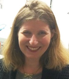 Aina E. Cohen合作论文数Stanford Synchrotron Radiation Laboratory14
 Jinhu Song合作论文数12Soltis S Michael合作论文数11
 Ana M Gonzalez合作论文数Cátedra de Microbiología General;Facultad de Bioquímica y Ciencias Biológicas;Universidad Nacional del Litoral;Facultad de Bioquímica y Ciencias Biológicas, Universidad Nacional del Litoral7
 Artem Y Lyubimov合作论文数6Irimpan I. Mathews合作论文数5
 Nicholas Sauter合作论文数5Penjit Moorhead合作论文数4Tsai Yingssu合作论文数4
 Elizabeth L Baxter合作论文数4Hollenbeck Michael合作论文数4
 James Holton合作论文数University of California, San Francisco;Molecular Biophysics and Integrated Bioimaging Division, A U.S. Department of Energy National Laboratory, University of California4
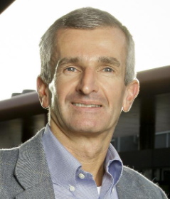 Axel Brunger合作论文数Department of Molecular and Cellular Physiology, Stanford University4
 Timothy M. Mcphillips合作论文数Genome Center, University of California Davis4Thomas Eriksson合作论文数4
 Clyde A. Smith合作论文数School of Biological Sciences, University of Auckland4
 Roberto Alonso-Mori合作论文数SLAC National Accelerator Laboratory, Stanford University;Biochemistry and Condensed Phase Chemistry Group, Chemical Sciences Department, Stanford University3
 Monarin Uervirojnangkoorn合作论文数3
 William Weis合作论文数School of Medicine, Stanford University3Mitchell D. Miller合作论文数Department of BioSciences, Rice University3
 Henrik Till Lemke合作论文数Laboratory for Condensed Matter, Center for Photon Science, Paul Scherrer Institute3Mike Soltis合作论文数2hsui chui合作论文数2
 Christopher Barnes合作论文数CALTECH, Div Biol & Biol Engn, Pasadena, CA 91125 USA2Keable Stephen合作论文数2
 Aashish Manglik合作论文数Department of Pharmaceutical Chemistry, School of Pharmacy, University of California San Francisco;Department of Anesthesia, School of Pharmacy, University of California San Francisco2
 Aaron S. Brewster合作论文数Physical Biosciences Division, Lawrence Berkeley National Laboratory;Physical Biosciences Division, Lawrence Berkeley National Laboratory2Dunten Pete W合作论文数2
 Oleg A Zadvornyy合作论文数Department of Chemistry and Biochemistry, Montana State University2Aguila Laura合作论文数2
 Woldeyes Rahel A合作论文数Chemistry and Chemical Biology Graduate Program, University of California2Mendez Derek合作论文数Department of Applied Physics2
 Henry Van Den Bedem合作论文数2Ashley M. Deacon合作论文数2
 Keith Hodgson合作论文数SLAC National Accelerator Laboratory, School of Humanities and Sciences, Stanford University2
 Britt Hedman合作论文数Stanford Synchrotron Radiation Laboratory, Stanford University, SLAC, M.S. 69, P.O. Box 4349, Stanford, CA 94309 USA2
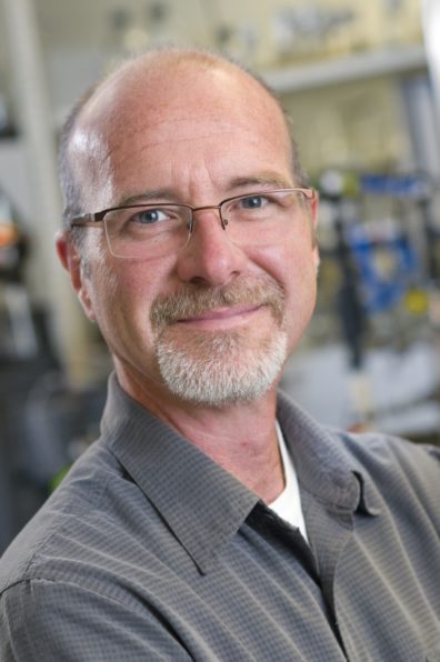 John W. Peters合作论文数Department of Chemistry and Biochemistry, Dodge Family College of Arts and Sciences, University of Oklahoma2
 Brian Kobilka合作论文数Department of Molecular and Cellular Physiology, School of Medicine, Stanford University2Guillermo Calero合作论文数2
 R. Paul Phizackerley合作论文数SLAC Faculty, Stanford University2
 James S. Fraser合作论文数Department of Molecular and Medical Genetics, University of Toronto2Irina Tsyba合作论文数2Guenter Wolf合作论文数2
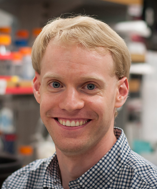 Andrew Kruse合作论文数The Department of Biological Chemistry and Molecular Pharmacology, Harvard Medical School2Paul J. Ellis合作论文数1S. Garrett Williams合作论文数1Neeraj Kumar合作论文数1
 Mohamed Ibrahim合作论文数1Julia Narevicius合作论文数1




