Glycation is a non-enzymatic process where reducing sugars react with the free amino groups of proteins, a phenomenon that occurs under hyperglycemic conditions such as diabetes. Ajwa dates are widely consumed for their health benefits, but limited studies have investigated their potential to inhibit glycation. In this study, we evaluate the antiglycation effects of Ajwa date pulp extracts (ADEs) using the glyoxal (GO)-catalase (CAT) glycation model. Phytochemical analysis revealed that the dry ADE contains significantly higher total phenolic content (2619 ± 121 mg GAE/100 g dry weight [DW] [p < 0.0001]) compared to fresh Ajwa dates. Moreover, the acid hydrolysis method proved more effective for extracting bound phenolic acids (1035 ± 353 mg/100 g DW) than the alkaline hydrolysis method (446 ± 18 mg/100 g DW) from dry Ajwa dates. Using ultrahigh-performance liquid chromatography with a photodiode array detector, p-coumaric and ferulic acid were identified as the primary polyphenols in the ADE. The study demonstrated that GO-induced CAT glycation and the resulting advanced glycation end-products (AGEs) were significantly inhibited by ADE, as measured by AGE-specific fluorescence. While glycation caused conformational changes in CAT, ADE treatment effectively reduced these alterations, as observed through ultraviolet-visible absorption, circular dichroism, and tryptophan spectroscopies. Molecular docking analysis showed that Ajwa date constituent; caffeic acid, p-coumaric acid and ferulic acid had estimated binding affinities of 6.9, 7.1 and 6.1 kcal/mol, respectively with CAT. Multiple hydrogen bonds were seen between these phenolic acids and amino acid residues in addition to other weak interactions. In conclusion, this study highlights the preventive effects of ADE against glycation, offering potential pathways for addressing diabetes-related complications with increased efficacy, selectivity and safety in humans.
L-asparaginase is a remarkable antineoplastic enzyme used in medicine for the treatment of acute lymphoblastic leukemia (ALL) as well as in food industries. In this work, the L-asparaginase-II gene from Salmonella paratyphi was codon-optimized, cloned, and expressed in E. coli as a His-tag fusion protein. Then, using a two-step chromatographic procedure it was purified to homogeneity as confirmed by SDS-PAGE, which also showed its monomeric molecular weight to be 37 kDa. This recombinant L-asparaginase II from Salmonella paratyphi (recSalA) was optimally active at pH 7.0 and 40 degrees C temperature. It was highly specific for L-asparagine as a substrate, while its glutaminase activity was low. The specific activity was found to be 197 U/mg and the kinetics elements K-m, V-max, and k(cat) were determined to be 21 mM, 28 mu M/min, and 39.6 S-1, respectively. Thermal stability was assessed using a spectrofluorometer and showed T-m value of 45 degrees C. The in-vitro effects of recombinant asparaginase on three different human cancerous cell lines (MCF7, A549 and Hep-2) by MTT assay showed remarkable anti-proliferative activity. Moreover, recSalA exhibited significant morphological changes in cancer cells and IC50 values ranged from 28 to 45.5 mu g/ml for tested cell lines. To investigate the binding mechanism of SalA, both substrates L-asparagine and l-glutamine were docked with the protein and the binding energy was calculated to be -4.2 kcal mol(-1) and - 4.4 kcal mol(-1), respectively. In summary, recSalA has significant efficacy as an anticancer agent with potential implications in oncology while its in-vivo validation needs further investigation.
Hexokinases (HKs) convert hexose sugars to hexose-6-phosphate, thus trapping them inside cells to meet the synthetic and energetic demands. HKs participate in various standard and altered physiological processes, including cancer, primarily through the reprogramming of cellular metabolism. Four canonical HKs have been identified with different expression patterns across tissues. HKs 1–3 play a role in glucose utilization, whereas HK 4 (glucokinase, GCK) also acts as a glucose sensor. Recently, a novel fifth HK, hexokinase domain containing 1 (HKDC1), has been identified, which plays a role in whole-body glucose utilization and insulin sensitivity. Beyond the metabolic functions, HKDC1 is differentially expressed in many forms of human cancer. This review focuses on the role of HKs, particularly HKDC1, in metabolic reprogramming and cancer progression.
Glycation is the non-enzymatic adduct formation between reducing sugars or dicarbonyls with proteins and is a crucial molecular event under hyperglycaemic conditions of diabetes. The accumulation of advanced glycation end products (AGEs) due to glycation of proteins has been implicated in several diseases associated with ageing and diabetes. Thus, investigating the antiglycation potential of some trace metal ions (Manganese; Mn2+, and Zinc; Zn2+) and polyphenolic extract of chickpea seeds (PEC) on the methylglyoxal (MGO) induced glycation of a phytocystatin isolated from chickpea was taken up to find an inexpensive and non-toxic therapeutic means of medicating protein glycation and associated diabetic complications. The current study focused on the comparative analyses of these micronutrients and herbal extracts in inhibiting protein glycation and AGEs formation in a quest to develop nutraceuticals for managing diabetes. The effect of metals (Mn2+, Zn2+) and PEC on protein glycation was assessed by different techniques, i.e., glycation-specific AGE fluorescence and absorbance, thiol protease inhibitory activity assay, and conformational alterations by spectroscopic assays. This study revealed the significant anti-glycation potencies of Mn2+, Zn2+, and PEC against the MGO-induced glycation of CPC, which might pave the way for resolving pathological complications of diabetes by combining higher levels of efficacy, selectivity, and safety in humans. Moreover, characterization and identification of different AGEs formed during the glycation process in diabetics was done to apply the same for determining the onset of glycation at the early stage so that appropriate steps be taken to address the menace of diabetic complications.
Hepatocellular carcinoma (HCC) is the primary form of liver cancer. It causes ∼ 800 000 deaths per year, which is expected to increase due to increasing rates of obesity and metabolic dysfunction associated steatotic liver disease (MASLD). Current therapies include immune checkpoint inhibitors, tyrosine kinase inhibitors, and monoclonal antibodies, but these therapies are not satisfactorily effective and often come with multiple side effects and recurrences. Metabolic reprogramming plays a significant role in HCC progression and is often conserved between tumor types. Thus, targeting rewired metabolic pathways could provide an attractive option for targeting tumor cells alone or in conjunction with existing treatments. Therefore, there is an urgent need to identify novel targets involved in cancer-mediated metabolic reprogramming in HCC. In this review, we provide an overview of molecular rewiring and metabolic reprogramming of glucose metabolism in HCC to understand better the concepts that might widen the therapeutic window against this deadly cancer.
Objective: To isolate and characterize a novel phytocystatin from walnut and investigate it for biopesticide development. Methods: A battery of methodology was employed. Initially, phytocystatin was extracted and purified from walnut using ammonium sulfate saturation (60-80%), followed by gel filtration chromatography on the Sephacryl S-100 HR column. Further characterization studies including pH and temperature stability, molecular weight, secondary structure, protease inhibitory assay and antimicrobial activity were carried using various techniques viz: spectroscopy, electrophoresis, and circular dichroism (CD) techniques. Results: Thiol protease inhibitor from walnut (WCPI) was isolated and purified with high (71.4%) yield and 184-fold purification. The molecular weight of the purified inhibitor was found to be around 11.2 kDa. Kinetic tests revealed that the inhibitor competitively inhibited papain and other cysteine proteases such as ficin and bromelain. It also exhibited significant antimicrobial activity against bacterial species. Conclusion: Walnut cysteine protease inhibitor (WCPI) from Juglans regia (Kashmiri walnut) was isolated to homogeneity and had features of other members of the phytocystatin family. It demonstrated potential antimicrobial activity and may serve as an initial step towards developing enhanced pest control methods based on natural molecules. (C) 2022 The Authors. Published by Elsevier B.V. on behalf of King Saud University.
Methylglyoxal (MG) is a potent glycating agent which reacts with proteins to form advanced glycation end products (AGEs). These chemically stable AGEs crosslink with proteins and could lead to amyloid formation that has the role in several diseases including Alzheimer's and Parkinson's. In this piece of work, glycation-induced conformational changes in HSA were observed with quenching of tryptophan fluorescence by 73.8% (41 nm red shift) and loss of hydrophobicity of HSA. CD spectroscopy result reaffirmed secondary structure changes in HSA. Moreover, MG-induced changes in HSA, proceeds to amyloid structure as characterized by an increase in thioflavin (ThT) fluorescence and transmission electron microscopy (TEM) images of HSA aggregates. Quercetin was found to inhibit both AGEs production and amyloid formation. Viability of MCF-7 cells was found to be increased with AGEs treatment, illustrating proliferation of cancer cells. Wound healing assay also revealed increased proliferation and migration of cells in the presence of AGEs. Additionally, molecular docking analyses were performed to demonstrate interactions involved in the stabilization of HSA-quercetin complex. The binding affinities of quercetin were found to be (K-d = 10(5) M-1) much higher compared with MG (K-d = 10(2) M-1). From this study, it is quite clear that quercetin reverses the effect of MG by sterically inhibiting the interaction between HSA and MG. Communicated by Ramaswamy H. Sarma
The continuous loss of human life due to the paucity of effective drugs against different forms of cancer demands a better/noble therapeutic approach. One possible way could be the use of nanostructures-based treatment methods. In the current piece of work, we have synthesized silver nanoparticles (AgNPs) using plant (Heliotropiumbacciferum) extract using AgNO3 as starting materials. The size, shape, and structure of synthesized AgNPs were confirmed by various spectroscopy and microscopic techniques. The average size of biosynthesized AgNPs was found to be in the range of 15 nm. The anticancer potential of these AgNPs was evaluated by a battery of tests such as MTT, scratch, and comet assays in breast (MCF-7) and colorectal (HCT-116) cancer models. The toxicity of AgNPs towards cancer cells was confirmed by the expression pattern of apoptotic (p53, Bax, caspase-3) and antiapoptotic (BCl-2) genes by RT-PCR. The cell viability assay showed an IC50 value of 5.44 and 9.54 µg/mL for AgNPs in MCF-7 and HCT-116 cell lines respectively. We also observed cell migration inhibiting potential of AgNPs in a concentration-dependent manner in MCF-7 cell lines. A tremendous rise (150–250%) in the production of ROS was observed as a result of AgNPs treatment compared with control. Moreover, the RT-PCR results indicated the difference in expression levels of pro/antiapoptotic proteins in both cancer cells. All these results indicate that cell death observed by us is mediated by ROS production, which might have altered the cellular redox status. Collectively, we report the antimetastasis potential of biogenic synthesized AgNPs against breast and colorectal cancers. The biogenic synthesis of AgNPs seems to be a promising anticancer therapy with greater efficacy against the studied cell lines.
Protein aggregation leads to vast conformational changes and plays a key role in the pathogenesis of various neurodegenerative diseases including Alzheimer's and Parkinson's. In the current piece of work, we have explored the interaction of quinoline yellow(QY) with myoglobin (Mb) at two different pH (3.5 and 7.4). Various spectroscopic techniques such as turbidity, Rayleigh light scattering (RLS), UV-Vis absorbance, fluorescence resonance energy transfer (FRET), far UV-CD along with transmission electron microscopy (TEM) and molecular docking have been utilized to characterize dye-induced aggregation in Mb. Binding results showed that interaction between QY and myoglobin is spontaneous and static in nature with high K-SV value of 2.14 x 10(4) M-1. On the other hand, thermodynamics studies (Delta H & Delta S) revealed that complex formation was driven by hydrogen and Van der Walls forces. Molecular docking analysis showed strong binding affinity (K-d = 4.95 x 10(4) M-1) between QY and Mb at Pro100, Ile101, Lys102, Glu105, Glu136, Arg139, Lys140, and Ala143 residues. The intrinsic fluorescence and circular dichroism studies indicated that QY induced conformational changes in Mb at pH 3.5. Turbidity and RLS studies showed aggregation of Mb in the presence of QY (0.2-5 mM). Moreover, kinetics data revealed nucleation independent aggregation of myoglobin in the presence of QY. TEM analysis further established amorphous nature of Mb aggregate induced by QY. At pH (7.4), QY was unable to induce aggregation in myoglobin; it might be due to repulsive nature of negatively charged dye and myoglobin or partially altered states of protein could be pre-requisite for binding and aggregation. (C) 2019 Published by Elsevier B.V.
Hyperglycaemic conditions facilitate the glycation of serum proteins which may have predisposition to aggregation and thus lead to complications. The current study investigates the glycation induced structural and functional modifications of chickpea cystatin (CPC) as well as biological toxicity of the modified protein forms, using CPC-glucose as a model system. Several structural intermediates were formed during the incubation of CPC with glucose (day 4, 8, 12, & 16) as revealed by circular dichroism (CD), altered intrinsic fluorescence, and high ANS binding. Further incubation of CPC with glucose (day 21) formed abundant β structures as revealed by Fourier transform infrared spectroscopy and CD analysis which may be due to the aggregation of protein. High thioflavin T fluorescence intensity and increased Congo red absorbance together with enhanced turbidity and Rayleigh scattering by this modified form confirmed the aggregation. Electron microscopy finally provided the valid physical authentication about the presence of aggregate structures. Functional inactivation of glucose incubated CPC was also observed with time. Single cell electrophoresis of lymphocytes and plasmid nicking assays in the presence of modified CPC showed the DNA damage which confirmed its biological toxicity. Hence, our study suggests that glycation of CPC not only leads to structural and functional alterations in proteins but also to biotoxic AGEs and aggregates.
Reactive dicarbonyl species such as methylglyoxal (MGO) and glyoxal (GO) have recently received extensive attention due to their high reactivity and ability to modify biological substances such as proteins, phospholipids, and DNA. In case of proteins these reactive species mainly react with lysine and arginine residues to form AGEs, oxidative products, and aggregates. Chickpea cystatin (CPC) was incubated with varying concentrations of glyoxal and methylglyoxal which caused, along with altered secondary and tertiary structures, glycation, functional inactivation, altered redox state, cross-linking and high-molecular-mass aggregation. All these processes were examined and characterized by UV-Vis, fluorescence, and CD spectroscopies. Further characterization of CPC modified by reactive dicarbonyls was done by polyacrylamide gel electrophoresis which also showed alterations in the CPC molecules. Thus, in addition to describing the effects of GO and MGO on structure, conformation and function of CPC, this study also shows the relatively superior modifying effect of methylglyoxal for CPC in terms of glycation, oxidation and aggregation. This model system could shed some more light on the role of the reactive dicarbonyls in the specific alterations of proteins with different biological consequences having implications to ageing and disease such as diabetes.
Amyloid fibrils are highly ordered protein assemblies known to contribute to the pathology of a variety of genetic and aging-associated diseases. Here, we have investigated the aggregation propensity of lysozyme in the presence of a negatively charged surfactant (SDS) and evaluated the anti-aggregation activity of rutin. Multiple approaches such as turbidity measurements, dye binding assays, intrinsic fluorescence, circular dichroism (CD), transmission electron microscopy (TEM), MTT and comet assays have been used for this purpose. We inferred that SDS induces aggregation of lysozyme in 0.2-0.6 mM concentration range while at higher concentration range (0.8-1.0 mM), it leads to solubilization/stabilization of protein. Intrinsic/extrinsic fluorescence and CD analysis confirmed significant conformational changes in lysozyme at 0.2 mM SDS. Thioflavin T (ThT), congo red binding and TEM analysis further reaffirmed the formation of lysozyme fibrils. Moreover, MTT assay demonstrated cytotoxicity of these fibrils towards neuroblastoma cell lines (SH-SY5Y) and their attenuation by rutin. Comet assay supported the cytotoxicity mechanism via DNA damage. Molecular docking results also advocate a strong interaction between lysozyme and rutin. The current study indicates a mechanistic approach assuming structural constraints and specific aromatic interactions of rutin with HEWL aggregates. (C) 2018 Published by Elsevier B.V.
Several mammalian proteins form pathological deposits under nonphysiological conditions that are associated with many degenerative diseases. Protein aggregation is associated with aging, as well as a variety of diseases, including cystic fibrosis, amyotrophic lateral sclerosis (ALS), and hypertrophic cardiomyopathy. There is a lack of any potential anti-amyloidogenic agents and therapeutics till date. Polyphenols have been accredited with myriad biological effects. An analysis of the effects of natural agents like baicalin (BC) and gallocatechin (GC) on aggregation process can open new avenues for the treatment of protein misfolding diseases. Thus, investigation of the effects of these flavonoids on Buffalo Heart Cystatin (BHC) aggregation induced by a reactive metabolic dialdehyde, glyoxal (GO), was taken up. Results have shown that elevated concentration of GO forms aggregates of BHC, which was characterized by an increase in the ANS fluorescence intensity, an increase in ThT fluorescence intensity, red shift in Congo red absorbance, negative ellipticity peak at 217 nm in the far-UVCD and BHC aggregates displaying by TEM. Using fluorescence spectroscopic analysis with Thioflavin T, CD and electron microscopic studies, anti-aggregation effects of polyphenols, BC and GC were analyzed. The study showed that BC and GC produced concentration-dependent anti-aggregation effects with GC producing a more pronounced effect than BC. The study proposed a mechanistic approach assuming structural constraints and specific aromatic interactions of polyphenols with sheets of BHC aggregates.
The maintenance of health requires successful cell functioning, which in turn depends upon the proper and active conformation of proteins besides other biomolecules. However, occasionally these proteins may misfold and lead to the appearance and progression of protein conformational diseases. These diseases apart from others include several neurodegenerative disorders (NDDs) such as Alzheimer's disease, Parkinson disease, Huntington's disease, multiple sclerosis, amyotrophic lateral sclerosis, and other lesser known diseases. Although much knowledge has been gained, these NDDs still warrant advance research in the elucidation of their mechanisms as well as effective therapeutic interventions and proper management. There is an ever-growing and urgent need to improve the diagnosis and management of NDDs due to their devastating nature, serious social impact and neuropsychiatric symptoms. It is also envisioned that we may be able to encourage, develop, and strengthen the cell defenses against amyloid toxicity and prevent neuronal destruction and consequently neurodegeneration. In this review, the implications of protein misfolding and aggregation in NDDs are discussed along with some of the most recent findings on the curative and beneficial effects of natural molecules such as polyphenols. This paper also reviews the anti-aggregation and protective effects of some organic and peptidic compounds duly supported experimentally, as prospective future therapeutics for NDDs. The synopses presented in this review shall prove helpful in further understanding of the causes, cures and management of lethal NDDs.
Metals such as Cu-2+, Fe-3+,Fe- and Zn2+ are major contributors to the biology of a brain in stages of health, aging, and disease because of their unique effects on both protein structures (misfolding) and oxidative stress. The relationship between metal ions and neurodegenerative diseases is very complicated. Our study highlights how metal ions influence amyloid formation at low pH and on preformed amyloid fibrils. By using thioflavin T assay, ANS fluorescence, Congo red assay, circular dichroism, and microscopy to elucidate the effects of Cu-2+,Cu- Fe3+, and Zn2+ on goat brain cystatin (GBC) aggregation at low pH. Results showed that Cu2+ and Fe3+ inhibit fibril formation of GBC by promoting amorphous aggregates. However, Zn2+ exclusively promotes fibril formation at low pH, leading to the formation of more ordered aggregates. Furthermore, the combined results of these complementary methods also suggested that Cu2+ and Fe3+ destabilize the beta-sheet secondary structure of preformed amyloid fibrils of GBC.
α-Crystallin, a member of small heat shock proteins, is the major structural protein within the eye lens and is believed to play an exceptional role in the stability of lens proteins and its transparency. In the current manuscript, we have investigated the effect of an organic solvent, trifluoroethanol (TFE), on the structure and function of α-crystallin isolated from camel eye lens. Incubation of this protein with TFE changed the secondary and tertiary structures, which resulted in the aggregation of α-crystallin as evidenced by intrinsic fluorescence, Rayleigh's scattering, Thioflavin T assay, and circular dichroism spectroscopic studies. The treatment with different concentrations of TFE led to increased exposure of hydrophobic domains of α-crystallin, which was observed by 8-anilino 1-napthalene sulfonic acid extrinsic fluorescence assay. These results clearly indicate that TFE induced significant changes in the secondary and tertiary structures of α-crystallin, leading to aggregation and amyloid formation. Furthermore, 3-(4,5-dimethylthiazol-2-yl)-2,5-diphenyl tetrazolium bromide assay established the cytotoxicity of the aggregated α-crystallin towards HepG2 cell lines through reactive oxygen species production. In conclusion, α-crystallin protein was found to be susceptible to conformational changes by TFE, suggesting that α-crystallin, although basically acting like a heat shock protein and functionally displaying chaperone-like activity, might capitulate to change in lens environment induced by diseased conditions or age-related changes, resulting in cataract formation.
The binding study of pesticides with proteins is of great importance in ecotoxicology. (a) Chlorpyrifos (CPF), an insecticide, (b) Mancozeb (MCZ), a fungicide, and (c) Glyphosate (GPS), a herbicide.
-Crystallin is a member of small heat shock proteins and is believed to play an exceptional role in the stability of eye lens proteins. The disruption or denaturation of the protein arrangement or solubility of the crystallin proteins can lead to vision problems including cataract. In the present study, we have examined the effect of chemical denaturants urea and guanidine hydrochloride (GdnHCl) on -crystallin aggregation, with special emphasis on protein conformational changes, unfolding, and amyloid fibril formation. GdnHCl (4M) induced a 16nm red shift in the intrinsic fluorescence of -crystallin, compared with 4nm shift by 8M urea suggesting a major change in -crystallin structure. Circular dichroism analysis showed marked increase in the ellipticity of -crystallin at 216nm, suggesting gain in -sheet structure in the presence of GdnHCl (0.5-1M) followed by unfolding at higher concentration (2-6M). However, only minor changes in the secondary structure of -crystallin were observed in the presence of urea. Moreover, 8-anilinonaphthalene-1-sulfonic acid fluorescence measurement in the presence of GdnHCl and urea showed changes in the hydrophobicity of -crystallin. Amyloid studies using thioflavin T fluorescence and congo red absorbance showed that GdnHCl induced amyloid formation in -crystallin, whereas urea induced aggregation in this protein. Electron microscopy studies further confirmed amyloid formation of -crystallin in the presence of GdnHCl, whereas only aggregate-like structures were observed in -crystallin treated with urea. Our results suggest that -crystallin is susceptible to unfolding in the presence of chaotropic agents like urea and GdnHCl. The destabilized protein has increased likelihood to fibrillate. Copyright (c) 2016 John Wiley & Sons, Ltd.
Fib having intrinsically disordered αC domains is involved in coagulation cascade and thrombosis. Fib molecules forms prefibrillar oligomers at 30%, and associate in 40 and 50% TFE to proceed α to β transition, suggesting the formation of an intermolecular β-structure. AFM images confirmed the nature of Fib aggregates at 40 and 50% TFE to be prefibrillar and fibrillar respectively. These aggregates possess high thioflavin T fluorescence with a shifted Congo red absorbance. Kinetics of Fib aggregation data at 50% TFE supports nucleation-dependent polymerization mechanism. At 60 and 70% TFE, no aggregation was observed. The inhibition of protein aggregation appears due to weakening of the hydrophobic interactions that were initially stabilizing the intermolecular β-sheet structure in the protein aggregation. The loss of hydrophobic contacts seems to favor the formation of intramolecular hydrogen bonds over intermolecular hydrogen bonds leading to helix formation. To conclude, protein aggregation is accompanied by the formation of β-sheet conformation, and induction of non-native helical segments in the protein inhibits aggregation. The discrepancy of the secondary structures on aggregation is proposed to stem from the disparity in the nature of the hydrogen bonds and packing of hydrophobic residues of the side chains in the β-sheet and α-helix conformation.
ZnO-NPs have been widely used in biomedical fields such as therapeutics, cellular imaging, and drug delivery. However, the risk of exposure of nanoparticles to the biological system is not well understood. Nanoparticle-protein interaction is pivotal to understand their biological behavior and predict nanoparticle toxicity that is crucial for its safer applications. In the present study zinc oxide nanoparticles (ZnO-NPs) were synthesized and subjected to interact with buffalo heart cystatin (BHC), purified from buffalo heart, to assess the effect(s) of ZnO-NPs on the structure and function of BHC. In vitro toxicity assessments revealed that BHC, upon interaction with ZnO-NPs, led to the altered protein conformation and perturbed function. A decrease in the anti-papain activity of BHC was observed. Spectroscopic studies demonstrated that formation of BHC-ZnO-NPs complex accompanied by structural changes in BHC along with a significant decrease in its α-helical content. ITC determined the thermodynamic parameters of binding between ZnO-NPs and BHC quantitatively. Increased surface hydrophobicity (change in the tertiary structure) was observed by ANS fluorescence that demonstrated the formation of molten globular intermediates that were found to be stable without any signs of aggregation as depicted by ThT fluorescence. TEM images gave the physical evidence of the formation of ZnO-NPs-BHC corona.
 Bano Bilqees合作论文数Faculty of Life Sciences, Aligarh Muslim University16
 Mohd Shahnawaz Khan合作论文数Faculty of Life Sciences, Aligarh Muslim University11
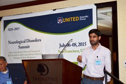 Bhat Waseem Feeroze合作论文数Faculty of Life Sciences, Aligarh Muslim University9
 Aamir Sohail合作论文数8
 Rehman Md T合作论文数5
 Shams Tabrez合作论文数4
 Azad Alam Siddiqui合作论文数4
 Mohammad Afsar合作论文数The authors wish it to be known that, in their opinion, the first 2 authors should be regarded as joint first authors.3Alokail Majed S合作论文数Dept Biochem, Biomarkers Res Program, King Saud Univ3
 Aabgeena Naeem合作论文数3
 Mohamed Alajmi合作论文数College of Pharmacy, King Saud University;Ministry of Environment Water and Agriculture3
 Fohad Mabood Husain合作论文数Faculty of Food and Agricultural Sciences, King Saud University3Aaliya Shah合作论文数2Hagar Ismail合作论文数2
 Zeenat Farooq合作论文数These authors contributed equally to this work.2
 Rizwan Hasan Khan合作论文数Interdisciplinary Biotechnology Unit, Aligarh Muslim University2
 Iftekhar Hassan合作论文数2
 Tabrez Shams合作论文数Kissimmee, Florida and is affiliated with multiple hospitals in the area2
 Mohammad Alsenaidy合作论文数2
 Hussain Afzal合作论文数2
 Salman Alamery合作论文数2
 Mohammad Furkan合作论文数Faculty of Life Sciences, Aligarh Muslim University2
 Imtiyaz Ahmad Bhat合作论文数2Gouse M Shaik合作论文数Department of Signal;Institute of Molecular Genetics;Academy of Sciences of the Czech Republic;Institute of Molecular Genetics, Academy of Sciences of the Czech Republic2
 Md Wasim Khan合作论文数Faculty of Life Sciences, Aligarh Muslim University2
 Afshin Iram合作论文数Faculty of Life Science, Aligarh Muslim University2Mohamed A. Ismael合作论文数1Ashraf Ali Khan合作论文数1Shams Uzzaman合作论文数1
 Rupavarshini Manoharan合作论文数1Ejlal Mohamed Abdullah合作论文数1Alya Alomari合作论文数1Rizwan Wahab合作论文数1Nouf Omar Alafaleq合作论文数1Abu Bakr Ahmad Fazili合作论文数1Nojood Altwaijry合作论文数College of Science, King Saud University1
 Bigyan Ranjan Jali合作论文数Dept Chem, Veer Surendra Sai Univ Technol1
 Arshad Mohammed合作论文数Department of Research and Scientific Communications, Isthmus Research and Publishing House1
 M. Tarique合作论文数Dept Child Hlth, Univ Missouri1
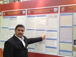 Ibrahim Mohammed Aziz合作论文数Dept Bot & Microbiol, King Saud Univ1
 Mohd Faraz合作论文数Department of Physics, Indian Institute of Technology Delhi1
 Brian T Layden合作论文数1
 Khan Rais Ahmad合作论文数Department of Chemistry, Aligarh Muslim University1
 Farid Shokry Ataya合作论文数College of Science, King Saud University;;Department of Molecular Biology, National Research Center;College of Science, King Saud University1
 Ghulam Md Ashraf合作论文数Department of Biosciences and Bioinformatics, Xi’an Jiaotong-Liverpool University;Glocal University1
 Syed Kashif Zaidi合作论文数Ctr Excellence Genom Med Res, King Abdulaziz Univ1
 Gufran Ahmed Siddiqui合作论文数1
 Khan Yusuf合作论文数National Institute of Plant Genome Research (NIPGR), Aruna Asaf Ali Marg, New Delhi 110 067, India.1
 Mohammad A Kamal合作论文数ABMMA.com.au1




