Introduction: Psychological stress is known to affect dermatological conditions, but its impact during national crises is not well established. Objectives: To assess the association between crisis-induced stress and the incidence of infectious and inflammatory skin diseases, focusing on herpes zoster. Methods: A retrospective cross-sectional study was performed using emergency room records from Hadassah Medical Center, Israel. Skin-related visits were compared between October and December 2023 (national crisis), and the same months in 2014–2022: between the COVID-19 lockdowns (March–May 2020, December 2020–February 2021) and pre-pandemic years (2014–2022). Logistic regression models, adjusted for age and sex, evaluated disease incidence. Results: Among 1,644 patients, infectious skin diseases increased significantly during the October–December 2023 crisis (odds ratio (OR)=2.106; P<0.001), mainly due to herpes zoster (OR=2.616; P=0.009). No significant change was observed for inflammatory skin diseases. During COVID-19 lockdowns, no significant difference was found, likely reflecting reduced healthcare utilization. Conclusions: National crisis-related stress was associated with higher incidence of infectious skin diseases, particularly herpes zoster, but not with inflammatory conditions. These findings support targeted interventions, including vaccination and stress management, during crises.
BACKGROUND:Cutaneous graft-versus-host disease (GVHD) is a common complication of allogeneic hematopoietic stem cell transplantation. Phototherapy has been used to treat cutaneous GVHD, but data on its safety and efficacy are sparse. AIM:Review the current medical literature regarding the efficacy, dosing, and safety of various types of phototherapies for the treatment of cutaneous GVHD. METHODS:A systematic review of PubMed, Embase, Cochrane, and ClinicalTrials databases was performed. Publications were screened according to the PRISMA guidelines. Exclusion criteria comprised case reports and case series reporting less than five patients, review articles, and articles not published in English. RESULTS:A total of 28/1304 (2.5%) studies were included. Fifteen studies (n = 267 patients) focused on psoralen and ultraviolet (UV) A (PUVA), in which 65.5% of patients received concomitantly other systemic treatments. The response rate was 89.9%, with a mean of 33.2 treatments. Adverse events were recorded in 54% but were mainly mild. Eight studies, encompassing 95 patients, focused on narrow-band (NB) UVB. A response was observed in 94%, with a mean number of 26 treatments and 8.6% adverse effects. UVA1 was reported in six studies (n = 132 patients). A response was recorded in 89.3% with a mean of 26.2 treatments. Adverse events were noted in 70.1%, with a discontinuation rate of 10.9%. It should be noted that adverse events were recorded during the follow-up period of the studies, which varied significantly, ranging from no follow-up to 31 months. CONCLUSIONS:Current data regarding the use of phototherapy for the treatment of cutaneous GVHD are based on retrospective studies and case series. The present report advocates the use of one of the three modalities of phototherapy as an effective and safe adjunctive treatment for cutaneous GVHD, especially NB UVB phototherapy.
One effort to combat the rising incidence of malignant melanoma is focused on early detection by the clinical and dermoscopic screening of melanocytic nevi. However, the interaction between nevi, which are congenital or acquired benign melanocytic proliferations, and melanoma is still enigmatic. On the one hand, the majority of melanomas are thought to form de novo, as only a third of primary melanomas are associated with a histologically identifiable nevus precursor. On the other hand, an increased number of melanocytic nevi is a strong risk factor for developing melanoma, including melanomas that do not derive from nevi. The formation of nevi is modulated by diverse factors, including pigmentation, genetic risk factors, and environmental sun exposure. While the molecular alterations that occur during the progression of a nevus to melanoma have been well characterized, many unanswered questions remain surrounding the process of nevus to melanoma evolution. In this review, we discuss clinical, histological, molecular, and genetic factors that influence nevus formation and progression to melanoma.
Acral necrotic ulcers in infancy are rare but have been described in type I interferonopathies. Herein, we present a case of an 8-year-old child who presented at the age of one month with severe ulceronecrotic lesions on the face and limbs with exacerbations following exposure to cold weather. Despite extensive investigation the case remains undiagnosed to this day. We hypothesize that this case represents a novel and yet unknown autoinflammatory disease.
Melanoma is widely treated with programmed cell death-1 (PD-1) inhibitors. As part of their anti-tumor immunity effect, they increase the susceptibility to cutaneous immune-related adverse events (cIRAE) among other autoimmune effects. To characterize the manifestations of cIRAE in melanoma patients treated with PD-1 inhibitors, and evaluate the correlation with tumor response. A retrospective study of 95 metastatic malignant melanoma patients treated with PD-1 inhibitors at the Hadassah Medical Center during 2013-2016. The most common cIRAE was pruritus reported by 39 (41%) patients. All other cIRAE were noted in 34 patients (35.8%), of which the most common cutaneous manifestation was vitiligo, demonstrated in 17 patients (17.9%) followed by various rashes (7.4%, including erythema multiforme, oral lichen planus, photosensitive rash, insect bite-like reaction, and urticaria), psoriasiform rash (3.2%), bullous pemphigoid (3.2%), and eczema (1%). Interestingly, higher response rates to immunotherapy were demonstrated in patients who developed pruritus (85%) and cIRAE (88%), with lower mortality rates in the cIRAE group (38.2%) versus the non-cIRAE group (70.5%, p = 0.002). cIRAE are common among malignant melanoma patients treated with PD-1 inhibitors and may be a marker for favorable prognosis.
Immune checkpoint receptors (ICR) modulate the immune response and are critical hubs for immunotherapy. However, data on their role in T lymphoid malignancies, such as cutaneous T cell lymphoma (CTCL), is sparse. We aimed to explore the role of ICR in the malignant features of transformed T lymphocytes and evaluate the effect of ICR-targeting monoclonal antibodies, often used as immunotherapy for solid tumors. We used the CTCL cell line HH and the Sézary cell line Hut78 to examine ICR expression and the effects of ICR inhibition on cell viability and proliferation. Despite their shared T cell progeny, the different CTCL cell lines exhibit markedly different ICR expression profiles. Programmed cell death-ligand 1 (PD-L1) was expressed by both cell lines, while programmed death-1 (PD-1) was expressed only by the HH cell line. Common to all malignant T cells was an autonomous hyper-proliferative state that did not require T cell receptor stimulation. A monoclonal antibody blocking PD-1 had a small but statistically significant augmenting effect on T cell proliferation. Of note, when the cells were exposed to ionizing radiation, healthy lymphocytes and those derived from the HH cell line were salvaged by anti-PD-L1. We show a regulatory role of ICR, mainly PD-1 and its ligand PD-L1, on cutaneous T cell malignancy.
To the Editor: Dupilumab is a monoclonal antibody approved for moderate-to-severe atopic dermatitis (AD), asthma, and chronic rhinosinusitis with nasal polyposis.1Eshtiaghi P. Gooderham M.J. Dupilumab: an evidence-based review of its potential in the treatment of atopic dermatitis.Core Evid. 2018; 13: 13-20Crossref PubMed Google Scholar Ocular surface disease was reported as an adverse event in AD dupilumab clinical trials, but not in non-AD clinical trials.1Eshtiaghi P. Gooderham M.J. Dupilumab: an evidence-based review of its potential in the treatment of atopic dermatitis.Core Evid. 2018; 13: 13-20Crossref PubMed Google Scholar Since then, numerous published studies have reported dupilumab-induced ocular surface disease (DIOSD) in patients with AD. This systematic review aims to summarize the current available data regarding DIOSD, including its risk factors, course, and treatment options. A literature review was conducted using the PubMed, Embase, Cochrane, and ClinicalTrials databases. Publications up to July 29, 2020 were screened according to the Preferred Reporting Items for Systematic Reviews and Meta-Analyses guidelines (Supplemental Fig 1 available via Mendeley at https://doi.org/10.17632/y4yxmmjmzs.1). A total of 77 clinical studies met the inclusion criteria, including 13 case reports, 11 case series, 28 retrospective studies, 11 prospective studies, 13 randomized controlled trials (RCTs), and a single meta-analysis of RCTs, encompassing 11,189 patients treated with dupilumab. Because the detailed characteristics of DIOSD were described primarily in case reports and series and less often in retrospective studies, results are presented separately for each category of clinical studies (Tables I and II).Table ICharacteristics of patients with dupilumab-induced ocular surface disease in case reports and case series (n = 86)CharacteristicNo. (% or range)Mean age in years (range)40.3 (18-83)∗For 11 patients, ages were not given; for 6 patients, only the median age was reported.Female:male ratio (numerical)2:3 (30:45)†Eleven patients were not reported with a male:female ratio.Indication Atopic dermatitis84 (97.6%) Allergic contact dermatitis1 (1.1%) Chronic actinic dermatitis1 (1.1%)Severity of atopic dermatitis Mild to moderate8 (9.3%) Severe‡Fourteen patients were described as having moderate-to-severe atopic dermatitis.43 (50%)Comorbidities Ophthalmologic predisposition§Including dryness, atopic blepharoconjunctivitis, herpes uveitis and secondary glaucoma, keratoconus, corneal transplantation, marginal keratitis, bacterial keratitis, iridocyclitis, and blepharoplasty.9 (10.4%) Other atopic conditions30 (34.8%)Course Conjunctivitis83 (96.5%) Keratitis4 (4.6%) Blepharitis21 (24.4%) Dry eyes5 (5.8%) Watery eyes4 (4.6%) Other4 (4.6%) Severe conjunctivitis20 (23.2%) Mean latency period to onset of conjunctivitis in weeks (range)14.75 (1-55) Patients referred to ophthalmologists33 (38.7%) Termination of dupilumab treatment‖In 1 patient, dupilumab was temporarily discontinued.12 (13.9%)Treatment¶Some patients received a combination of treatments. Only efficacious treatments are included. Anti-inflammatory agents included corticosteroids, calcineurin inhibitors, and nonsteroidal anti-inflammatory drugs. Moisturizers included artificial tears, trehalose, and petroleum jelly. Antibacterial agents included antibiotics and antiviral agents. Other treatments included topical (12 patients) and systemic (1 patient) antihistamines, lifitergast (1 patient), and eye cleansers (4 patients). Topical anti-inflammatory agents43 (50%) Systemic anti-inflammatory agents3 (3.4%) Topical moisturizers23 (26.7%) Topical antibacterial agents15 (17.4%) Systemic antibacterial agents6 (6.9%) Other17 (19.7%)∗ For 11 patients, ages were not given; for 6 patients, only the median age was reported.† Eleven patients were not reported with a male:female ratio.‡ Fourteen patients were described as having moderate-to-severe atopic dermatitis.§ Including dryness, atopic blepharoconjunctivitis, herpes uveitis and secondary glaucoma, keratoconus, corneal transplantation, marginal keratitis, bacterial keratitis, iridocyclitis, and blepharoplasty.‖ In 1 patient, dupilumab was temporarily discontinued.¶ Some patients received a combination of treatments. Only efficacious treatments are included. Anti-inflammatory agents included corticosteroids, calcineurin inhibitors, and nonsteroidal anti-inflammatory drugs. Moisturizers included artificial tears, trehalose, and petroleum jelly. Antibacterial agents included antibiotics and antiviral agents. Other treatments included topical (12 patients) and systemic (1 patient) antihistamines, lifitergast (1 patient), and eye cleansers (4 patients). Open table in a new tab Table IIRates of DIOSD reported in clinical studies, including retrospective, prospective, open-label studies, and RCTsStudyTotal treated; nPatients with DIOSD; nRate of DIOSD; %Latency to DIOSD; weeksSevere DIOSD; n (% of patients with DIOSD)Real life retrospective186038520.610.114 (15.2)∗Of the total number of patients for which data on severity and discontinuation were known.Prospective and open label4735118725.1-38 (3.6)∗Of the total number of patients for which data on severity and discontinuation were known.RCTTotal treated; nPatients and rate of DIOSD in treatment group; n (%)Total treated with placebo; nPatients and rate of ocular surface disorders in placebo group; n (%)Severe DIOSD; n (% of patients with DIOSD) Atopic dermatitis indication2,532276 (10.9)132559 (4.5)8 (6)∗Of the total number of patients for which data on severity and discontinuation were known. Nonatopic dermatitis indication203012 (0.59)9537 (0.73)0DIOSD, Dupilumab-induced ocular surface disease; RCT, randomized controlled trial.∗ Of the total number of patients for which data on severity and discontinuation were known. Open table in a new tab DIOSD, Dupilumab-induced ocular surface disease; RCT, randomized controlled trial. DIOSD was unique to AD, but was also reported in AD-related dermatologic disorders, such as nummular and dyshidrotic eczema, prurigo nodularis, and allergic contact dermatitis. The incidence of DIOSD was higher in prospective (25.1%) and real-life retrospective trials (20.6%) than in RCTs (10.9%). Most DIOSD cases appeared in the first weeks to months of treatment, were mild to moderate, and resolved with continued dupilumab treatment. Various ophthalmic disorders were described, other than conjunctivitis, including dry or watery eyes, blepharitis, keratitis, eyelid dermatitis, and palpebral edema. Cessation of dupilumab due to DIOSD was reported in 86 patients (4.5% of patients with DIOSD). Permanent sequelae were even rarer, involving several cases of cicatricial changes.2Barnes A.C. Blandford A.D. Perry J.D. Cicatricial ectropion in a patient treated with dupilumab.Am J Ophthalmol Case Rep. 2017; 7: 120-122Crossref PubMed Scopus (45) Google Scholar,3Levine R.M. Tattersall I.W. Gaudio P.A. et al.Cicatrizing blepharoconjunctivitis occurring during dupilumab treatment and a proposed algorithm for its management.JAMA Dermatol. 2018; 154: 1485-1486Crossref PubMed Scopus (26) Google Scholar Possible DIOSD risk factors were previous ophthalmic disorders, high baseline AD severity, and elevated IgE levels. Several beneficial treatment options for DIOSD included warm compresses, artificial tears, topical antihistamines/mast cell stabilizers, topical antibiotics, topical corticosteroids, cyclosporine eye drops, tacrolimus ointment, and lifitegrast eye drops. The main limitation of this systematic review is the vast heterogeneity among the included studies. Nevertheless, this review confirms that an increased incidence of DIOSD is observed almost exclusively in patients with AD and AD-related dermatologic disorders. The higher incidence of DIOSD in prospective and real-life retrospective trials than in RCTs may be due to the increased awareness of this adverse effect, which may have been overlooked in earlier RCTs. The pathogenic mechanisms responsible for DIOSD remain obscure. By blocking interleukin 13, dupilumab may cause goblet cell hypoplasia, resulting in decreased mucin secretion and mucosal epithelial barrier dysfunction, leading to DIOSD.4Tukler Henriksson J. Coursey T.G. Corry D.B. et al.IL-13 stimulates proliferation and expression of mucin and immunomodulatory genes in cultured conjunctival goblet cells.Invest Ophthalmol Vis Sci. 2015; 56: 4186-4197Crossref PubMed Scopus (65) Google Scholar Alternatively, upregulation of Th1 response due to the effect of dupilumab on Th2 signaling may contribute to DIOSD pathogenesis.5Maudinet A. Law-Koune S. Duretz C. et al.Ocular surface diseases induced by dupilumab in severe atopic dermatitis.Ophthalmol Ther. 2019; 8: 485-490Crossref PubMed Scopus (39) Google Scholar In conclusion, DIOSD is a common adverse effect of dupilumab in AD, which tends to resolve with topical treatment and continuation of dupilumab. Long-term sequelae are rare. Predisposing factors of DIOSD and its pathomechanisms should be further investigated. None disclosed.
BACKGROUND:Ultraviolet (UV) A1 phototherapy is considered a beneficial treatment for various inflammatory, sclerotic, malignant, and other skin conditions. However, the available data regarding its efficacy for different indications, the potential side effects, and the recommended treatment protocols are sparse. OBJECTIVES:To assess the efficacy of UVA1 phototherapy and identify correlation between different indications and treatment protocols to response rates. METHODS:We performed a retrospective study of a cohort of 335 patients treated with UVA1 phototherapy at the Department of Dermatology at Hadassah Medical Center, Jerusalem, Israel, between 2008 and 2018. RESULTS:The study population included 163 patients with inflammatory diseases (mainly atopic dermatitis and other types of eczema), 67 patients with sclerotic diseases (morphea and graft versus host disease), nine patients with neoplastic diseases (cutaneous T cell lymphoma), and 188 patients with other cutaneous disorders. Response rates ranged between 85% and 89% across indications, without differences in response rates among the indication groups (p = .941). In a multivariant logistic regression model, increased number of treatments and higher maximal dosages were associated with response to treatment (p < .001). Using ROC analysis, a cut-off of 8 UVA1 phototherapy treatments was chosen as predictive for beneficial response (86.4% sensitivity, 78% specificity). A cut-off of 40 J/cm2 was chosen as an optimal maximal dosage for differentiating between responders and non-responders (51.1% sensitivity, 83.1% specificity). CONCLUSIONS:UVA1 phototherapy is an effective treatment for a variety of skin conditions. In most patients, at least eight treatments of a medium-high dosage are required for clinical response.
Systemic sclerosis (scleroderma, SSc) is an incurable autoimmune disease with high morbidity and mortality rates. Here, we conducted a population-scale single-cell genomic analysis of skin and blood samples of 56 healthy controls and 97 SSc patients at different stages of the disease. We found immune compartment dysfunction only in a specific subtype of diffuse SSc patients but global dysregulation of the stromal compartment, particularly in a previously undefined subset of LGR5+-scleroderma-associated fibroblasts (ScAFs). ScAFs are perturbed morphologically and molecularly in SSc patients. Single-cell multiome profiling of stromal cells revealed ScAF-specific markers, pathways, regulatory elements, and transcription factors underlining disease development. Systematic analysis of these molecular features with clinical metadata associates specific ScAF targets with disease pathogenesis and SSc clinical traits. Our high-resolution atlas of the sclerodermatous skin spectrum will enable a paradigm shift in the understanding of SSc disease and facilitate the development of biomarkers and therapeutic strategies.
Pityriasis rubra pilaris induced by PD-1 inhibitor nivolumab Dear Editor, A 28-year-old with relapsed Hodgkin lymphoma was treated with PD-1 inhibitor nivolumab for 2 years and 6 months. The treatment was halted because of pneumonitis and was resumed 6 months later. Four months after the renewal of immunotherapy, the patient developed a widespread rash, consisting of widespread reddish-orange colored scaling plaques, scattered follicular papules with islands of spared skin (Fig. 1). A punch biopsy from a scaling plaque demonstrated alternating orthokeratosis and parakeratosis, keratotic plugs, hypergranulosis, and mild acanthosis (Fig. 2). Differential diagnosis included other papulosquamous eruptions, such as psoriasis. However, the clinical and histological features were typical for pityriasis rubra pilaris (PRP)-like eruption, which was secondary to PD-1 inhibitor treatment. The rash was classified as grade 3 skin toxicity. Accordingly, nivolumab was stopped, and the patient was treated with topical and systemic corticosteroids and with methotrexate. The patient was intolerant to phototherapy and was not interested in acitretin because of planned future pregnancies. Over the following 3 months, the rash gradually faded. However, the lymphoma continued to progress until the patient passed away because of a severe infection.
Few studies have reported an association between psoriasis and atopic comorbidity in adults. A population-based cross-sectional study was performed to investigate the possible association of psoriasis with allergic rhinitis or asthma among adolescents. Adolescents (16-18 years of age) medically evaluated for military service between 1999 and 2014 were included. Medical records were obtained from the database of the Israeli Defense Forces. Of the 887,765 adolescents studied, 3,112 patients had psoriasis (56.1% mild; 43.9% moderate-to-severe). Psoriasis was significantly associated with allergic rhinitis (adjusted odds ratio (aOR) 1.3; 95% confidence interval (CI) 1.2-1.5) and asthma (aOR 1.2; 95% CI 1.0-1.3), compared with controls without psoriasis. Moderate-to-severe psoriasis was associated with allergic rhinitis (aOR 1.3; 95% CI 1.1-1.5) and asthma (aOR 1.5; 95% CI 1.2-1.7), while mild psoriasis was only associated with allergic rhinitis (aOR 1.4; 95% CI 1.2-1.6). In conclusion, amongst adolescents, psoriasis was found to be associated with allergic rhinitis and asthma.
Background and Aims: Although psoriasis can develop at any age, the data regarding its characteristics in adolescents are sparse. This study was designed to determine the psoriasis prevalence and its associations with the body mass index (BMI), lipid profile, and comorbidities in adolescents. Methods: This was a nationwide population-based cross-sectional retrospective study of adolescents (16–18 years old) evaluated for military service between January 1999 and January 2014. Results: Our database included 887,765 adolescents (57.1% males), of whom 3,112 (0.35%) were diagnosed with psoriasis. During the 15-year study period, the psoriasis prevalence increased by 1.4-fold, from 0.3 to 0.42% (1.25-fold for the males and 1.63-fold for the females). Certain comorbidities, such as contact dermatitis, hyperhidrosis, and arthritis, were significantly associated with psoriasis (odds ratios [ORs] of 2.26, 1.51, and 5.3, respectively). The adolescents with psoriasis had significantly elevated BMI and triglyceride values. We found increased ORs of 1.34 (95% confidence interval [CI] = 1.25–1.56) and 1.56 (95% CI = 1.32–1.83) for the overweight and obese adolescents, respectively, while a lower BMI (<20) had an opposite effect with psoriasis (OR = 0.8). Conclusions: Based on our results, the psoriasis prevalence in Israeli adolescents is rising. Dermatological comorbidities and an increased BMI were associated with psoriasis in these adolescents. A better understanding of the distinctive epidemiological characteristics of juvenile psoriasis may allow for the early detection of comorbidities and improve its management.
Chronic graft versus host disease (cGVHD) is a complication of allogeneic haematopoietic stem cell transplantation (HSCT). The aim of this study was to clinically characterize childhood cutaneous cGVHD. A retrospective study of children treated with HSCT at 2 tertiary medical centres in Israel between 2011 and 2014 was performed. A total of 112 children were included. Cutaneous cGVHD developed in 18% of subjects. Risk factors were older age, HSCT from peripheral blood and acute lymphoblastic leukaemia. The eruption was lichenoid in 90% of subjects, of whom one-third progressed to sclerosis. Topical treatments were usually sufficient in localized disease. Widespread eruption necessitated phototherapy, extracorporeal photopheresis and/or systemic immunosuppressants. Patients presenting with palmoplantar keratoderma, developed sclerosis. To the best of our knowledge, this is the first study describing childhood cutaneous cGVHD. Lichenoid eruption is the most common cutaneous pattern of cGVHD in children. Sclerotic changes may be associated with prior keratoderma. cGVHD poses a therapeutic challenge and better treatments should be sought.
Objectives Confidentiality of health information is an important aspect of the physician patient relationship. The use of digital medical records has made data much more accessible. To prevent data leakage, many countries have created regulations regarding medical data accessibility. These regulations require a unique user ID for each medical staff member, and this must be protected by a password, which should be kept undisclosed by all means. Methods We performed a four-question Google Forms-based survey of medical staff. In the survey, each participant was asked if he/she ever obtained the password of another medical staff member. Then, we asked how many times such an episode occurred and the reason for it. Results A total of 299 surveys were gathered. The responses showed that 220 (73.6%) participants reported that they had obtained the password of another medical staff member. Only 171 (57.2%) estimated how many time it happened, with an average estimation of 4.75 episodes. All the residents that took part in the study (45, 15%) had obtained the password of another medical staff member, while only 57.5% (38/66) of the nurses reported this. Conclusions The use of unique user IDs and passwords to defend the privacy of medical data is a common requirement in medical organizations. Unfortunately, the use of passwords is doomed because medical staff members share their passwords with one another. Strict regulations requiring each staff member to have it's a unique user ID might lead to password sharing and to a decrease in data safety.
The skin has an important role as a barrier, protecting the organism from its environment. Therefore, it participates significantly in several immunological processes, and this novel role is gradually revealed in recent studies. We overview the main components of the cutaneous immunological response, divided into contact and systemic antigen exposures. The major players of the immunopathological skin reactions are the keratinocytes, Langerhans cells and dermal dendritic cells. In addition, we discuss the use of animal models for exploring the complexity of these reactions.
BackgroundRecent data have shown an increasing occurrence of atopic dermatitis (AD) in children and adolescents, as well as in adults. Most of the epidemiologic research on AD is limited to pediatric and youth populations and is based on self-reported questionnaires.MethodsA nationwide, population-based, cross-sectional retrospective study of adolescents with AD was performed to estimate its prevalence, trends, and association with demographic factors and comorbidities. The study included all Israeli teens going through medical evaluation as part of the assessment before being conscripted into the military from 1998 to 2013.ResultsA total of 1,187,757 adolescents were included in the study population, with an overall prevalence of AD of 0.64% in boys and 0.9% in girls. Over the study period, the prevalence of AD steadily increased, especially in the mild disease group. A greater risk of AD was found in subjects with high predicted socioeconomic status (male: odds ratio [OR] 1.14 [95% confidence interval {CI} 1.11, 1.16]; female: OR 1.08, [95% CI 1.05, 1.10]) and Israeli-born subjects (male: OR 1.34 [95% CI 1.21, 1.48]; female: OR 1.12 [95% CI 1.01, 1.23]). Allergic conditions such as asthma, conjunctivitis, and contact dermatitis were more prevalent in subjects with AD. There was a significantly higher prevalence of migraine in patients with AD (male: OR 1.35 [95% CI 1.18, 1.54]; female: OR 1.51 [95% CI 1.30, 1.74]).ConclusionThis large cross-sectional study demonstrates the increasing prevalence of AD in adolescents and its relation to other allergic diseases and migraine. It is hoped that greater awareness of the distinctive epidemiologic characteristics of this population will lead to better recognition and management of the disease and its comorbidities.
Background: Janus kinase (JAK) inhibitors are emerging as a promising new treatment modality for many inflammatory conditions.Objective: Our aim was to systematically review the available data on the use of JAK inhibitors in cutaneous diseases.Methods: This is a systematic review of PubMed and ClinicalTrials.gov.Results: One hundred thirty-four articles matched our search terms, of which 78 were original articles and 12 reports on adverse events. Eighteen clinical trials were found. JAK inhibitors have been extensively studied for psoriasis, showing beneficial results that were comparable to the effects achieved by etanercept. Favorable results were also observed for alopecia areata. Promising preliminary results were reported for vitiligo, dermatitis, graft versus host disease, cutaneous T cell lymphoma, and lupus erythematosus. The most common adverse events reported were infections, mostly nasopharyngitis and upper respiratory tract infections.Limitations: It was not possible to perform a meta-analysis of the results.Conclusions: This systematic review shows that while JAK inhibitors hold promise for many skin disorders, there are still gaps regarding the correct dosing and safety profile of these medications for dermatologic indications. Additional trials are necessary to address these gaps.
Journal of the European Academy of Dermatology and VenereologyVolume 30, Issue 12 p. e232-e234 Letter to the Editor Squamous cell carcinoma in situ in association with HPV 11 in Netherton's syndrome patient: a case report R. Shreberk-Hassidim, R. Shreberk-Hassidim Department of Dermatology, Hadassah - Hebrew University Medical Center, Jerusalem, IsraelSearch for more papers by this authorA. Hassidim, A. Hassidim Department of Plastic surgery, Hadassah - Hebrew University Medical Center, Jerusalem, IsraelSearch for more papers by this authorN. Adler, N. Adler Department of Plastic surgery, Hadassah - Hebrew University Medical Center, Jerusalem, IsraelSearch for more papers by this authorL. Horev, L. Horev Department of Dermatology, Hadassah - Hebrew University Medical Center, Jerusalem, IsraelSearch for more papers by this authorA. Maly, A. Maly Department of Pathology, Hadassah - Hebrew University Medical Center, Jerusalem, IsraelSearch for more papers by this authorA. Zlotogorski, Corresponding Author A. Zlotogorski zloto@cc.huji.ac.il Department of Dermatology, Hadassah - Hebrew University Medical Center, Jerusalem, IsraelCorrespondence: A. Zlotogorski. E-mail: zloto@cc.huji.ac.ilSearch for more papers by this authorY. Ramot, Y. Ramot Department of Dermatology, Hadassah - Hebrew University Medical Center, Jerusalem, IsraelSearch for more papers by this author R. Shreberk-Hassidim, R. Shreberk-Hassidim Department of Dermatology, Hadassah - Hebrew University Medical Center, Jerusalem, IsraelSearch for more papers by this authorA. Hassidim, A. Hassidim Department of Plastic surgery, Hadassah - Hebrew University Medical Center, Jerusalem, IsraelSearch for more papers by this authorN. Adler, N. Adler Department of Plastic surgery, Hadassah - Hebrew University Medical Center, Jerusalem, IsraelSearch for more papers by this authorL. Horev, L. Horev Department of Dermatology, Hadassah - Hebrew University Medical Center, Jerusalem, IsraelSearch for more papers by this authorA. Maly, A. Maly Department of Pathology, Hadassah - Hebrew University Medical Center, Jerusalem, IsraelSearch for more papers by this authorA. Zlotogorski, Corresponding Author A. Zlotogorski zloto@cc.huji.ac.il Department of Dermatology, Hadassah - Hebrew University Medical Center, Jerusalem, IsraelCorrespondence: A. Zlotogorski. E-mail: zloto@cc.huji.ac.ilSearch for more papers by this authorY. Ramot, Y. Ramot Department of Dermatology, Hadassah - Hebrew University Medical Center, Jerusalem, IsraelSearch for more papers by this author First published: 02 February 2016 https://doi.org/10.1111/jdv.13568Citations: 1Read the full textAboutPDF ToolsRequest permissionExport citationAdd to favoritesTrack citation ShareShare Give accessShare full text accessShare full-text accessPlease review our Terms and Conditions of Use and check box below to share full-text version of article.I have read and accept the Wiley Online Library Terms and Conditions of UseShareable LinkUse the link below to share a full-text version of this article with your friends and colleagues. Learn more.Copy URL Share a linkShare onFacebookTwitterLinkedInRedditWechat No abstract is available for this article.Citing Literature Volume30, Issue12December 2016Pages e232-e234 RelatedInformation
 Abraham Zlotogorski合作论文数Department of Dermatology and Venereology, Hadassah Medical Center12
 Yuval Ramot合作论文数Hadassah Medical Center, Faculty of Medicine, Hebrew University of Jerusalem10
 Alexander Maly合作论文数9Ayal Hassidim合作论文数8
 Vered Molho合作论文数8Merims Sharon合作论文数4Liran Horev合作论文数3Sivan Sheffer合作论文数2
 Elena Thomaidou合作论文数2N. Astman合作论文数2Eran Galili合作论文数Department of Dermatology, Sheba Medical Center2Aviv Barzilai合作论文数2Michal Lotem合作论文数2Emilia Hodak合作论文数1Dana Hirsch合作论文数1Sivan Sheffer-Levi合作论文数1Efrat Hagai合作论文数1Dalit Amar合作论文数1Adam Yalin合作论文数1Anna Elia合作论文数1Adi Moshe合作论文数1
 Hagit Peleg合作论文数1Julius Golender合作论文数1Suhail Aamar合作论文数1Tal Goldberger合作论文数1Gil Armoni合作论文数1
 Mor Zada合作论文数1
 Batia Avni合作论文数1Tahel Fachler合作论文数1Shir Shlomi-Loubaton合作论文数1
 Jonathan E Cohen合作论文数1Danny Daniely合作论文数1Adiel Cohen合作论文数1Lilach Aizenbud合作论文数1
 Fadi Sheban合作论文数Dept Syst Immunol, Weizmann Inst Sci1
 Liat Fellus Alyagor合作论文数Dept Vet Resources, Weizmann Inst Sci1
 Oren Barboy合作论文数Dept Immunol, Weizmann Inst Sci1
 Georgina Maria Sarika合作论文数1Sigal Grisariu合作论文数1
 Amir Giladi合作论文数Department of Immunology, Weizmann Institute of Science;Hubrecht Institute for Developmental Biology and Stem Cell Research1Diego Adhemar Jaitin合作论文数1Lilach Moyal合作论文数1
 Tomer M Salame合作论文数Weizmann Inst Sci, Dept Biol Serv, Rehovot, Israel1Emma Hajaj合作论文数1Fadi Kharouf合作论文数1
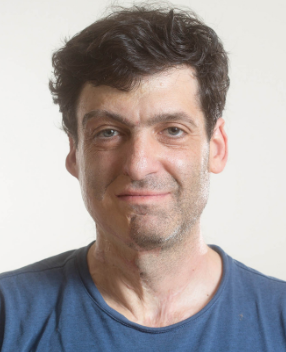 Dan Ariely合作论文数The Fuqua School of Business, Duke University;Center for Advanced Hindsight, Duke University;Accumulator;Irrational Capital;Kayma Labs;Epilog;BEworks;Shapa Health;Genie1
 Zvi Gilula合作论文数Chicago Booth School of Business, The University of Chicago1Twig Gilad合作论文数Dept Med B, Chaim Sheba Med Ctr1Balbir-Gurman Alexandra合作论文数1




