PURPOSE:To improve the imaging efficiency of 3D carotid simultaneous noncontrast angiography and intraplaque hemorrhage (SNAP) MRI by reconstruction of under-sampled images and concurrent optimization of sampling masks for the two shots of SNAP respectively. METHODS:A model-based deep learning architecture (deepSNAP) was proposed to recover under-sampled 3D carotid SNAP MRI. Sampling locations on the ky-kz plane were parameterized to enable respective optimization of the sampling masks for the two shots. A dataset of 100 3D carotid SNAP MRI scans was utilized (80 training, 20 test). Image recovery performance was compared with established techniques under different acceleration factors. Lumen area measurement accuracy and intraplaque hemorrhage (IPH) identification were evaluated at 6× acceleration. Prospective feasibility was assessed in 10 healthy volunteers with quantitative comparison against established methods. RESULTS:deepSNAP exhibited superior image recovery performance on the test set, surpassing all comparison methods. Optimized masks generated by deepSNAP improved reconstruction performance across all comparison methods. High agreement between reconstructed images and original images was observed for lumen area measurement (ICC = 0.995, 95% CI: 0.993-0.996) and IPH detection (Cohen's κ = 0.976, 95% CI: 0.943-1.000). In the prospective experiment, deepSNAP achieved promising image quality and structural fidelity. CONCLUSION:The deepSNAP model achieved under-sampled image reconstruction and simultaneous sampling mask optimization for SNAP, ensured the clinical practicability of the reconstructed images, and demonstrated preliminary technical feasibility in a prospective setting.
Background Supracardiac atherosclerosis is a major cause of stroke. Although vessel wall MRI (vwMRI) can assess plaques, current techniques lack full head-neck-aortic arch coverage and efficiency. Purpose To develop and evaluate a head-neck-aortic vwMRI protocol for supracardiac atherosclerosis, compare its performance with that of supra-aortic CT angiography (CTA), and determine its feasibility in helping identify causes of embolic stroke of undetermined source (ESUS). Materials and Methods We developed a noncontrast head-neck-aortic vwMRI protocol integrated with neural network-based acceleration and multichannel coils, enabling a scan time of approximately 15 minutes. Then, participants with acute ischemic stroke or transient ischemic attack were recruited from October 2024 to March 2025. They received routine supra-aortic CTA and this new vwMRI protocol. Plaque vulnerabilities, vascular stenosis, and stroke etiologies were compared between the modalities. The accuracy of vwMRI for detecting calcification was evaluated using CTA as the reference standard, and the McNemar test was used to compare the findings between CTA and vwMRI. Results Among 108 participants who underwent imaging with both modalities (mean age, 56 years ± 14 [SD]; 89 male participants), the plaque detection rate was higher with vwMRI than CTA (88 of 108 participants [81.5%] vs 75 of 108 participants [69.4%]; P = .002). Moreover, vwMRI helped achieve 91% (98 of 108 participants) accuracy for calcification detection. vwMRI helped identify intraplaque hemorrhage in 27.8% (30 of 108) of participants and demonstrated an ulceration rate comparable with that of CTA (28 of 108 participants [25.9%] vs 25 of 108 participants [23.1%]; P = .51). CTA helped identify 49 participants with vascular stenosis, whereas vwMRI helped identify 47 participants with causes including atherosclerosis (n = 41), thrombus (n = 3), and dissection (n = 3). For 38 participants initially classified with ESUS via CTA, vwMRI enabled reclassification of 16, reducing the percentage of participants with ESUS from 35.2% (38 of 108 participants) to 20.4% (22 of 108 participants) (P < .001). Conclusion This head-neck-aortic vwMRI protocol was feasible and demonstrated a higher plaque detection rate than supra-aortic CTA. © RSNA, 2026 Supplemental material is available for this article. See also the editorial by von Kummer in this issue.
BACKGROUND:It has been shown that rupture of vulnerable plaques of atherosclerosis (AS) is one of the main causes of ischemic stroke. Increasing evidence suggests that the inflammatory changes of perivascular adipose tissue (PVAT) were independently associated with both vulnerable plaque characteristics and cerebrovascular symptoms. Therefore, it is essential to conduct non-invasive joint assessment of carotid AS plaques and PVAT characteristics for better stratifying the risk of ischemic cerebrovascular events. PURPOSE:To develop a simultaneous magnetic resonance (MR) imaging technique for carotid artery PVAT and vessel wall, and determine its feasibility and repeatability. METHODS:This study developed an MR sequence for simultaneous imaging carotid vessel wall and PVAT. Seventeen healthy subjects and nine patients with carotid AS were recruited for MR imaging experiments, of whom five healthy subjects were selected for the repeatability test. All participants underwent bilateral carotid three-dimensional MR imaging by acquiring the proposed iMSDE-mDIXON, MERGE, and mDIXON sequences. To evaluate the reliability of the proposed iMSDE-mDIXON sequence, we analyzed its agreement in measuring morphology (lumen area, wall area, mean wall thickness, and normalized wall index) of carotid wall with the reference sequence of MERGE, as well as its agreement in quantifying carotid PVAT (PVAT area, proton density fat fraction [PDFF], area index, and volume index) with the reference sequence of mDIXON. The interclass correlation coefficient (ICC) and Bland-Altman plots were conducted in statistical analysis. RESULTS:The proposed iMSDE-mDIXON technique demonstrated high reliability in quantifying carotid vessel wall morphology (healthy subjects: ICC = 0.903-0.997; patients: ICC = 0.928-0.999) and PVAT morphology (healthy subjects: ICC = 0.906-0.988; patients: ICC = 0.957-0.996). Although iMSDE-mDIXON sequence showed potential in assessing carotid AS, there was a substantial bias (>20%) in PDFF quantification. Nevertheless, moderate to excellent agreement was maintained between iMSDE-mDIXON and mDIXON in measuring PVAT PDFF both in healthy subjects (ICC: left, 0.782; right, 0.740) and AS patients (ICC: left, 0.790; right, 0.628). In addition, the proposed sequence showed excellent agreement in quantifying carotid vessel wall (ICC = 0.845-0.999) and PVAT morphology (ICC = 0.841-0.989) between the repeated scans. CONCLUSIONS:This study proposed an iMSDE-mDIXON sequence that enables simultaneous imaging of the carotid vessel wall and PVAT in a single scan with high efficiency, reliability, and repeatability. This technique has considerable potential for jointly characterizing the changes of PVAT and pathology of the vessel wall in carotid artery.
Background:Pseudo-continuous arterial spin labeling (PCASL) at 7T benefits from increased signal-to-noise ratio (SNR) and prolonged T1, but suffers from field inhomogeneities and increased specific absorption rate (SAR). We proposed that 5T magnetic resonance imaging (MRI) system may be a balanced choice for PCASL imaging. The aim of this study was to achieve whole-cerebrum PCASL imaging at ultra-high field 5T MRI system, assess the reproducibility and preliminarily explore its clinical application in moyamoya disease/syndrome. Methods:Twenty healthy volunteers were prospectively recruited for the reproducibility analysis. Both single-delay and multi-delay PCASL sequences were scanned twice on the 5T MRI scanner separated by a 10-minute period. Uncorrected cerebral blood flow (uCBF) from single-delay arterial spin labeling (ASL), corrected cerebral blood flow (cCBF) and arterial transit time (ATT) from multi-delay ASL were computed. The reproducibility of uCBF, cCBF and ATT were evaluated by calculating intraclass correlation coefficient (ICC), within-subject coefficient of variation (wsCV) and Pearson correlation coefficients between twice scans in grey matter regions and white matter (WM). Also, 26 patients diagnosed with moyamoya disease/syndrome were included and underwent multi-delay PCASL. The severity of intracranial arteries was graded as magnetic resonance angiography (MRA) score using time-of-flight (TOF) MRA. The relationship between MRA score and cCBF/ATT were assessed by one-way analysis of variance and Pearson correlation analysis. Results:uCBF, cCBF and ATT showed excellent reliability in all regions with ICCs ranging from 0.856 to 0.962, wsCVs ranging from 2.39% to 6.76% and Pearson correlation coefficients ranging from 0.865 to 0.966. Multi-delay ASL demonstrated superior reproducibility of CBF quantification compared to single-delay ASL in regions with heterogeneous transit time, including WM, occipital lobe, limbic system and subcortical region. In patients with moyamoya disease/syndrome, those with higher anterior cerebral artery (ACA) or middle cerebral artery (MCA) scores exhibited lower cCBF (P<0.05). Correlation analysis showed that MRA score was negatively associated with cCBF (r=-0.540, P<0.001) and positively associated with ATT (r=0.515, P<0.001). Conclusions:Whole-cerebrum PCASL imaging at 5T ultra-high field was achieved with good reproducibility and applied well in patients with moyamoya disease/syndrome, which offers a promising tool in the assessment of hemodynamic conditions in cerebrovascular diseases.
BACKGROUND:Multi-parametric imaging of the carotid artery enables quantitative characterization of vulnerable atherosclerotic plaques, which is crucial for preventing ischemic stroke. However, the existing sequential acquisition-based multi-parametric imaging techniques of carotid artery lack bright blood imaging, which is essential for plaque components identification and boundary delineation. This study aims to develop a joint bright, gray, and black blood imaging technique for carotid artery multi-parametric imaging and validate its accuracy and feasibility. METHODS:The proposed technique incorporated variable flip angles, variable duration of improved motion-sensitized driven equilibrium prepulse, and variable time of echo mapping strategies with three-dimensional multi-shot spoiled gradient-recalled echo acquisition, generating T1, T2, and T2* maps. Bright, gray, and black blood images were sequentially acquired in six scans covering the entire extracranial artery with isotropic resolution (0.7 mm) when natural inflow blood enhancement and imposed blood suppression module were alternatively performed. A B1-specific dictionary was simulated and matched to the measured signal for T1 and T2 estimation while least square fitting was applied for T2* estimation. The proposed technique was compared against reference sequences and validated in healthy volunteers (n = 8) and patients (n = 4) with carotid atherosclerotic plaques. RESULTS:The proposed technique achieved an agreement of R2 = 0.99 in T1, T2, and T2* measurements with standard sequences in phantom study. In healthy volunteer study, the proposed technique reached high intraclass correlation coefficients (ICC: 0.906-0.956) with reference sequences in measuring T1, T2, and T2* of cervical muscle, but overestimation and underestimation were observed in T1 (against modified Look-Locker inversion-recovery, bias = 4.8%) and T2 (against gradient and spin echo sequence, bias = -3.3%), respectively. No significant difference was found in the measurement of morphology and quantitative parameters between scan and rescan, while excellent intra- (ICC: 0.804-0.999) and inter-observer (ICC: 0.816-0.982) repeatability was reached. In patient study, the proposed technique demonstrated reliable performance in analyzing vascular morphology and characterizing plaque components with distinctive signal characteristics and quantitative values. CONCLUSION:The proposed technique enables joint bright, gray, and black blood imaging technique for carotid artery multi-parametric imaging with large coverage and isotropic resolution, indicating clinical potential for comprehensive characterization of carotid vulnerable plaque.
BACKGROUND:The feasibility of renal multi-delay arterial spin labeling (ASL) imaging at 5 T remains unclear. PURPOSE:To evaluate the feasibility of the saturated multi-delay renal ASL (SAMURAI) sequence at 5 T by comparing image quality and perfusion quantification with 3 T. STUDY TYPE:Prospective, cross-sectional. POPULATION:Twenty healthy volunteers (28.6 ± 7.8 years, 9 males) for primary comparison; 6 volunteers (24.2 ± 1.5 years, 5 males) for reproducibility study at 5 T. FIELD STRENGTH/SEQUENCE:SAMURAI sequence at 3 T and 5 T. ASSESSMENT:The SAMURAI sequence was optimized at 5 T with renal-specific B1 shimming and an optimized saturation scheme by numerical simulation. Each participant underwent 3 T and 5 T scans in randomized order. Cortical T1 value, renal blood flow (RBF), arterial and tissue bolus arrival times were measured. The signal-to-noise ratio (SNR) and cortico-medullary contrast-to-noise ratio (CNR) were calculated from perfusion-weighted images. Short-term repeatability (n = 20) and reproducibility (n = 6) tests of quantitative parameters were performed at 5 T. STATISTICAL TESTS:Differences and agreement between 3 T and 5 T were analyzed using the Wilcoxon signed-rank test, intraclass correlation coefficients (ICC) and linear correlation analysis (R 2). The repeatability at 5 T was assessed by ICC. A p < 0.05 was considered statistically significant. RESULTS:Renal cortical T1 values were significantly higher at 5 T than 3 T (1417.9 ± 75.7 ms vs. 1184.2 ± 84.4 ms), with R 2 = 0.509. Cortical RBF showed an insignificant difference between 5 T and 3 T: 324.4 (interquartile range [IQR]: 310.1-366.4) vs. 329.7 (IQR: 309.5-368.1) [mL/100 g/min] (p = 0.333), with R 2 = 0.914. 5 T showed significantly higher mean SNR (4.6 vs. 3.9) and CNR (3.2 vs. 2.0) than 3 T across all inversion times, with excellent repeatability and reproducibility of quantitative parameters (ICC = 0.855-0.973). DATA CONCLUSIONS:Renal quantitative imaging with SAMURAI sequence at 5 T is feasible and repeatable, with significantly higher SNR and CNR than 3 T and strong interfield agreement of cortical RBF measurements. LEVEL OF EVIDENCE: 2: TECHNICAL EFFICACY STAGE:1.
Cerebral perfusion plays a crucial role in maintaining brain function and is tightly coupled with neuronal activity. While previous studies have examined cerebral perfusion trajectories across development and aging, precise characterization of its lifespan dynamics has been limited by small sample sizes and methodological inconsistencies. In this study, we construct the first comprehensive normative model of cerebral perfusion across the human lifespan (birth to 85 years) using a large multi-site dataset of over 12,000 high-quality arterial spin labeling (ASL) MRI scans. Leveraging generalized additive models for location, scale, and shape (GAMLSS), we mapped nonlinear growth trajectories of cerebral perfusion at global, network, and regional levels. We observed a rapid postnatal increase in cerebral perfusion, peaking at approximately 7.1 years, followed by a gradual decline into adulthood. Sex differences were evident, with distinct regional maturation patterns rather than uniform differences across all brain regions. Beyond normative modeling, we quantified individual deviations from expected CBF patterns in neurodegenerative and psychiatric conditions, identifying disease-specific perfusion abnormalities across four brain disorders. Using longitudinal data, we established typical and atypical cerebral perfusion trajectories, highlighting the prognostic value of perfusion-based biomarkers for detecting disease progression. Our findings provide a robust normative framework for cerebral perfusion, facilitating precise characterization of brain health across the lifespan and enhancing the early identification of neurovascular dysfunction in clinical populations.
Background Accurate assessment of the vulnerability of carotid atherosclerotic plaques is crucial for stroke prevention. The three-dimensional (3D) magnetic resonance (MR) vessel wall imaging (VWI) has been increasingly employed to evaluate carotid plaques due to its extensive coverage and isotropic high spatial resolution. However, the accuracy of such technique lacks validation by histology. Objective This study aims to validate the accuracy of 3D multi-contrast MR VWI used variable-flip-angle (VFA) and turbo spin echo (TSE) readout in identifying vulnerable carotid plaques, using histological analysis as a reference. Methods Twenty-one male patients (mean age: 64.4 ± 7.2 years) scheduled for carotid endarterectomy (CEA) were recruited for this study. All patients underwent carotid multi-contrast MR VWI, including 3D T1- and T2-weighted variable flip angle-based turbo spin echo (VFA-TSE) sequences, as well as 3D time of flight (TOF) MR angiography (MRA), using a 3.0T MR system. Histological processing was performed for carotid plaque specimens. The presence or absence, along with the area measurements, of lipid-rich necrotic core (LRNC), intraplaque hemorrhage (IPH), and calcifications (CA) were independently evaluated on both MR images and histological sections. Cohen’s kappa (κ) analysis was utilized to determine the agreement between 3D multi-contrast MR VWI and histology in identifying carotid plaque compositions before and after excluding compositions bellow certain size threshold. Spearman’s correlation analysis was also conducted to assess the agreement in quantifying plaque compositions. Results A total of 81 slices of MR images were successfully matched with histological sections. Moderate to almost perfect agreements were observed between 3D MR VWI and histology in the identification of LRNC (κ: 0.85 and 0.89), IPH (κ: 0.65 and 0.69), and CA (κ: 0.46 and 0.62) before and after excluding compositions smaller than 0.79 mm2. Strong to very strong correlations were found in the quantification of plaque compositions including LRNC (r=0.88), IPH (r=0.80), and CA (r=0.74) between MR imaging and histology. Conclusion The 3D VFA-TSE multi-contrast MR VWI is capable of accurately characterizing vulnerable carotid atherosclerotic plaques.
Feed-forward 3D generative models like the Large Reconstruction Model (LRM) have demonstrated exceptional generation speed. However, the transformer-based methods do not leverage the geometric priors of the triplane component in their architecture, often leading to sub-optimal quality given the limited size of 3D data and slow training. In this work, we present the Convolutional Reconstruction Model (CRM), a high-fidelity feed-forward single image-to-3D generative model. Recognizing the limitations posed by sparse 3D data, we highlight the necessity of integrating geometric priors into network design. CRM builds on the key observation that the visualization of triplane exhibits spatial correspondence of six orthographic images. First, it generates six orthographic view images from a single input image, then feeds these images into a convolutional U-Net, leveraging its strong pixel-level alignment capabilities and significant bandwidth to create a high-resolution triplane. CRM further employs Flexicubes as geometric representation, facilitating direct end-to-end optimization on textured meshes. Overall, our model delivers a high-fidelity textured mesh from an image in just 10 seconds, without any test-time optimization.
Background: Accurate assessment of the vulnerability of carotid atherosclerotic plaques is crucial for stroke prevention. The three-dimensional (3D) magnetic resonance (MR) vessel wall imaging (VWI) has been increasingly employed to evaluate carotid plaques due to its extensive coverage and isotropic high spatial resolution. However, the accuracy of such techniques lacks validation by histology. Therefore, this study aims to validate the accuracy of 3D multi-contrast MR VWI with variable-flip-angle (VFA) and turbo spin echo (TSE) readout in identifying vulnerable carotid plaques, using histological analysis as a reference. Methods: Twenty-one male patients (mean age: 64.4 +/- 7.2 years old) scheduled for carotid endarterectomy (CEA) were recruited in this study. All patients underwent carotid multi-contrast MR VWI, including 3D T1- and T2-weighted VFA-TSE sequences, as well as 3D time of flight (TOF) MR angiography (MRA), using a 3.0T MR system before surgery. Histological processing was performed for carotid plaque specimens. The presence or absence, along with the area measurements, of lipid-rich necrotic core (LRNC), intraplaque hemorrhage (IPH), and calcifications (CA) were independently evaluated on both MR images and histological sections. Cohen's kappa (kappa) analysis was utilized to determine the agreement between 3D multi-contrast MR VWI and histology in identifying carotid plaque compositions before and after excluding compositions bellow certain size threshold. Spearman's correlation analysis was also conducted to assess the agreement in quantifying plaque compositions. Results: A total of 81 slices of MR images were successfully matched with histological sections. Moderate to almost perfect agreements were observed between 3D MR VWI and histology in the identification of LRNC (kappa: 0.85 and 0.89), IPH (kappa: 0.65 and 0.69), and CA (kappa: 0.46 and 0.62) before and after excluding compositions smaller than 0.79 mm2. Strong to very strong correlations were found in the quantification of plaque compositions including LRNC (r=0.88), IPH (r=0.80), and CA (r=0.74) between MR imaging and histology. Conclusion: The 3D VFA-TSE multi-contrast MR VWI is capable of accurately characterizing vulnerable carotid atherosclerotic plaques.
Aneurysm inflow angle has been shown to be associated with hemodynamic changes by computational fluid dynamics. However, these studies were based on single aneurysm model and were limited to side‐wall aneurysms.
PURPOSE:Atherosclerotic plaques of carotid artery (CA) and middle cerebral artery (MCA) are important causes of acute ischemic stroke (AIS). This study was designed to jointly assess the plaque distribution and features of CA and MCA in AIS patients with pial infarction (PI) and perforating artery infarction (PAI), and to investigate the associations between plaque characteristics and ischemic infarction patterns. METHODS:Imaging data of sixty-five patients from a cross-sectional study were reviewed. All the patients had acute infarction in the MCA territory on diffusion weighted imaging (DWI) and underwent CA and MCA vessel wall imaging (VWI). The CA and MCA plaque presence and high-risk features on the ipsilateral side of infarction were analyzed. The brain infarction lesions were divided into PI group vs. non-PI group, and PAI group vs. non-PAI group. Different plaque distribution types and plaque features were compared in each two groups, and their associations were investigated using binary logistic regression. RESULTS:Sixty-five patients (mean age, 54.6 ± 10.1 years; 61 men) were included. The CA high-risk plaque (OR: 5.683 [1.409-22.929], P = 0.015) and MCA plaque presence (OR: 3.949 [1.397-11.162], P = 0.010) were significantly associated with PI. MCA plaques that involved the orifice of the perforating arteries were significantly associated with PAI (OR: 15.167 [1.851-124.257], P = 0.011). CONCLUSION:CA and MCA plaques show distinct distribution and high-risk features in patients with PI and PAI. Combined intracranial and extracranial arteries imaging should be considered for the evaluation of the symptomatic ischemic patients.
Background: Magnetic resonance imaging (MRI) has the potential in assessing the inflammation of perivascular adipose tissue (PVAT) due to its excellent soft tissue contrast. However, evidence is lacking for the association between carotid PVAT measured by MRI and carotid vulnerable atherosclerotic plaques. This study aimed to investigate the association between signal intensity of PVAT and vulnerable plaques in carotid arteries using multi-contrast magnetic resonance (MR) vessel wall imaging. Methods: In this cross-sectional study, a total of 104 patients (mean age, 64.9±7.0 years; 86 men) with unilateral moderate-to-severe atherosclerotic stenosis referred to carotid endarterectomy (CEA) were recruited from April 2018 to December 2020 at Department of Neurosurgery of Peking University Third Hospital. All patients underwent multi-contrast MR vessel wall imaging including time-of-flight (ToF) MR angiography, black-blood T1-weighted (T1w) and T2-weighted (T2w) and simultaneous non-contrast angiography and intraplaque hemorrhage (IPH) imaging sequences. Patients with contraindications to endarterectomy or MRI examinations were excluded. The signal-to-noise ratio (SNR) and contrast-to-noise ratio (CNR) of PVAT were measured on ToF images and vulnerable plaque characteristics including IPH, large lipid-rich necrotic core (LRNC), and fibrous cap rupture (FCR) were identified. The SNR and CNR of PVAT were compared between slices with and without vulnerable plaque features using Mann-Whitney U test and their associations were analyzed using the generalized linear mixed model (GLMM). Results: Carotid artery slices with IPH (30.93±14.56 vs. 27.34±10.02; P<0.001), FCR (30.35±13.82 vs. 27.53±10.37; P=0.006), and vulnerable plaque (29.15±12.52 vs. 27.32±10.05; P=0.016) had significantly higher value of SNR of PVAT compared to those without. After adjusting for clinical confounders, the SNR of PVAT was significantly associated with presence of IPH [odds ratio (OR) =0.627, 95% confidence interval (CI): 0.465–0.847, Puncorr=0.002, PFDR=0.016] and vulnerable plaque (OR =0.762, 95% CI: 0.629–0.924, Puncorr=0.006, PFDR=0.020). However, no significant association was found between the CNR of PVAT and presence of vulnerable plaque features (all P>0.05). Conclusions: The SNR of carotid artery PVAT measured by ToF MR angiography is independently associated with vulnerable atherosclerotic plaque features, suggesting that the signal intensity of PVAT might be an effective indicator for vulnerable plaque.
The purpose of the current study was to develop and evaluate a three-dimensional single Breath-hOLd cardiac T2 mapping sequence (3D BOLT) with low-rank plus sparse (L + S) reconstruction for rapid whole-heart T2 measurement. 3D BOLT collects three highly accelerated electrocardiogram-triggered volumes with whole-heart coverage, all within a single 12-heartbeat breath-hold. Saturation pulses are performed every heartbeat to prepare longitudinal magnetization before T2 preparation (T2 -prep) or readout, and the echo time of T2 -prep is varied per volume for variable T2 weighting. Accelerated volumes are reconstructed jointly by an L + S algorithm. 3D BOLT was optimized and validated against gradient spin echo (GraSE) and a previously published approach (three-dimensional free-breathing cardiac T2 mapping [3DFBT2]) in both phantoms and human subjects (11 healthy subjects and 10 patients). The repeatability of 3D BOLT was validated on healthy subjects. Retrospective experiments indicated that 3D BOLT with 4.2-fold acceleration achieved T2 measurements comparable with those obtained with fully sampled data. T2 measured in phantoms using 3D BOLT demonstrated good accuracy and precision compared with the reference (R2 > 0.99). All in vivo imaging was successful and the average left ventricle T2 s measured by GraSE, 3DFBT2, and 3D BOLT were comparable and consistent for all healthy subjects (47.0 ± 2.3 vs. 47.7 ± 2.7 vs. 48.4 ± 1.8 ms) and patients (50.8 ± 3.0 vs. 48.6 ± 3.9 vs. 49.1 ± 3.7 ms), respectively. Myocardial T2 measured by 3D BOLT had excellent agreement with 3DFBT2 and there was no significant difference in mean, standard deviation, and coefficient of variation. 3D BOLT showed excellent repeatability (intraclass correlation coefficient: 0.938). The proposed 3D BOLT achieved whole-heart T2 mapping in a single breath-hold with good accuracy, precision, and repeatability on T2 measurements.
Background:Arterial compliance (AC) and vascular resistance (VR) are crucial for the regulation capacity of the vascular system. However, alterations of these features and hemodynamics due to atherosclerosis in a single intracranial artery territory have not been extensively investigated. Thus this study aimed to examine the AC, VR, and hemodynamic variations due to plaque and infarction in the middle cerebral artery (MCA).Methods:Patients with symptomatic MCA atherosclerosis were recruited. Both sides of the MCA were assessed and then classified according to the following scheme: group 0, without plaque; group 1, with plaque but without infarct; group 2, with plaque and infarct in the supplying territories. Data on AC, VR, blood flow, and pulsatility index (PI) were obtained based on 4D flow magnetic resonance imaging (MRI) and the Windkessel model.Results:A total of 63 patients were recruited. After 17 MCAs were excluded (occlusion, n=6; poor image quality, n=11), datasets on 109 MCAs were finally collected and classified into group 0 (n=39), group 1 (n=40), and group 2 (n=30). From groups 0 to 2, there was a decrease in AC (0.0060±0.0031 vs. 0.0052±0.0029 vs. 0.0026±0.0020 mL/mmHg) and an increase in VR [28.65±16.11 vs. 42.59±27.53 vs. 63.21±40.37 mmHg/(mL/s)]. Compared to group 1, group 2 had significantly decreased AC (0.0052±0.0029 vs. 0.0026±0.0020 mL/mmHg; P=0.003) and increased VR [42.59±27.53 vs. 63.21±40.37 mmHg/(mL/s); P=0.021]. From group 0 to group 2, there was a decrease in blood flow (179.29±73.57 vs. 125.11±59.04 vs. 92.05±48.79 mL/min; P<0.001). The PI varied significantly among the 3 groups (0.86±0.20 vs. 1.12±0.50 vs. 0.79±0.16; P<0.001), with group 1 having the highest PI.Conclusions:With the occurrence of plaque and infarct, AC and blood flow progressively decrease while VR increases. The PI was the highest in the group with plaque and without infarct. Assessments of vascular function and hemodynamics in a single artery territory can clarify comprehensive alterations in the cerebral vascular system (CVS).
Hemodynamics may play an important role in border zone infarct (BZI), but macroscopic and microscopic hemodynamics of BZI still remain unclear. This study aims to investigate arterial flow and tissue perfusion differences between BZI and non-BZI in patients with unilateral middle cerebral artery (MCA) territory infarcts. Subacute ischemic stroke patients with unilateral infarcts at MCA territory were included. Imaging protocols included 4D flow, ASL (arterial spin labeling), and routine clinical brain MRI scan. A total of 56 patients (56.1 ± 11.9 years, 39 male) were included and divided as BZI (n = 26) and non-BZI (n = 30). BZI was further subdivided as cortical BZI (CBZI, n = 9), internal BZI (IBZI, n = 11), and mixed BZI (n = 6). Average blood flow (Flowavg), regional average cerebral blood flow (CBFavg) were compared between infarct and contralateral sides to test hemodynamic lateralization. Flow-index and CBF-index (infarct sides/contralateral sides) were compared between groups and subgroups. Flowavg and CBFavg showed significant lateralization in both BZI and non-BZI as well as CBZI and IBZI. Flow-index (0.51 ± 0.37 vs. 0.87 ± 0.36, p < 0.01) and CBF-index (0.70 ± 0.21 vs. 0.90 ± 0.19, p < 0.01) were significantly different between BZI and non-BZI but were not significantly different between CBZI and IBZI. In summary, hemodynamic lateralization can occur in subacute stroke patients with BZI and non-BZI and the one that occurs in BZI tends to be more severe in view of arterial flow and tissue perfusion.
Background and Purpose The association between risk factors and intracranial atherosclerosis disease (ICAD) determined by magnetic resonance (MR) vessel wall imaging in Chinese population has not been investigated. The aim of this study was to investigate the associations of conventional vascular risk factors with asymptomatic and symptomatic ICAD using MR vessel wall imaging in Chinese population. Methods The study population was recruited from two cohort studies of ICASMAP and CAMERA comprised 104 symptomatic ICAD subjects (57.1 ± 11.1 years; 35.6% females), 51 asymptomatic ICAD subjects (70.1 ± 8.4 years; 50.0% females) and 418 controls (58.0 ± 13.3 years; 61.0% females) defined as asymptomatic subjects without ICAD on MR vessel wall imaging. We compared the vascular risk factors between the three groups using a multivariate logistic regression analysis. Results Compared with controls, there was a significant positive association between age (OR: 1.07, 95% CI: 1.03–1.10, p < 0.001) and hypertension (OR: 3.03, 95% CI: 1.45–6.36, p = 0.003) and asymptomatic ICAD. There was a positive association of smoking (OR: 3.41, 95% CI: 1.57–7.42, p = 0.001), hypertension (OR: 7.43, 95% CI: 3.81–14.49, p < 0.001) and diabetes (OR: 3.54, 95% CI: 1.93–6.49, p < 0.001) and an inverse association of high-density lipoprotein (HDL) (p < 0.017) with symptomatic ICAD. Compared to asymptomatic ICAD, there was a significant inverse association of age (OR: 0.86, 95% CI: 0.81–0.92, p < 0.001) and HDL (p < 0.001) with symptomatic ICAD. Conclusion Old age and hypertension are associated with asymptomatic ICAD and smoking, hypertension, diabetes and lower HDL are associated with an increased risk of symptomatic ICAD in Chinese population. Clinical Trial Registration URL: http://www.clinicaltrials.gov. Unique identifier: NCT03417063.
Background Inflammation and hemodynamics are interrelated risk factors for intracranial aneurysm rupture. This study aimed to identify the relationship between these risk factors from an individual-patient perspective using biomarkers of aneurysm wall enhancement (AWE) derived from high-resolution magnetic resonance imaging (HR-MRI) and hemodynamic parameters by four-dimensional flow MRI (4D-flow MRI). Methods A total of 29 patients with 29 unruptured intracranial aneurysms larger than 4 mm were included in this prospective cross-sectional study. A total of 24 aneurysms had AWE and 5 did not have AWE. A three-dimensional (3D) vessel model of each individual aneurysm was generated with 3D time-of-flight magnetic resonance angiography (3D TOF-MRA). Quantification of AWE was sampled with HR-MRI. Time-averaged wall shear stress (WSS) and oscillatory shear index (OSI) were calculated from the 4D-flow MRI. The correlation between spatial distribution of AWE and hemodynamic parameters measured at pixel-level was evaluated for each aneurysm. Results In aneurysms with AWE, the spatial distribution of WSS was negatively correlated with AWE in 100% (24/24) of aneurysms, though 2 had an absolute value of the correlation coefficient <0.1. The OSI was positively correlated with AWE in 91.7% (22/24) of aneurysms; the other 2 aneurysms showed a negative correlation with AWE. In aneurysms with no AWE, there was no correlation between WSS (100%, 5/5), OSI (80%, 4/5), and wall inflammation. Conclusions The spatial distribution of WSS was negatively correlated with AWE in aneurysms with AWE, and OSI was positively correlated with AWE in most aneurysms with AWE. While aneurysms that did not contain AWE showed no correlation between hemodynamics and wall inflammation.
In the Funding information, the Grant/Award number of the first funding should be changed from 2017YFC010802 to 2017YFC0108702. The updated funding information section appears below: National Key R&D Program of China, Grant/Award Number: 2017YFC0108702; National Natural Science Foundation of China, Grants/Award Numbers: 81371540, 81571667, and 61801026. We regret this error in our manuscript. The authors do not have any conflicts of interest to report. Research reported in this publication was supported by the National Key R&D Program of China, Grant/Award Number: 2017YFC0108702; National Natural Science Foundation of China, Grants/Award Numbers: 81371540, 81571667, and 61801026.
Both stenosis rate and intraplaque hemorrhage (IPH) are important predictors of stroke risk. Simultaneous non-contrast angiography and intraplaque hemorrhage (SNAP) cardiovascular magnetic resonance (CMR) imaging can detect both stenosis rate and IPH. We aimed to evaluate consistency between SNAP and digital subtraction angiography (DSA) to assess symptomatic patients with stroke and explore the performance of SNAP to identify IPH and the clinical factors associated with IPH. Eighty-one symptomatic patients with stroke, admitted to Wuhan Union Hospital who underwent CMR high-resolution vessel wall imaging (HR-VWI) and SNAP, were retrospectively identified. For patients who received interventional therapy, the imaging functions of SNAP and HR-VWI were compared with DSA. The diameters of the intracranial and carotid vessels were measured, and stenotic vessels were identified. The consistency of SNAP and HR-VWI in identifying IPH was also examined, and the correlations between IPH and clinical factors were analyzed. SNAP was more consistent with DSA than HR-VWI in measuring vascular stenosis (intraclass correlation coefficient [ICC]SNAP-DSA = 0.917, ICC HR-VWI-DSA = 0.878). Regarding the diameter measurements of each intracranial and carotid vessel segment, SNAP was superior or similar to HR-VWI, and both were consistent with DSA in the measurement of major intracranial vascular segments. HR-VWI and SNAP exhibited acceptable agreement in identifying IPH (Kappa = 0.839, 95% confidence interval [CI]: 0.704–0.974). Patients who underwent interventional therapy had a higher plaque burden (P < 0.001). Patients with IPH had lower levels of high-density lipoprotein cholesterol (HDL) (P = 0.038) and higher levels of blood glucose (P = 0.007) and cystatin C (P = 0.040). CMR SNAP is consistent with DSA in measuring vessel diameters and identifying atherosclerosis stenosis in each intracranial and carotid vessel segment. SNAP is also a potential alternative to HR-VWI in identifying stenosis and IPH.
 Xihai Zhao (赵锡海)合作论文数Center for Biomedical Imaging Research, Department of Biomedical Engineering, School of Medicine, Tsinghua University32
 Rui Li (李睿)合作论文数School of Biomedical Engineering, Tsinghua University17
 Huiyu Qiao合作论文数16
 Huijun Chen (陈慧军)合作论文数Department of Biomedical Engineering, School of Medicine, Tsinghua University14
 Zihan Ning合作论文数12
 Zechen Zhou合作论文数10
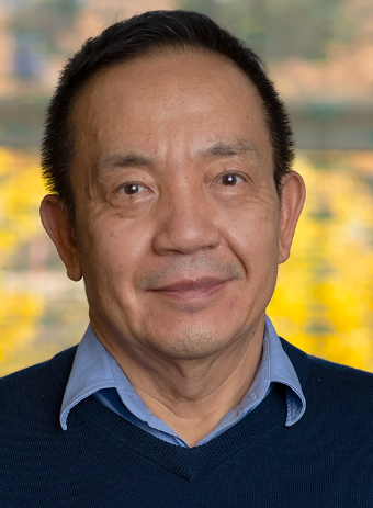 Chun Yuan合作论文数Department of Biomedical Engineering, John and Marcia Price College of Engineering, The University of Utah;Department of Radiology, The University of Utah;Department of Biomedical Informatics, The University of Utah10
 Zhensen Chen合作论文数9Hualu Han合作论文数8Rui Shen合作论文数8Dandan Yang合作论文数7
 Dongye Li (李东野)合作论文数徐州医学院附属医院心血管内科7Jinnan Wang合作论文数6
 Kui Ying (应葵)合作论文数Department of Engineering Physics, Tsinghua University6
 Niranjan Balu合作论文数Department of Radiology, University of Washington5
 Jie Sun合作论文数Univ. Washington, University of Washington5
 Shi Wang (王石)合作论文数Department of Engineering Physics, Tsinghua University5Haikun Qi (齐海坤)合作论文数4Ran Huo合作论文数4Tao Wang合作论文数4Qiang Zhang合作论文数4Jia Ning合作论文数4Mingzhu Fu合作论文数4Yongjun Han合作论文数4
 Rui Guo合作论文数School of medical technology, Beijing Institute of Technology4Shi Xinwei合作论文数4
 Feiyu Chen合作论文数4
 Thomas S. Hatsukami合作论文数Division of Vascular Surgery, Department of Surgery, School of Medicine, University of Washington;Vascular Imaging Laboratory, Department of Radiology, School of Medicine, University of Washington4Miaoqi Zhang合作论文数3Jingli Cao合作论文数3Mi Shen合作论文数3
 Yishi Wang合作论文数3Xing Wei合作论文数3
 Gaifen Liu (刘改芬)合作论文数Tiantan Hospital, Capital Medical University3
 Binbin Sui (隋滨滨)合作论文数Beijing Tiantan Hospital, Capital Medical University3
 Xiaowei Song合作论文数Surrey Memorial Hospital, Fraser Health Authority3
 Peiyi Gao (高培毅)合作论文数Beijing Tiantan Hospital, Capital Medical University3Bingyao Chen (陈炳耀)合作论文数3Haining Liu合作论文数2Hanyu Wei (魏寒宇)合作论文数2Chenming Wei合作论文数2Chenlin Du合作论文数2Ying Liao合作论文数2Jifan Li合作论文数2Boya Xu合作论文数2Dongyue Si合作论文数2
 Nan Zhang合作论文数2Aiqi Sun合作论文数Sch Med, Tsinghua Univ2




