Background:JAK/STAT interferon signaling interacts with the PI3K/AKT/mTOR pathway to drive hepatocellular carcinoma (HCC) progression and metastasis. RSAD2, an interferon-inducible gene, is upregulated by the PI3K/AKT/mTOR pathway and serves as a key factor for metabolic reprogramming to promote stem-like properties of cancer stem cells and tumor proliferation. In patients with resected HCC, RSAD2 upregulation showed an association with microvascular invasion, which is a proven risk factor for developing HCC metastasis. This clinical observation was compatible with preclinical findings. On the other hand, RSAD2 upregulation has been reported to confer poor prognosis in breast and gastric cancers. However, further clinical study of RSAD2 in HCC is lacking. As a result, we investigated the clinical implications of RSAD2 gene expression in HCC patients, in terms of its associations with survival, the presence of extra-hepatic metastasis, and other clinical manifestations. Methods: We studied 309 treatment-naïve HCC patients, as well as data from the TCGA and GTEx databases. Results:RSAD2 gene expression was differentially upregulated in HCC tumors when compared to normal liver tissues (p < 0.01). Elevated RSAD2 mRNA levels in the blood and the presence of extra-hepatic metastasis were independent prognostic factors for poor overall survival (OS) (p < 0.01). The median OS of patients with high RSAD2 expression vs. low expression were 5.4 vs. 14.2 months, respectively (p < 0.01). A high RSAD2 mRNA level was significantly correlated with the presence of extra-hepatic metastasis, nutritional disturbance, and functional impairment after controlling for confounding clinical factors (p < 0.05). Conclusions: High RSAD2 gene expression is associated with poorer OS, the presence of extra-hepatic metastasis, and quality-of-life disturbances in HCC patients.
Lumbar injection is a minimally invasive spinal procedure that requires accurate needle targeting in the spinal region under image guidance. Contrary to X-ray imaging commonly used for this procedure, magnetic resonance imaging (MRI) offers high soft tissue contrast and no ionizing radiation exposure. However, most existing magnetic resonance (MR)-safe robotic needle manipulators feature short stroke, low joint stiffness, and complex mechanism. This work introduces an intrinsically MR-safe robotic needle for the spine procedure, consisting of a 2-degree-of-freedom (DOF) compact remote center of motion (RCM) mechanism and 1-DOF needle driver operating under bellows-based hydraulic actuation. It is equipped with hydraulically activated locks and customized encoders for enhanced joint stiffness and closed-loop control. Communicating bellows are introduced in the teleoperation setup, minimizing the risk of fluid leakage. A geometry-based static model was developed and verified for the communicating bellows actuation system. The robotic system was shown to achieve subdegree and submillimeter motion accuracy in its RCM DOFs, and negligible signal-to-noise ratio reduction during the MR imaging quality test. This is to the authors' knowledge, the first robotic manipulator with both motorized RCM and needle translation using MR-safe hydraulic bellows actuators, potentially enabling real-time needle orientation and placement control under continuous MR scanning with optimal image quality.
ObjectiveThe purposes of this study were to retrospectively review mid-term outcomes of stent-assisted coil embolization for acutely ruptured cerebral aneurysms, and to identify risk factors.MethodsBetween April 2013 and October 2018, thirty-four consecutive patients had stent-assisted coil embolization for treatment of acutely ruptured cerebral aneurysms that were not amendable by simple coiling. Hospital subarachnoid hemorrhage registry and notes on hospital electronic medical systems were retrospectively reviewed. All patients had a dose of intravenous abciximab 10 mg and heparin 2000 units before stenting and started dual antiplatelet therapy after procedure.ResultsOf the thirty-four consecutive patients, twenty-six (76 %) were treated with braided stents and eight (24 %) were treated with laser-cut stents. There was no rebleeding in thirty-two patients who achieved total aneurysmal occlusion or residual neck only. Rebleeding occurred in two patients who only achieved partial embolization, resulting in mortality. There were three (9 %) thromboembolic complications, in which two were ADL-independent upon hospital discharge and the one with WFNS Grade V succumbed. Twenty (59 %) patients achieved clinical favorable outcomes (mRS 0–2) at follow-up (mean+/-SD: 17+/-15 months). The need of EVD insertion was a statistically significant risk factor for procedure-related cerebral infarction, intracerebral hemorrhage and 30-day mortality. The GOS and mRS were closely related to the need of EVD insertion and sidewall location of aneurysm.ConclusionsThe study established the efficacy and safety profile of stent-assisted embolization for treatment acutely ruptured cerebral aneurysms. The embolization goal should be total aneurysmal occlusion or with minimal residual neck to prevent rebleeding and reduce mortality. The use of such technique should be considered when simple coiling or microsurgical clipping is not feasible or suitable. The need of EVD insertion is an independent risk factor for intracerebral hematoma and 30-day mortality. When needed EVD insertion shall be considered before antiplatelet agents, and require exceptionally meticulous hemostasis intraoperatively.
Micro/nanorobots provide a promising approach for intravascular therapy with high precision. However, blood vessel is a highly complex system, and performing interventional therapy in those submillimeter segments remains challenging. While micro/nanorobots can enter submillimeter segments, they may still comprise nonbiodegradable parts, posing a considerable challenge for post-use removal. Here, we developed a retrievable magnetic colloidal microswarm, composed of tPA-anchored Fe 3 O 4 @mSiO 2 nanorobots (tPA-nbots), to archive tPA-mediated thrombolysis under balloon catheter-assisted magnetic actuation with x-ray fluoroscopy imaging system (CMAFIS). By deploying tPA-nbot transcatheter to the vicinity of the thrombus, the tPA-nbot microswarms were magnetically actuated to the blood clot at the submillimeter vessels with high precision. After thrombolysis, the tPA-nbots can be retrieved via the CMAFIS, as demonstrated in ex vivo organ of human placenta and in vivo carotid artery of rabbit. The proposed colloidal microswarm provides a promising robotic tool with high spatial precision for enhanced thrombolysis with low side effects.
Micro/nanorobotic swarms consisting of numerous tiny building blocks show great potential in biomedical applications because of their collective active delivery ability, enhanced imaging contrast, and environment-adaptive capability. However, in vivo real-time imaging and tracking of micro/nanorobotic swarms remain a challenge, considering the limited imaging size and spatial-temporal resolution of current imaging modalities. Here, we propose a strategy that enables real-time tracking and navigation of a microswarm in stagnant and flowing blood environments by using laser speckle contrast imaging (LSCI), featuring full-field imaging, high temporal-spatial resolution, and noninvasiveness. The change in dynamic convection induced by the microswarm can be quantitatively investigated by analyzing the perfusion unit (PU) distribution, offering an alternative approach to investigate the swarm behavior and its interaction with various blood environments. Both the microswarm and surrounding environment were monitored and imaged by LSCI in real time, and the images were further analyzed for simultaneous swarm tracking and navigation in the complex vascular system. Moreover, our strategy realized real-time tracking and delivery of a microswarm in vivo, showing promising potential for LSCI-guided active delivery of microswarm in the vascular system.
The recent rise of swarming microrobotics offers great promise in the revolution of minimally invasive embolization procedure for treating aneurysm. However, targeted embolization treatment of aneurysm using microrobots has significant challenges in the delivery capability and filling controllability. Here, we develop an interventional catheterization-integrated swarming microrobotic platform for aneurysm on-demand embolization in physiological blood flow. A pH-responsive self-healing hydrogel doped with magnetic and imaging agents is developed as the embolic microgels, which enables long-term self-adhesion under biological condition in a controllable manner. The embolization strategy is initiated by catheter-assisted deployment of swarming microgels, followed by the application of external magnetic field for targeted aggregation of microrobots into aneurysm sac under the real-time guidance of ultrasound and fluoroscopy imaging. Mild acidic stimulus is applied to trigger the welding of microgels with satisfactory bio-/hemocompatibility and physical stability and realize complete embolization. Our work presents a promising connection between the design and control of microrobotic swarms toward practical applications in dynamic environments.
Precipitating hydrophobic injectable liquid (PHIL; MicroVention, Aliso Viejo, CA, USA) and Squid (Balt, Irvine, CA, USA) are 2 newer liquid embolic agents used in endovascular embolization of cerebral arteriovenous malformation (AVM). This study aims to investigate and compare the effectiveness and safety profile of the 2 newer liquid embolic agents in the embolization of cerebral AVM. This is a retrospective study on all patients diagnosed with cerebral AVM undergoing endovascular embolization with liquid embolic agents PHIL and Squid admitted to the Division of Neurosurgery, Department of Surgery in Prince of Wales Hospital from January 2014 to June 2021. Twenty-three patients with cerebral AVM were treated with 34 sessions of endovascular embolization with either PHIL or Squid (17 sessions each) liquid embolic agents with a male to female ratio of 2.3:1 (male 16; female 7) and mean age of 44.6 (range, 12 to 67). The mean total nidus obliteration rate per session was 57% (range, 5% to 100%). Twenty-one patients (91.3%) received further embolization, stereotactic radiosurgery, or surgical excision after initial endovascular embolization. There were 2 morbidities (1 neurological and 1 non-neurological, 6%) and no mortalities (0%). All patients had static or improvement in modified Rankin Scale at 3 to 6 months at discharge. PHIL and Squid are effective and safe liquid embolic agents for endovascular embolization of cerebral AVM, achieving satisfactory nidal obliteration rates and patient functional outcomes.
Arterial feeders supplying a hepatocellular carcinoma are known to be interconnected through the sinusoid-like tumor vasculature. It was observed angiographically that when one of the feeders is selectively catheterized for drug delivery in transarterial chemoembolization (TACE), the whole tumor vasculature will be filled up, if the arterial inflow from all the other feeders is temporarily arrested with selective occlusion of feeding arteries (SOFA) using an additional catheter (balloon catheter). The feasibility of using the SOFA technique in TACE (SOFA-TACE) is discussed. In this prospective, monocentric feasibility study, with informed consent obtained, 8 consecutive patients of median age 64.5 years (60–68.8) and tumor dimension 4.7 cm (3.2–6.1), having specific tumor features (solitary, hypervascularity, well-defined, ≤ 7 cm, multiple tumor feeders), received SOFA-TACE using ethiodized oil–cisplatin suspension. Tumor response was assessed with 3-monthly CT using modified RECIST. A single tumor feeder was catheterized for drug delivery (8 cases). All other tumor feeders were successfully occluded with a balloon at one site (8 cases). Complete filling of the vasculature of the whole tumor was achieved in 7 of 8 cases with the SOFA technique as shown on arteriogram and CT, except in a case with an intratumoral septum. There was no complication. Surveillance CT (median 25 months, range 22–28) showed complete response in all cases. SOFA-TACE is feasible with reasonable safety and favorable treatment outcome; it may be a valuable technical option that may facilitate the procedures of selective TACE in technically challenging cases.
We propose ultrasound Doppler imaging and magnetic navigation of collective cell microrobots in whole blood. Cell microrobots are cultured using stem cells and iron microparticles, they have spheroidal structures and can be actuated under external magnetic fields. A collective of cell microrobots can be reversibly gathered and spread due to the tunable magnetic interaction, and are able to exhibit collective motion in whole blood under rotating magnetic fields. Simulation results indicate that the induced blood flow around the collective pattern affects the motion of red blood cells (RBCs), and experimental results show that Doppler signals are observed when emitting ultrasound waves to the microrobots. The induced Doppler signals are affected by the input field frequency and the ultrasound parameters (pulse repetition frequency). Due to the induced three-dimensional blood flow, Doppler signals can be observed when the imaging plane is above the collective microrobots, which enables indirect localization when performing navigation on an uneven surface. Our study investigates a strategy for pattern formation and navigation of collective microrobots under ultrasound Doppler imaging, demonstrating that the integration of collective control approach and medical imaging holds great potential for real-time active delivery tasks.
Introduction: This investigator-initiated clinical trial aims to study the efficacy and safety of administering selective internal radiation therapy with resin yttrium-90 microspheres (SIRT) followed by standard chemotherapy in unresectable intrahepatic cholangiocarcinoma (ICC). Methods: A phase 2 single-arm multicenter study was conducted in patients with unresectable ICC (NCT02167711). SIRT was administered at dose of 120 Gy targeted at tumor followed by commencement of gemcitabine 1,000 mg/m2 and cisplatin 25 mg/m2 on days one and eight of a 21-day cycle. The primary endpoint was overall survival (OS), and the secondary endpoints include progression-free survival (PFS), response rate according to Response Evaluation Criteria in solid tumors 1.1, toxicity, and time from SIRT to commencement of chemotherapy. Results: Total 31 patients were screened and twenty-four were recruited. All patients completed SIRT and 16 of them underwent subsequent chemotherapy. The median cycle of chemotherapy was 5 (range: 1–8). The median OS was 13.6 months (95% CI: 5.4–21.6) for the intent-to-treat population. Among 16 patients undergoing chemotherapy, the median OS was 21.6 months (95% CI: 7.3–25.2) and the median PFS was 9 months (95% CI: 3.2–13.1). The response rate was 25% (95% CI: 3.8–46.2%), and the disease control rate was 75% (95% CI: 53.8–96.2%). No new safety signal was observed, with fewer than 10% of patients suffering from grade 3 or higher treatment-related adverse events. The median time from SIRT to chemotherapy was 29 (range: 7–42) days. Eight patients could not receive chemotherapy due to rapid progressive disease (n = 4), underlying treatment unrelated comorbidities (n = 2), and withdrawal of consent due to personal reasons (n = 2). Conclusions: Treatment of SIRT followed by standard gemcitabine and cisplatin chemotherapy is feasible and effective for unresectable ICC. Further studies are required to study the optimal sequence of SIRT and chemotherapy.
BACKGROUND:Bleeding from carotid artery pseudoaneurysms is an emergency condition with high morbidity and mortality. We aimed to identify risk factors predicting pseudoaneurysmal bleeding as the cause of profuse epistaxis in irradiated head and neck cancer patients with suspect carotid blowout or pseudoaneurysms.METHODS:We retrospectively reviewed consecutive patients with history of radiation therapy for head and neck cancers and with nasal, oral or ear bleeding requiring in-patient treatment from hospital database. Pseudoaneurysms were subgrouped into internal carotid artery (ICA) pseudoaneurysms, and external carotid artery (ECA) pseudoaneurysms. The treatment outcomes were evaluated using 30-day mortality rate, recurrent bleeding, and cerebral infarction.RESULTS:There were 41 admissions for suspected carotid blowout or pseudoaneurysms from 1 July 2016 to 30 June 2020 with 17 bleeding pseudoaneurysms identified, including 11 internal carotid arteries (ICA) pseudoaneurysms and 6 external carotid arteries (ECA) pseudoaneurysms. Among ICA pseudoaneurysms, six patients passed Balloon occlusion test with embolization and parent artery occlusion (trapping) of ICA performed, and all ECA pseudoaneurysms were embolized with parent artery occlusion (trapping). Baseline hypertension and hypotension on arrival were predictive for pseudoaneurysmal bleeding. The degree of haemoglobin drop was not significantly different between pseudoaneurysmal bleeding and non-pseudoaneurysmal bleeding (2.1 ± 1.4 g/dL vs. 1.6 ± 1.4 g/dL, p = 0.234).CONCLUSIONS:We identified baseline hypertension and hypotension on arrival as predictive factors for pseudoaneurysmal bleeding in patients with irradiated head and neck cancer. Presence of these risk factors should alert the clinicians to the possibility of carotid pseudoaneurysms.
To compare the survival outcomes of patients treated with transarterial ethanol ablation (TEA) with those treated with liver resection (LR) for solitary HCC less than 5 cm in diameter, in patients stratified according to liver function using ALBI grade. This retrospective study approved by the Institutional Committee included all treatment-naïve patients with solitary HCC (≤ 5 cm) and Child–Pugh score 5, and who had received TEA (33 patients) or LR (192 patients) between 2004 and 2012. Treatment outcomes were compared between patients treated with TEA and LR after a period of at least 7 years of follow-up. Comparison was repeated for those patients with ALBI grade 2 or 3. Both overall survival (OS, months) and recurrence-free survival (RFS months) were significantly longer in the LR group (OS: LR 129.7[119.5, 140], TEA 69.1[55.9, 82.3], P < 0.0001; RFS: LR 91.3[43.5, 139.1], TEA 13.8 [11, 16.5], P < 0.0001). In patients with ALBI grade 2 or 3, there was no significant difference between the groups in OS or RFS (OS: LR 43.1[0, 91.2], TEA 55.4 [43.7, 67.2], P = 0.65; RFS: LR 17.8 [11.4, 24.2], TEA 11.9 [6.7, 17.1], P = 0.132). Transient epigastric discomfort and low-grade fever without consequence occurred in 8 patients (8/33 or 24.2%) in the TEA group. The overall survival after LR for HCCs ≤ 5 cm was superior to that after TEA but similar when compared in patients with ALBI grade 2 or 3, the ALBI grade is useful for patient selection for TEA or LR for HCCs ≤ 5 cm.
Swarming micro/nanorobots offer great promise in performing targeted delivery inside diverse hard-to-reach environments. However, swarm navigation in dynamic environments challenges delivery capability and real-time swarm localization. Here, we report a strategy to navigate a nanoparticle microswarm in real time under ultrasound Doppler imaging guidance for active endovascular delivery. A magnetic microswarm was formed and navigated near the boundary of vessels, where the reduced drag of blood flow and strong interactions between nanoparticles enable upstream and downstream navigation in flowing blood (mean velocity up to 40.8 mm/s). The microswarm-induced three-dimensional blood flow enables Doppler imaging from multiple viewing configurations and real-time tracking in different environments (i.e., stagnant, flowing blood, and pulsatile flow). We also demonstrate the ultrasound Doppler-guided swarm formation and navigation in the porcine coronary artery ex vivo. Our strategy presents a promising connection between swarm control and real-time imaging of microrobotic swarms for localized delivery in dynamic environments.
The objective was to evaluate the local treatment efficacy of transarterial ethanol ablation (TEA) as compared to radiofrequency ablation (RFA) for small hepatocellular carcinoma (HCC). This was a retrospective study between January 2005 and April 2017, in which the treatment outcomes of all patients who received either percutaneous TEA or RFA for HCC tumors of size ≤ 3 cm, of Child–Pugh grade A or B, received no prior treatment other than surgical resection, were compared. For TEA, a mixture of absolute ethanol and ethiodized oil at a proportion of 1:2 by volume was administered superselectively into the tumor via a microcatheter placed at the feeding arteries. The TEA group and the RFA group consisted of 68 consecutive patients (88 treated target tumors) and 129 consecutive patients (129 tumors), respectively. Technical success was achieved in all the target tumors in both groups. Grade 3 complication (CIRSE Classification) of prolonged fever occurred in 3 cases with multi-focal and large tumors in the TEA group. There was no statistically significant difference in complete response rate between the TEA group (84/88 or 95.5%) and the RFA group (188/195 or 96.4%) (p = 0.7). Time to progression in the TEA group [median 11.9 months, interquartile range (IQR) 5.6–18 months] was not statistically different from that in the RFA group (median 9.5 months, IQR 3.5–18.7 months) (p = 0.773). TEA could be an effective alternative of RFA for the local treatment of small HCC; it is especially valuable for tumors of unfavorable location.
Purpose To evaluate the feasibility, safety, and treatment effectiveness of ablative chemoembolization (ACE) in the treatment of hepatocellular carcinoma (HCC) and compare with a similar patient cohort who underwent conventional transarterial chemoembolization (cTACE). Materials and Methods This was a prospective phase I nonrandomized study conducted between March 2013 and October 2016 in accordance to the Declaration of Helsinki and Declaration Good Clinical Practice with written informed consent. There were 36 men and eight women (median age, 64 years [interquartile range, 58-74] and 74.5 years [interquartile range, 70-80], respectively). The primary end points were treatment safety and tumor response. The secondary end points were time to progression, progression-free survival, conversion to partial hepatectomy, and viable HCC within the tumor specimen. The end points of the study group (n = 22) were compared with those of a case-matched control group (n = 22) of patients who underwent conventional cTACE during the same period by using a Pearson χ2 test. Results Treatment with ACE was successfully completed in all patients without adverse effects. The complete response (CR) rates by patient or by tumor were both 100%. The median time to progression and median progression-free survival were significantly longer in the study group than in the control group (both were 28 months vs 10 months, respectively; P < .001). The number of patient conversions to hepatectomy was seven for ACE and three for cTACE. In the tumor specimens, viable tumor was found in two of eight specimens that underwent ACE and three of three that underwent cTACE. Conclusion ACE is a feasible, safe, and well-tolerated treatment for patients with HCC; it is highly effective and may be more effective than cTACE in achieving CR. © RSNA, 2017 Online supplemental material is available for this article.
Introduction: Patients with symptomatic high-grade intracranial atherosclerotic disease (ICAD) are at risk of stroke recurrence even under stringent medical therapy. Understanding how the plaque evolves in these patients may help prevent recurrent stroke. Methods: In a prospective, multicenter referral, longitudinal study, we recruited 40 patients (mean age = 61.1 years) with symptomatic high-grade ICAD (70-99% stenosis) confirmed by 3-dimensional rotational angiography (3DRA) from July 2007 to Feb 2016. All patients had 3DRA at baseline and 1 year. All patients received contemporary optimal medical treatment. We assessed the morphological features of qualifying ICAD lesions in the paired 3DRA images. A plaque was considered progressive if the plaque surface evolved from being smooth to irregular/ulcerated or from irregular to ulcerated, or the affected artery occluded at 1 year. We explored factors associated with plaque progression. Results: The qualifying ICAD lesions were in average 77.7% stenosis at baseline. The point of maximum stenosis was located at the distal end of the plaques in 21 (52.5%) cases, and 38 (95.0%) patients had eccentric plaques at baseline. The mean upstream plaque shoulder angulation was 46.4° at baseline. Eleven (27.5%) ICAD lesions progressed from baseline to 1 year. Longer lesions (p=0.038) with a smaller upstream plaque shoulder angulation (p=0.038) and the maximum stenosis locating at the distal end of the lesion (p=0.056) at baseline were more likely to progress in the plaque morphology over 1 year. Conclusions: Under modern medical treatment, although the majority of symptomatic ICAD lesions might regress over 1 year after an index stroke, lesions of a longer length, a smaller upstream angulation, or with the maximum stenosis located at the distal end were more likely to progress in terms of the plaque morphology.
Compressed sensing magnetic resonance imaging (CS-MRI) has attracted considerable attention due to its great potential in reducing scanning time and guaranteeing high-quality reconstruction. In conventional CS-MRI framework, the total variation (TV) penalty and L1-norm constraint on wavelet coefficients are commonly combined to reduce the reconstruction error. However, TV sometimes tends to cause staircase-like artifacts due to its nature in favoring piecewise constant solution. To overcome the model-dependent deficiency, a hybrid TV (TV1,2) regularizer is introduced in this paper by combining TV with its second-order version (TV2). It is well known that the wavelet coefficients of MR images are not only approximately sparse, but also have the property of tree-structured hierarchical sparsity. Therefore, a L0-regularized tree-structured sparsity constraint is proposed to better represent the measure of sparseness in wavelet domain. In what follows, we present our new CS-MRI framework by combining the TV1,2 regularizer and L0-regularized tree-structured sparsity constraint. However, the combination makes CS-MRI problem difficult to handle due to the nonconvex and nonsmooth natures of mixed constraints. To achieve solution stability, the resulting composite minimization problem is decomposed into several simpler subproblems. Each of these subproblems has a closed-form solution or could be efficiently solved using existing numerical method. The results from simulation and in vivo experiments have demonstrated the good performance of our proposed method compared with several conventional MRI reconstruction methods.
 George Kwok Chu Wong合作论文数Department of Surgery, Prince of Wales Hospital7
 Hui Joyce Wai Yi合作论文数7Kit Fai Lee合作论文数5
 Qianqian Wang (王乾乾)合作论文数School of Mechanical Engineering, Southeast University5Thomas Leung合作论文数5
 John D. Wong合作论文数School of Modern Languages & Cultures, Faculty of Arts, The University of Hong Kong5
 LAI Bo San Paul (賴寶山)合作论文数Division of Hepato-biliary and Pancreatic Surgery, Department of Surgery, Faculty of Medicine, The Chinese University of Hong Kong;Wong Tze Lam - Hing Tak Centre of Surgical Outcome Research, Faculty of Medicine, The Chinese University of Hong Kong;School of Biomedical Sciences, Faculty of Medicine, The Chinese University of Hong Kong;Department of Surgery, Prince of Wales Hospital;Hospital Authority5
 Li Zhang (張立)合作论文数Department of Mechanical and Automation Engineering, Faculty of Engineering, The Chinese University of Hong Kong;Department of Surgery, Faculty of Medicine, The Chinese University of Hong Kong;Shenzhen Institutes of Advanced Technology of the Chinese Academy of Sciences – CUHK Joint Laboratory of Robotics and Intelligent Systems5
 Bonaventure Ip合作论文数4
 Kai Fung Chan合作论文数Department of Biomedical Engineering, The Chinese University of Hong Kong4Liu Ryan Wen合作论文数China Unicom4
 Winnie Yeo (楊明明)合作论文数Department of Clinical Oncology, Faculty of Medicine, The Chinese University of Hong Kong4
 Lin Shi合作论文数Research Centre for Medical Image Computing, Faculty of Medicine, The Chinese University of Hong Kong;Division of Neurology, Department of Medicine and Therapeutics, The Chinese University of Hong Kong;Division of Biomedical Engineering, The Chinese University of Hong Kong4
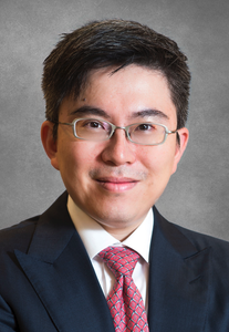 Stephen Lam Chan合作论文数Department of Clinical Oncology, Faculty of Medicine, The Chinese University of Hong Kong4
 Edwin Hui Pun (許斌)合作论文数Department of Clinical Oncology, The Chinese University of Hong Kong;Hong Kong Institute of Integrative Medicine, The Chinese University of Hong Kong4Tin Fong James Zhuang合作论文数3Carmen Chi Min Cho合作论文数3Qinglong Wang合作论文数3Sunny Cheung合作论文数3Li Leung合作论文数3
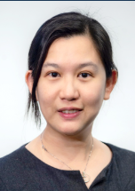 Ching Ning Charing Chong (莊清寧)合作论文数Department of Surgery, Faculty of Medicine, The Chinese University of Hong Kong;Department of Surgery, Prince of Wales Hospital, The Chinese University of Hong Kong3
 Wai Sang Poon (潘伟生)合作论文数Department of Surgery, Faculty of Medicine, The Chinese University of Hong Kong3Frankie Mo合作论文数Chinese University of Hong Kong3Yim Pan Chui合作论文数2rebecca yuenting ng合作论文数2Cheuk Man Chu合作论文数2Shuai Jiang合作论文数2Florence S Y Fan合作论文数2Yannie Soo合作论文数2Zhipeng Ning合作论文数2
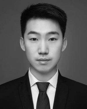 Haojin Yang合作论文数Chinese Univ Hong Kong, Dept Mech & Automat Engn, Shatin NT, Hong Kong 999077, Peoples R China2
 Xingzhou Du (杜星洲)合作论文数Institute of Robotics and Intelligent Systems, Dalian University of Technology2Tang Peggy合作论文数2Lee Janet Fung-yee合作论文数2
 Chi-Hang Yee合作论文数2
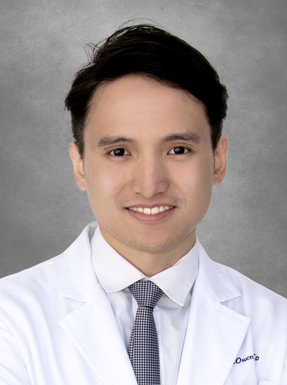 Owen H Ko (高浩)合作论文数Department of Medicine & Therapeutics, Faculty of Medicine, The Chinese University of Hong Kong2
 Defeng Wang (王德峰)合作论文数College of Medicine and Biological Information Engineering, Northeastern University;The Chinese University of Hong Kong2
 Cindy Leng合作论文数Nuffield Department of Clinical Neurosciences, University of Oxford;The Chinese University of Hong Kong2Ip Vincent H L合作论文数2
 Vincent Mok (莫仲棠)合作论文数Division of Neurology, Department of Medicine & Therapeutics, Faculty of Medicine, The Chinese University of Hong Kong;Medical Centre, The Chinese University of Hong Kong2
 Dongdong Jin (金东东)合作论文数School of Integrated Circuits, Harbin Institute of Technology, Shenzhen2
 Ben Wang (王奔)合作论文数College of Chemistry and Environmental Engineering, Shenzhen University2
 Pheng Ann Heng (王平安)合作论文数Department of Computer Science and Engineering, The Chinese University of Hong Kong2
 Tony Mok Shu Kam (莫樹錦)合作论文数Department of Clinical Oncology, Faculty of Medicine, The Chinese University of Hong Kong2Xinying Zou合作论文数2
 Wan-yee Joseph Lau (刘允怡)合作论文数Department of Surgery, Faculty of Medicine, The Chinese University of Hong Kong2
 Philip Wai Yan Chiu (趙偉仁)合作论文数Department of Surgery, Faculty of Medicine, The Chinese University of Hong Kong2Lawrence K. S. Wong合作论文数2




