Alterations in excitation/inhibition (E/I) balance and changes in motor neurons (MN) activity may contribute to MN vulnerability in ALS. The balance of pathogenic versus adaptive changes occurring in inhibitory synapses and affecting E/I balance remain unclear. Confocal microscopy of MN from P45 male SOD1G93A mice reveal downregulated GlyR but upregulated GABAR clusters at inhibitory synapses. GlyR and GABAR respond to PSAM and DREADD chemogenetic alterations of MN excitability, with increased activity driving increase in inhibitory clusters. An E3 ligase-conjugated intrabody (GFE3) degrades Gephyrin, decreases GABAR and GlyR clusters, increases net activity, and downregulates disease markers. However, simultaneous decrease of inhibition and increased activity by actPSAM and GFE3 shows no net beneficial effects on disease markers. Thus inhibitory synapses are involved in the early phases of ALS pathogenesis and respond to persistent homeostatic loops, and their suppression delivers a net activity increase, offering potential benefits on disease pathways.
Homeostatic feedback loops are essential to stabilize the activity of neurons and neuronal networks. It has been hypothesized that, in the context of Amyotrophic Lateral Sclerosis (ALS), an excessive gain in feedback loops might hyper- or hypo-excite motoneurons (MNs) and contribute to the pathogenesis. Here, we investigated how the neuromodulation of MN intrinsic properties is homeostatically controlled in presymptomatic adult SOD1(G93A) mice and in the age-matched control WT mice. First, we determined that Adrb2 and Adrb3 adrenergic receptors, which are Gs-coupled receptors and subject to tight and robust feedback loops, are specifically expressed in spinal MNs of both SOD1 and WT mice at P45. We then demonstrated that these receptors elicit a so-far overlooked neuromodulation of the electrical properties of MNs, in particular the frequency-current gain, a crucial determinant of excitability. These electrical properties are homeostatically regulated following receptor engagement, which triggers ion channel transcriptional changes and downregulates those receptors. These homeostatic feedbacks are not dysregulated in presymptomatic SOD1 mice, and they set the MN excitability upon β-adrenergic neuromodulation.
Abstract Glutamatergic excitatory synapses not only shape spiking activity and neuronal communication but also initiate activity-dependent signaling pathways that trigger transcriptional programs. Since glutamatergic excitatory synapses onto spinal motoneurons (MNs) are impaired presymptomatically in Amyotrophic Lateral Sclerosis, we investigated whether synapto-nuclear coupling is disrupted in MNs from mSOD1 mice and whether restoring it mitigates pathology. We developed an in vivo approach to selectively investigate the coupling between synaptic excitation and nuclear CREB phosphorylation in spinal MNs. Specific activation of Ia-MN synapses induces CREB phosphorylation in wild-type MNs but not in mSOD1 MNs at P50, indicating presymptomatic synapto-nuclear uncoupling. Enhancing cAMP/PKA signaling by pharmacological inhibition of cAMP degradation restored synapto-nuclear coupling, reduced misfolded SOD1, and slowed neuromuscular-junction denervation. Thus, activity-dependent synapto-nuclear signaling is impaired yet pharmacologically rescuable in mSOD1 MNs, supporting synapto-nuclear signaling as a determinant of MN resilience.
Rats exhibit significant recovery of locomotor function following incomplete spinal cord injuries, albeit with altered gait expression and reduced speed and stepping frequency. These changes likely result from and give insight into the reorganization within spared and injured spinal circuitry. Previously, we developed computational models of the mouse spinal locomotor circuitry controlling speed-dependent gait expression (Danner et al., 2017; Zhang et al., 2022). Here, we adapted these models to the rat and used the adapted model to explore potential circuit-level changes underlying altered gait expression observed after recovery from two different thoracic spinal cord injuries (lateral hemisection and contusion) that have roughly comparable levels of locomotor recovery (Danner et al., 2023). The model reproduced experimentally observed gait expression before injury and after recovery from lateral hemisection and contusion and suggests two distinct, injury-specific routes to restored function. First, recovery after lateral hemisection required substantial functional restoration of damaged descending drive and long propriospinal connections, suggesting compensatory plasticity through formation of detour pathways. Second, recovery after a moderate midline contusion predominantly relied on reorganization of spared sublesional networks and altered control of supralesional cervical circuits, compensating for weakened propriospinal and descending pathways. Despite these differences, sensitivity analysis revealed that restored activation of sublesional lumbar rhythm-generating circuits and appropriately balanced lumbar commissural connectivity are the key determinants of post-injury gait expression, suggesting that injury symmetry shapes how the cord reorganizes, but effective recovery in both cases depends on re-engaging these lumbar networks, which makes them prime targets for therapeutic intervention.
Within a year after a spinal cord injury (SCI), 75% of individuals develop spasticity. While normal movement relies on the ability to adjust reflexes appropriately, and on reciprocal inhibition of antagonistic muscles, spastic individuals display hyperactive spinal reflexes and involuntary muscle co-contractions. Current anti-spastic medications can suppress uncontrolled movements, but by acting on GABAergic signalling, these medications lead to severe side-effects and weakened muscle force, making them incompatible with activity-based therapies. We have previously shown that pharmacologically enhancing activity of KCC2, a chloride cotransporter, reduces signs of spasticity in anaesthetized chronic SCI rats. Here, we examine the effect of enhancing KCC2 in awake animals, using a battery of tests assessing hyperreflexia in multiple reflex pathways required for normal movement as well as locomotor function. Sprague-Dawley rats were implanted with chronic EMG electrodes bilaterally in ankle flexor and ankle extensor muscles and received a complete spinal transection at T12. Four weeks following SCI, the stretch reflex, the non-nociceptive cutaneous reflex pathway, the flexor withdrawal reflex and the crossed-extensor reflex pathway as well as locomotor function were evaluated before and after receiving the KCC2 enhancer, CLP290. Our results show that enhancing KCC2 activity normalizes reflex responses in multiple pathways and reduces muscle co-contraction without weakening motor output, thereby improving stepping ability. This work reveals the substantial potential for KCC2 enhancers as a novel anti-spastic treatment. KEY POINTS: After chronic spinal cord injury (SCI), the dysregulation of chloride homeostasis and subsequent decrease in spinal inhibition contributes to the development of spastic symptoms. We previously showed that enhancing the activity of the chloride co-transporter KCC2 after SCI decreases electrophysiological correlates of hyperreflexia and spasticity in deeply anaesthetized animals. Here, we examine the effect of enhancing KCC2 activity in awake animals on the excitability of multiple spinal reflex pathways required for normal movement including locomotion. We show that CLP290 decreases hyperreflexia, and reduces muscle co-contraction without weakening motor output, thereby improving stepping ability. Our results suggest that KCC2 enhancers such as CLP290 have the potential to be used as an anti-spastic treatment after SCI.
Mammals exhibit robust walking across diverse environments, a capability largely attributed to central pattern generators (CPGs) in the spinal cord. Afferent feedback modulates CPG output and plays a critical role in adaptive locomotion, yet its specific contributions remain poorly understood. To investigate this, we used a neuromusculoskeletal model to simulate hindlimb locomotion in spinalized cats encountering a hole and experiencing a sudden loss of ground support, as described in prior experimental studies. The model couples a trunk-and-hindlimb musculoskeletal system to a pair of two-level, half-center CPGs-one for each hindlimb. The model reproduced the observed adaptive interlimb coordination that allows cats to maintain walking after the sudden loss of ground support. Notably, the adaptive response emerged without re-optimizing parameters, which were tuned for steady walking in an environment without holes. Nullcline analysis based on dynamical systems theory revealed that afferent feedback mechanisms controlling the transitions between fast and slow neuronal dynamics facilitated adaptive interlimb coordination. These findings provide mechanistic insight into how spinal feedback circuits support robust locomotion through dynamic interactions between the nervous system, the musculoskeletal system, and the environment.
Rats exhibit significant recovery of locomotor function following incomplete spinal cord injuries, albeit with altered gait expression and reduced speed and stepping frequency. These changes likely result from and give insight into the reorganization within spared and injured spinal circuitry. Previously, we developed computational models of the mouse spinal locomotor circuitry controlling speed-dependent gait expression (Danner et al. 2017; Zhang et al. 2022). Here, we adapted these models to the rat and used the adapted model to explore potential circuit-level changes underlying altered gait expression observed after recovery from two different thoracic spinal cord injuries (lateral hemisection and contusion) that have roughly comparable levels of locomotor recovery (Danner et al., 2023). The model reproduced experimentally observed gait expression before injury and after recovery from lateral hemisection and contusion, and suggests two distinct, injury-specific routes to restored function. First, recovery after lateral hemisection required substantial functional restoration of damaged descending drive and long propriospinal connections, suggesting compensatory plasticity through formation of detour pathways. Second, recovery after a moderate midline contusion predominantly relied on reorganization of spared sublesional networks and altered control of supralesional cervical circuits, compensating for weakened propriospinal and descending pathways. Despite these differences, sensitivity analysis revealed that restored activation of sublesional lumbar rhythm-generating circuits and appropriately balanced lumbar commissural connectivity are the key determinants of post-injury gait expression, suggesting that injury symmetry shapes how the cord reorganizes, but effective recovery in both cases depends on re-engaging these lumbar networks, which makes them prime targets for therapeutic intervention.
Locomotion is a complex process involving specific interactions between the central neural controller and the mechanical components of the system. The basic rhythmic activity generated by locomotor circuits in the spinal cord defines rhythmic limb movements and their central coordination. The operation of these circuits is modulated by sensory feedback from the limbs providing information about the state of the limbs and the body. However, the specific role and contribution of central interactions and sensory feedback in the control of locomotor gait and posture remain poorly understood. We use biomechanical data on quadrupedal locomotion in mice and recent findings on the organization of neural interactions within the spinal locomotor circuitry to create and analyse a tractable mathematical model of mouse locomotion. The model includes a simplified mechanical model of the mouse body with four limbs and a central controller composed of four rhythm generators, each operating as a state machine controlling the state of one limb. Feedback signals characterize the load and extension of each limb as well as postural stability (balance). We systematically investigate and compare several model versions and compare their behaviour to existing experimental data on mouse locomotion. Our results highlight the specific roles of sensory feedback and some central propriospinal interactions between circuits controlling fore and hind limbs for speed-dependent gait expression. Our models suggest that postural imbalance feedback may be critically involved in the control of swing-to-stance transitions in each limb and the stabilization of walking direction.
Objective. Studying the neural components regulating movement in human locomotion is obstructed by the inability to perform invasive experimental recording in the human neural circuits. Neuromechanical simulations can provide insights by modeling the locomotor circuits. Past neuromechanical models proposed control of locomotion either driven by central pattern generators (CPGs) with simple sensory commands or by a purely reflex-based network regulated by state-machine mechanisms, which activate and deactivate reflexes depending on the detected gait cycle phases. However, the physiological interpretation of these state machines remains unclear. Here, we present a physiologically plausible model to investigate spinal control and modulation of human locomotion. Approach. We propose a bio-inspired controller composed of two coupled CPGs that produce the rhythm and pattern, and a reflex-based network simulating low-level reflex pathways and Renshaw cells. This reflex network is based on leaky-integration neurons, and the whole system does not rely on changing reflex gains according to the gait cycle state. The musculoskeletal model is composed of a skeletal structure and nine muscles per leg generating movement in sagittal plane. Main results. Optimizing the open parameters for effort minimization and stability, human kinematics and muscle activation naturally emerged. Furthermore, when CPGs were not activated, periodic motion could not be achieved through optimization, suggesting the necessity of this component to generate rhythmic behavior without a state machine mechanism regulating reflex activation. The controller could reproduce ranges of speeds from 0.3 to 1.9 m s-1. The results showed that the net influence of feedback on motoneurons (MNs) during perturbed locomotion is predominantly inhibitory and that the CPGs provide the timing of MNs' activation by exciting or inhibiting muscles in specific gait phases. Significance. The proposed bio-inspired controller could contribute to our understanding of locomotor circuits of the intact spinal cord and could be used to study neuromotor disorders.
Hiccups (singultus) comprise repeated episodes of sharp, large-volume inspiration followed by glottic closure, interspersed among normal inspiration. Hiccupping causes involuntary contractions of diaphragm and thoracic musculature. While generally short-lived and self-limiting, hiccupping can last hours, or days, or longer, and cause wide-ranging medical, quality of life, and economic burdens. A proposed mechanism for hiccups is a pathway comprising: 1) an afferent limb including ascending fibers of the vagus nerve, 2) a central processing component in the midbrain and brainstem, and 3) an efferent limb including motorneurons of the diaphragm, glottis, and intercostal muscles. We hypothesize that hiccups represent a disinhibition of vestigial neural circuitry in the central processing component, manifesting as a multi-stable state of abnormal respiratory rhythm: a "respiratory arrhythmia". This hypothesis applies the concept of multi-stable (chaotic) systems to the respiratory cycle and stable, but aberrant, breathing rhythms. A system is multi-stable if, for a set of inputs, it has multiple potential stable states, depending on the system's prior state and the effects of external perturbations. This hypothesis is predicated on Straus et al.'s suggestion that hiccups are an evolutionary preservation of gill ventilation, normally suppressed by neural circuitry controlling mammalian breathing. The respiratory system, it is proposed, could stably exhibit eupneic breathing motor patterns or a mix of eupneic and aquatic-like patterns (hiccups). That is, eupnea alone, or eupnea plus gill ventilation are alternative stable equilibrium points of a multi-stable system. Evidence supporting the hypothesis includes computational models of the respiratory control center manifesting hiccup-like patterns, and experimental data showing that medullary stimulation induces hiccups-like breathing in cats. Additional evidence includes clinical reports of therapeutic perturbations to, or near, the putative respiratory pattern generator resetting the breathing pattern to the default stable state, eupnea. These approaches, which provide strong afferent input to the brainstem regulatory network rather than disrupting aberrant afferent signaling, include elevating CO2 concentrations to excite central and peripheral chemoreceptors increasing respiratory drive. Other examples include nasopharyngeal stimulation or sudden administration of strong sweet or sour tastes. Viewing hiccups as a respiratory arrhythmia could prove helpful in developing targeted interventions.
Thoracic spinal cord injury affects long propriospinal neurons that interconnect the cervical and lumbar en-largements. These neurons are crucial for coordinating forelimb and hindlimb locomotor movements in a speed-dependent manner. However, recovery from spinal cord injury is usually studied over a very limited range of speeds that may not fully expose circuitry dysfunction. To overcome this limitation, we investigated overground locomotion in rats trained to move over an extended distance with a wide range of speeds both pre-injury and after recovery from thoracic hemisection or contusion injuries. In this experimental context, intact rats expressed a speed-dependent continuum of alternating (walk and trot) and non-alternating (canter, gallop, half-bound gallop, and bound) gaits. After a lateral hemisection injury, rats recovered the ability to locomote over a wide range of speeds but lost the ability to use the highest-speed gaits (half-bound gallop and bound) and predomi-nantly used the limb contralateral to the injury as lead during canter and gallop. A moderate contusion injury caused a greater reduction in maximal speed, loss of all non-alternating gaits, and emergence of novel alternating gaits. These changes resulted from weak fore-hind coupling together with appropriate control of left-right alternation. After hemisection, animals expressed a subset of intact gaits with appropriate interlimb coordination even on the side of the injury, where the long propriospinal connections were severed. These observations highlight how investigating locomotion over the full range of speeds can reveal otherwise hidden aspects of spinal locomotor control and post-injury recovery.
Crossed reflexes are mediated by commissural pathways transmitting sensory information to the contralateral side of the body, but the underlying network is not fully understood. Commissural pathways coordinating the activities of spinal locomotor circuits during locomotion have been characterized in mice, but their relationship to crossed reflexes is unknown. We show the involvement of two genetically distinct groups of commissural interneurons (CINs) described in mice, V0 and V3 CINs, in the crossed reflex pathways. Our data suggest that the exclusively excitatory V3 CINs are directly involved in the excitatory crossed reflexes and show that they are essential for the inhibitory crossed reflexes. In contrast, the V0 CINs, a population that includes excitatory and inhibitory CINs, are not directly involved in excitatory or inhibitory crossed reflexes but downregulate the inhibitory crossed reflexes. Our data provide insights into the spinal circuitry underlying crossed reflexes in mice, describing the roles of V0 and V3 CINs in crossed reflexes.
Mammalian locomotion is generated by central pattern generators (CPGs) in the spinal cord, which produce alternating flexor and extensor activities controlling the locomotor movements of each limb. Afferent feedback signals from the limbs are integrated by the CPGs to provide adaptive control of locomotion. Responses of CPG-generated neural activity to afferent feedback stimulation have been previously studied during fictive locomotion in immobilized cats. Yet, locomotion in awake, behaving animals involves dynamic interactions between central neuronal circuits, afferent feedback, musculoskeletal system, and environment. To study these complex interactions, we developed a model simulating interactions between a half-center CPG and the musculoskeletal system of a cat hindlimb. Then, we analyzed the role of afferent feedback in the locomotor adaptation from a dynamic viewpoint using the methods of dynamical systems theory and nullcline analysis. Our model reproduced limb movements during regular cat walking as well as adaptive changes of these movements when the foot steps into a hole. The model generates important insights into the mechanism for adaptive locomotion resulting from dynamic interactions between the CPG-based neural circuits, the musculoskeletal system, and the environment.
Speed-dependent interlimb coordination allows animals to maintain stable locomotion under different circumstances. The V3 neurons are known to be involved in interlimb coordination. We previously modeled the locomotor spinal circuitry controlling interlimb coordination (Danner et al., 2017). This model included the local V3 neurons that mediate mutual excitation between left and right rhythm generators (RGs). Here, our focus was on V3 neurons involved in ascending long propriospinal interactions (aLPNs). Using retrograde tracing, we revealed a subpopulation of lumbar V3 aLPNs with contralateral cervical projections. V3OFF mice, in which all V3 neurons were silenced, had a significantly reduced maximal locomotor speed, were unable to move using stable trot, gallop, or bound, and predominantly used a lateral-sequence walk. To reproduce this data and understand the functional roles of V3 aLPNs, we extended our previous model by incorporating diagonal V3 aLPNs mediating inputs from each lumbar RG to the contralateral cervical RG. The extended model reproduces our experimental results and suggests that locally projecting V3 neurons, mediating left–right interactions within lumbar and cervical cords, promote left–right synchronization necessary for gallop and bound, whereas the V3 aLPNs promote synchronization between diagonal fore and hind RGs necessary for trot. The model proposes the organization of spinal circuits available for future experimental testing.
 Ursula S. Hofstoetter合作论文数25
 Karen Minassian合作论文数Center for Biomedical Engineering and Physics Austria, Medical University Vienna24
 Winfried Mayr合作论文数Center for Biomedical Engineering and Physics Austria, Medical University Vienna17
 Ilya Rybak合作论文数Laboratory for Theoretical and Computational Neuroscience, Department of Neurobiology and Anatomy, College of Medicine, Drexel University16
 Frank Rattay合作论文数Institute for Analysis and Scientific Computing, Technische Universität Wien13
 Natalia A Shevtsova合作论文数School of Biomedical Engineering, Drexel University12
 Matthias Krenn合作论文数Center of Medical Physics and Biomedical Engineering, Medical University of Vienna10
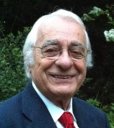 Milan R. Dimitrijevic合作论文数Division of Restorative Neurology and Human Neurobiology, Baylor College of Medicine/Department of Physical Medicine and Rehabilitation, Baylor College of Medicine8
 Jessica Ausborn合作论文数College of Medicine, Drexel University7
 Heinrich Binder合作论文数5
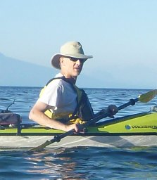 David S K Magnuson合作论文数Univ Louisville, Kentucky Spinal Cord Injury Res Ctr, Louisville, KY 40292 USA5Freundl Brigitta合作论文数5
 Sergey N. Markin合作论文数Department of Neurobiology and Anatomy, Drexel University College of Medicine5
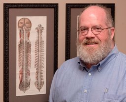 Tansey Keith E合作论文数Crawford Research Institute, Emory University School of Medicine4Caron Guillaume合作论文数UMR 7287 «Institut des Sciences du Mouvement: Etienne-Jules MAREY» (ISM-EJM),, Aix-Marseille Université4
 Auke Jan Ijspeert合作论文数Institute of Bioengineering, Ecole Polytechnique Fédérale de Lausanne4
 Shravan Tata Ramalingasetty合作论文数3
 Dylan Deska-Gauthier合作论文数3Ying Zhang合作论文数Dalhousie Univ, Dept Med Neurosci, Halifax, NS B3H 4R2, Canada3
 Daniel Zytnicki合作论文数Laboratoire de Neurophysiologie, Collège de France, Paris France3
 Alain Frigon合作论文数Dept Physiol .;Univ . Montreal;Dept Physiol ., Univ . Montreal3
 Shinya Aoi合作论文数Dept . of Aeronautics and Astronautics;Graduate School of Engineering;Kyoto University;Graduate School of Engineering, Kyoto University3
 Francesco Roselli合作论文数Department of Neurological and Psychiatric Sciences, University of Bari3
 Yaroslav I. Molkov合作论文数Department of Neurobiology & Anatomy, Drexel University College of Medicine3Kazuo TSUCHIYA合作论文数2
 Jonathan Arreguit合作论文数Ecole Polytech Fed Lausanne, STI BioRob, ME D1 1226,Stn 9, CH-1015 Lausanne, Switzerland2Dai Yanagihara合作论文数2
 Marcin Baczyk合作论文数University of Gothenburg, University School of Physical Education2
 Han Zhang合作论文数2
 Helmut Kern合作论文数2
 Soichiro Fujiki合作论文数2
 Julien Bouvier合作论文数Institut de Neurobiologie Alfred Fessard (FRC 2118, Centre de Recherche de Gif-sur Yvette (C.N.R.S.2Olivier D. Laflamme合作论文数1Aboud Tahanis合作论文数1Casey Hainline合作论文数1Colin Mackay合作论文数1Sruthi Krishnamurthy合作论文数1
 Charly G. Lecomte合作论文数1Barry McKay合作论文数1Andrea Toth合作论文数1
 Elizaveta Latash合作论文数1
 Cristina Peixoto Santos合作论文数1
 Diego Torricelli合作论文数1Guoning Yu合作论文数1Anushree Sabnis合作论文数1Joanna Borowska-Fielding合作论文数1
 Andrea Di Russo合作论文数1
 Jadwiga N Bilchak合作论文数1
 Dimitri Rodarie合作论文数Ecole Polytech Fed Lausanne EPFL, Blue Brain Project, Geneva, Switzerland1




