A wireless brain–spine interface is presented that enables macaques with a spinal cord injury to regain locomotor movements of a paralysed leg. Grégoire Courtine and colleagues show that a fully implantable, wireless brain–spine interface can be used to improve locomotion after a unilateral spinal lesion in monkeys without training. The authors implanted monkeys with an electrode array in the leg area of the motor cortex and a stimulator in the lumbar spinal cord, enabling real-time decoding and stimulation. Decoded activity from the motor cortex was used to stimulate 'hotspot' locations in the lumbar spinal cord that control hindlimb flexion and extension during locomotion. Stimulating these hotspots enhanced flexion and extension of the target muscles during locomotion in intact monkeys and restored weight-bearing locomotion of the paralysed leg in monkeys with a unilateral spinal cord lesion six days after the injury. This proof-of-principle study shows that a similar system may improve or restore locomotion in people with spinal cord injury. Spinal cord injury disrupts the communication between the brain and the spinal circuits that orchestrate movement. To bypass the lesion, brain–computer interfaces1,2,3 have directly linked cortical activity to electrical stimulation of muscles, and have thus restored grasping abilities after hand paralysis1,4. Theoretically, this strategy could also restore control over leg muscle activity for walking5. However, replicating the complex sequence of individual muscle activation patterns underlying natural and adaptive locomotor movements poses formidable conceptual and technological challenges6,7. Recently, it was shown in rats that epidural electrical stimulation of the lumbar spinal cord can reproduce the natural activation of synergistic muscle groups producing locomotion8,9,10. Here we interface leg motor cortex activity with epidural electrical stimulation protocols to establish a brain–spine interface that alleviated gait deficits after a spinal cord injury in non-human primates. Rhesus monkeys (Macaca mulatta) were implanted with an intracortical microelectrode array in the leg area of the motor cortex and with a spinal cord stimulation system composed of a spatially selective epidural implant and a pulse generator with real-time triggering capabilities. We designed and implemented wireless control systems that linked online neural decoding of extension and flexion motor states with stimulation protocols promoting these movements. These systems allowed the monkeys to behave freely without any restrictions or constraining tethered electronics. After validation of the brain–spine interface in intact (uninjured) monkeys, we performed a unilateral corticospinal tract lesion at the thoracic level. As early as six days post-injury and without prior training of the monkeys, the brain–spine interface restored weight-bearing locomotion of the paralysed leg on a treadmill and overground. The implantable components integrated in the brain–spine interface have all been approved for investigational applications in similar human research, suggesting a practical translational pathway for proof-of-concept studies in people with spinal cord injury.
Contrary to cats and primates, cortical contribution to hindlimb locomotor movements is not critical in rats. However, the importance of the motor cortex to regain locomotion after neurological disorders in rats suggests that cortical engagement in hindlimb motor control may depend on the behavioral context. To investigate this possibility, we recorded whole-body kinematics, muscle synergies, and hindlimb motor cortex modulation in freely moving rats performing a range of natural locomotor procedures. We found that the activation of hindlimb motor cortex preceded gait initiation. During overground locomotion, the motor cortex exhibited consistent neuronal population responses that were synchronized with the spatiotemporal activation of hindlimb motoneurons. Behaviors requiring enhanced muscle activity or skilled paw placement correlated with substantial adjustment in neuronal population responses. In contrast, all rats exhibited a reduction of cortical activity during more automated behavior, such as stepping on a treadmill. Despite the facultative role of the motor cortex in the production of locomotion in rats, these results show that the encoding of hindlimb features in motor cortex dynamics is comparable in rats and cats. However, the extent of motor cortex modulations appears linked to the degree of volitional engagement and complexity of the task, reemphasizing the importance of goal-directed behaviors for motor control studies, rehabilitation, and neuroprosthetics. SIGNIFICANCE STATEMENT We mapped the neuronal population responses in the hindlimb motor cortex to hindlimb kinematics and hindlimb muscle synergies across a spectrum of natural locomotion behaviors. Robust task-specific neuronal population responses revealed that the rat motor cortex displays similar modulation as other mammals during locomotion. However, the reduced motor cortex activity during more automated behaviors suggests a relationship between the degree of engagement and task complexity. This relationship emphasizes the importance of the behavioral procedure to engage the motor cortex during motor control studies, gait rehabilitation, and locomotor neuroprosthetic developments in rats.
Electrical neuromodulation of lumbar segments improves motor control after spinal cord injury in animal models and humans. However, the physiological principles underlying the effect of this intervention remain poorly understood, which has limited the therapeutic approach to continuous stimulation applied to restricted spinal cord locations. Here we developed stimulation protocols that reproduce the natural dynamics of motoneuron activation during locomotion. For this, we computed the spatiotemporal activation pattern of muscle synergies during locomotion in healthy rats. Computer simulations identified optimal electrode locations to target each synergy through the recruitment of proprioceptive feedback circuits. This framework steered the design of spatially selective spinal implants and real-time control software that modulate extensor and flexor synergies with precise temporal resolution. Spatiotemporal neuromodulation therapies improved gait quality, weight-bearing capacity, endurance and skilled locomotion in several rodent models of spinal cord injury. These new concepts are directly translatable to strategies to improve motor control in humans.
The mechanical mismatch between soft neural tissues and stiff neural implants hinders the long-term performance of implantable neuroprostheses. Here, we designed and fabricated soft neural implants with the shape and elasticity of dura mater, the protective membrane of the brain and spinal cord. The electronic dura mater, which we call e-dura, embeds interconnects, electrodes, and chemotrodes that sustain millions of mechanical stretch cycles, electrical stimulation pulses, and chemical injections. These integrated modalities enable multiple neuroprosthetic applications. The soft implants extracted cortical states in freely behaving animals for brain-machine interface and delivered electrochemical spinal neuromodulation that restored locomotion after paralyzing spinal cord injury.
Half of human spinal cord injuries lead to chronic paralysis. Here, we introduce an electrochemical neuroprosthesis and a robotic postural interface designed to encourage supraspinally mediated movements in rats with paralyzing lesions. Despite the interruption of direct supraspinal pathways, the cortex regained the capacity to transform contextual information into task-specific commands to execute refined locomotion. This recovery relied on the extensive remodeling of cortical projections, including the formation of brainstem and intraspinal relays that restored qualitative control over electrochemically enabled lumbosacral circuitries. Automated treadmill-restricted training, which did not engage cortical neurons, failed to promote translesional plasticity and recovery. By encouraging active participation under functional states, our training paradigm triggered a cortex-dependent recovery that may improve function after similar injuries in humans.
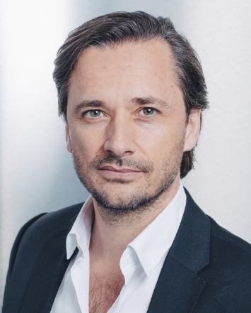 Grégoire Courtine合作论文数Interschool Institute Neuro-X, School of Life Sciences, Ecole Polytechnique Fédérale de Lausanne;ONWARD;Centre Hospitalier Universitaire Vaudois8
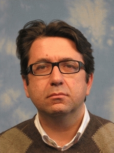 Silvestro Micera合作论文数School of Engineering, Ecole Polytechnique Federale de Lausanne7
 Quentin Barraud合作论文数7
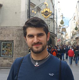 Eduardo Martin Moraud合作论文数Center for Neuroprosthetics and Institute of Bioengineering, EPFL7
 Jerome Gandar合作论文数5
 Pavel Musienko合作论文数Pavlov Institute of Physiology, Russian Academy of Sciences5
 Arthur Hirsch合作论文数4
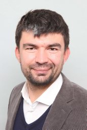 Tomislav Milekovic合作论文数Bernstein Center Freiburg, University of Freiburg4
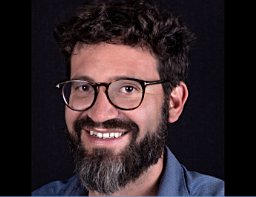 Marco Capogrosso合作论文数4
 Natalia Pavlova合作论文数Centre for Neuroprosthetics and Brain Mind Institute, EPFL;St. Petersburg, Pavlov Institute of Physiology;Centre for Neuroprosthetics and Brain Mind Institute, Pavlov Institute of Physiology4
 Leonie Asboth合作论文数4
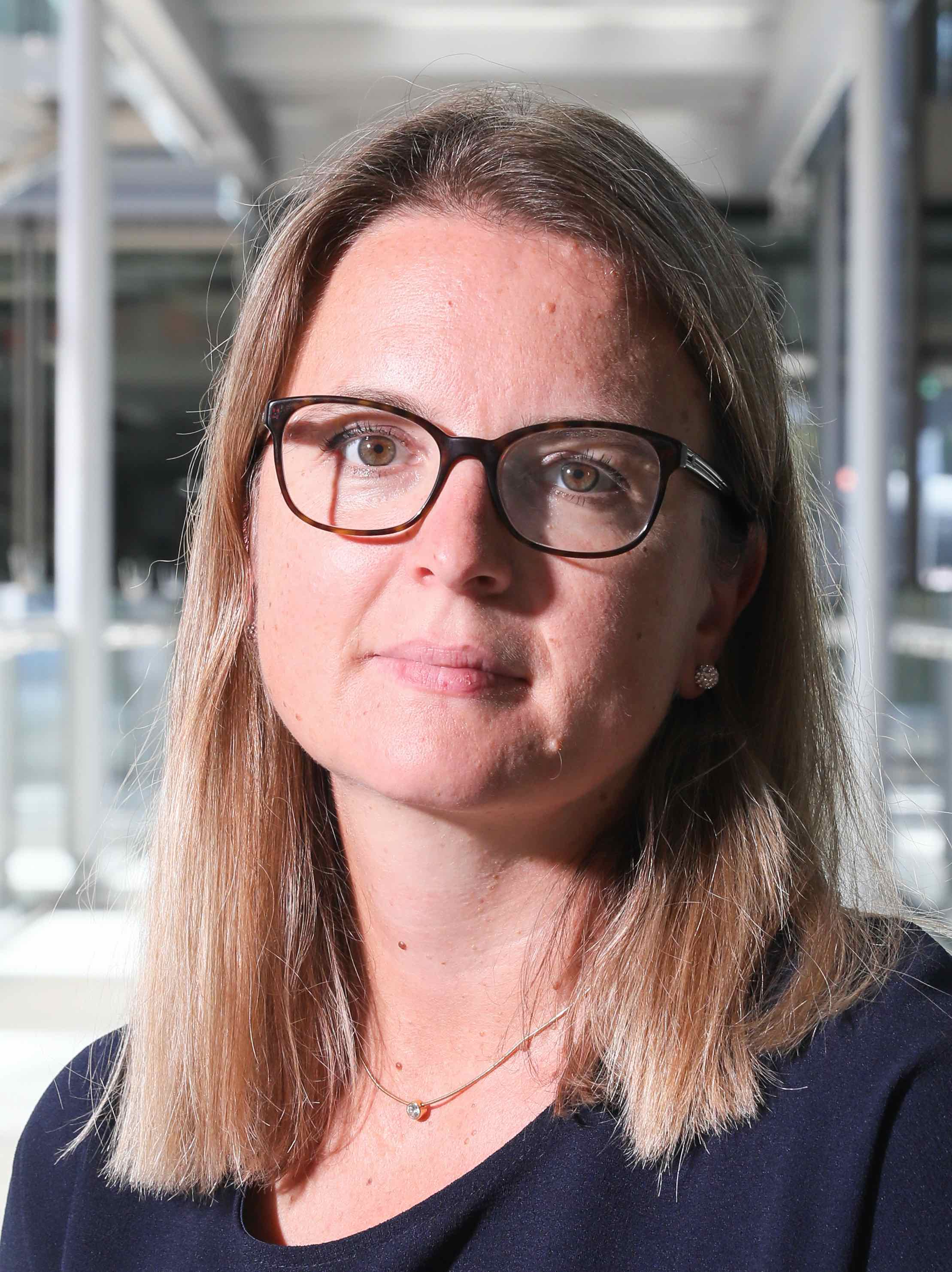 Stéphanie Lacour合作论文数Bertarelli Foundation, School of Engineering, Ecole Polytechnique Federale de Lausanne;Laboratory for Soft Bioelectronic Interfaces, Ecole Polytechnique Federale de Lausanne;Center for Neuroprosthetics, Ecole Polytechnique Federale de Lausanne;Neuro-X institute, Ecole Polytechnique Federale de Lausanne4
 Rubia Van Den Brand合作论文数4
 Ivan Minev合作论文数Department of Automatic Control and Systems Engineering, Faculty of Engineering, The University of Sheffield4
 Nadia Dominici合作论文数Laboratory of Neuromotor Physiology, Santa Lucia Foundation4
 Jack Digiovanna合作论文数Department of Biomedical Engineering, University of Florida4
 Lucia Friedli合作论文数3
 Qihan Liu合作论文数University of Science and Technology of China, Univ. of Sci. and Technol. of China3
 Nicolas Vachicouras合作论文数Institute of Microengineering and Institute of Bioengineering, Ecole Polytechnique Fédérale de Lausanne (EPFL;Centre for Neuroprosthetics and Brain Mind Institute, EPFL;Institute of Microengineering and Institute of Bioengineering, EPFL3Nikolaus Wenger合作论文数Center for Neuroprosthetics and Brain Mind Institute, School of Life Sciences, Swiss Federal Institute of Technology (EPFL), Lausanne, Switzerland3Torres Rafael Fajardo合作论文数3
 Zhigang Suo (锁志刚)合作论文数John A. Paulson School of Engineering and Applied Sciences, Harvard University;Kavli Institute for Bionano Science & Technology, Harvard University3Alexandre Larmagnac合作论文数3Kay Bartholdi合作论文数2Julie Kreider合作论文数2peter detemple合作论文数2
 Janos Vörös合作论文数Laboratory of Biosensors and Bioelectronics, Institute for Biomedical Engineering, Department of Information Technology and Electrical Engineering, Eidgenössische Technische Hochschule Zürich2
 Erwan Bezard合作论文数Centre Broca Nouvelle-Aquitaine, Université de Bordeaux;SE-Therapeutics;Institut des Maladies Neurodégénératives CNRS;Motac Neuroscience Ltd;TreeFrog Therapeutics2
 Isabel Vollenweider合作论文数2Heutschi Janine合作论文数2Huerlimann Michèle合作论文数2
 Jocelyne Bloch合作论文数Department of Neurosurgery, University Hospital of Lausanne2silvio kraus合作论文数1P. Musienko合作论文数1Janos Voeroes合作论文数1David Xing合作论文数1felix schmitz合作论文数1Nicolas Buse合作论文数1
 Camille G. Le Goff合作论文数Center for Neuroprosthetics and Brain Mind Institute, Swiss Federal Institute of Technology Lausanne1Laetitia Baud合作论文数Swiss Fed Inst Technol EPFL, Ctr Neuroprosthet, Int Parapleg Fdn Chair Spinal Cord Repair, Lausanne, Switzerland1Elodie Rey合作论文数1
 David Borton合作论文数Center for Biomedical Engineering, Department of Molecular Pharmacology, Physiology, and Biotechnology, Brown University1
 Jean-Baptiste Mignardot合作论文数1Wai Kin D. Ko合作论文数1Capogrosso Michela合作论文数1Yang Jianzhong合作论文数1
 Wagner Fabien合作论文数Remote Sensing Division, National Institute for Space Research - INPE;UMR 93, Cirad;Ecologie des Forêts de Guyane;Kourou;Cirad, National Institute for Space Research - INPE1
 Janine Beauparlant合作论文数1




