An optical vortex (OV) beam is a singular beam with a helical wavefront and a spatially varying phase profile, whereas spontaneous parametric down conversion (SPDC) is a well-established process of generating two-photon state of light. Here, we report photon generation via SPDC using a supercontinuum (SC) beam and a SC OV beam as pump. Specifically, we report generation of an integer charge SC OV beam with a spectral range of 420-480 nm by illuminating a narrow-band spiral phase plate (SPP) designed for 467 nm with a SC light beam of 390-500 nm spectral range. The broad bandwidth of the generated SC OV beam is confirmed using bandpass filters, and cylindrical lens based measurement is adapted to estimate the topological charge of the beam. The mode purity of the generated SC OV beam is determined by analysing its complex field, measured using a Mach–Zehnder interferometer. Besides, generation of fractional OV beam is observed around 400 nm, and the results are compared to the case of using a CW laser operating at 405 nm. In addition, we report generation of photon pairs via SPDC by pumping a β-BBO crystal with the SC beam and the integer charge SC OV beam. The down-conversion of the SC beam with a spectral range of 60 nm is confirmed using bandpass filters in the pump beam and recording the SPDC signal. A similar measurement is also conducted using the generated CW fractional OV beam. This investigation is expected to be useful in quantum imaging applications, where exploring new ways of generating photons and entanglement is of increasing interest.
Surface-Enhanced Raman Spectroscopy (SERS) is a powerful technique for molecular identification, based on the correspondence between Raman spectral peaks and the characteristic vibrational modes of the analyte. To intensify the Raman response, SERS relies on nanostructured metallic surfaces, like plasmonic nanopores, that can amplify the electromagnetic field in a highly confined space. When applied to sequencing applications, this approach overcomes the limitations of traditional electrical sensing in biological and solid-state nanopores, offering higher molecular discrimination and enabling fast optical readout. In this work, we propose an experimental setup for protein and DNA sequencing exploiting a Single-Photon Avalanche Diode (SPAD)-based detection system coupled with a custom software application. Our current setup includes plasmonic nanopores, a continuous-wave laser for excitation, gratings to resolve spectral components and a microscope equipped with a SPAD camera for detection. We provide a detailed description of the camera and custom software, followed by preliminary results demonstrating a 10-mu s translocation time per nucleotide and highlighting the potential for biomolecules sequencing applications, while outlining the remaining steps needed for single-residue resolution. Future steps include fluorescence background suppression exploiting the SPAD array time-gating feature, and also nanopore and SPAD arrays performance improvement to better control the molecule position and translocation, thus leading to full sequencing capabilities.
Traditional spontaneous parametric down-conversion (SPDC) generally has a broad spectral band, while quantum optical coherence tomography (QOCT) aims to achieve an ultra-broadband joint spectrum to ensure good axial resolution. Ultra-broadband supercontinuum (SC) sources enable axial resolutions of approximately 1 µm in OCT. This study investigates the impact of ultra-broadband SPDC using an SC source to generate entangled photon pairs within the 700-1000 nm range. By examining the tuning capabilities and dimensional design of the Beta Barium Borate (BBO) crystal, we explore the combination of the OCT SC source and SPDC for QOCT. The findings contribute to future developments in QOCT.
We present the most compact (4 cm 3 ) time domain diffuse optical spectroscopy system, hosting 8 laser sources emitting at different wavelengths, a large area time-gated detector, and a time-to-digital converter. We tested the optode using standardized protocols, enlightening a high light harvesting capability and large penetration depth in detecting realistic optical inhomogeneities. We tested it also in vivo for the non-invasive assessment of blood parameters in a vascular occlusion test and for the detection of brain activation, demonstrating the optode capability to follow task-related hemodynamic changes even through a single measurement, with no need for averaging or filtering as instead needed with most available systems. The optode is thus a good candidate for next generation home-monitoring devices, thanks to its good performances, small dimension and potential low-cost.
A new integrated SPAD detector is presented. The detector features increased collection efficiencies due to the use of an immersion lens system. Attainable collection improvement factors and application examples are presented.
SOLUS is a multimodal imaging system comprising the first miniaturized handheld device to perform time domain Diffuse Optical Tomography at 8 visible and near infrared wavelengths. The hand-held probe also includes B -mode ultrasounds, Shear Wave Elastography and Color Doppler sonography, being its first goal the multiparametric non-invasive diagnosis of breast cancer. This work aims at presenting the system and its main capabilities, focusing on the optical characterization carried out to assess the overall performance of the developed photonics technologies (picosecond pulsed lasers, high -sensitive time -gated sensors and integrated electronics) and of the software for tomographic reconstructions (perturbative model based on Born approximation). Systematic measurements performed on tissue -mimicking phantoms, reproducing a perturbation (e.g., a lesion) in a homogenous background, helped understand the system efficiency range. Variations in absorption are tracked with acceptable quality, which is key to estimate tissue composition, up to 0.25 cm -1 for the bulk (relative error on average of 16 %) and 0.16 cm -1 for sufficiently big perturbations (relative error on average of 26 % for 6 cm3 inhomogeneities). Instead, the system showed low sensitivity to a localized perturbation in scattering and a relative error on average of 17 % for the scattering bulk assessment. An example case of clinical measurement is also discussed.
We evaluate analytical and artificial intelligence strategies to enhance the informative content of the SOLUS multimodal database (Diffuse Optical Tomography, B-mode ultrasounds, Color-Doppler and Shear Wave Elastography images) to discriminate benign and malignant breast lesions.
We investigate two different approaches for imprinting orbital angular momentum (OAM) on different spectral components of a broadband ultraviolet beam with wavelength 350-500 nm for application in quantum optical coherence tomography. Two different approaches using a spiral phase plate (SPP) are studied to achieve this goal. The first approach involves using only a SPP, calibrated for a particular wavelength, for broadband application. However, this approach leads to the presence of unmodulated components in the output beam. In the second approach, combination of SPP and grating is used to remove the unmodulated part and to filter out the imprinted OAM beam. ACKNOWLEDGEMENTS This work is supported by Villum Fonden (Villum Investigator project Table-Top Synchrotrons, No. 00037822) and Horizon Europe, the European Union's Framework Programme for Research and Innovation, under Grant Agreement No. 101070062 (SEQUOIA). Views and opinions expressed are however those of the authors only and do not necessarily reflect those of the European Union. The European Union cannot be held responsible for them.
We present initial evidence of the SOLUS potential for the multimodal non-invasive diagnosis of breast cancer by describing the correlation between optical and standard radiological data and analyzing a case study.
Quantum microscopy requires efficient detectors able to identify temporal correlations among photons. Photon coincidences are usually detected by postprocessing their timestamps measured by means of time-to-digital converters (TDCs), through a time and power-consuming procedure, which impairs the overall system performance. In this article, we propose an innovative single-photon sensitive imager based on single-photon avalanche diodes (SPADs), able to signal coincident photon pairs along with their position through a TDC-free, event-driven architecture. The result is a highly efficient detector (25.8%) with a 100% duty cycle and minimized data throughput. The modular architecture and the 330 ns readout time, independent of pixel number, pave the way to large format imagers based on the same paradigm. The detector enabled quantum imaging at extremely low, microwatt-level optical pump powers, four orders of magnitude lower than previous experiments with similar optical setups.
A machine learning classification algorithm is applied to the SOLUS database to discriminate benign and malignant breast lesions, based on absorption and composition properties retrieved through diffuse optical tomography. The Mann-Whitney test indicates oxy-hemoglobin (p-value = 0.0007) and lipids (0.0387) as the most significant constituents for lesion classification, but work is in progress for further analysis. Together with sensitivity (91%), specificity (75%) and the Area Under the ROC Curve (0.83), special metrics for imbalanced datasets (27% of malignant lesions) are applied to the machine learning outcome: balanced accuracy (83%) and Matthews Correlation Coefficient (0.65). The initial results underline the promising informative content of optical data.
The optode is an innovative, ultra-compact (few cm3) multiwavelength system for time-domain diffuse optics. We present here the first in-vivo measurements that benefit from this technological breakthrough.
Quantum techniques can be used to enhance the signal-to-noise ratio in optical imaging. Leveraging the latest advances in single-photon avalanche diode array cameras and multiphoton detection techniques, here, we introduce a supersensitive phase imager, which uses space-polarization hyperentanglement to operate over a large field of view without the need of scanning operation. We show quantum-enhanced imaging of birefringent and nonbirefringent phase samples over large areas, with sensitivity improvements over equivalent classical measurements carried out with equal number of photons. The potential applicability is demonstrated by imaging a biomedical protein microarray sample. Our technology is inherently scalable to high-resolution images and represents an essential step toward practical quantum-enhanced imaging.
To improve non-invasively the specificity in the diagnosis of breast cancer after a positive screening mammography or doubt/suspicious ultrasound examination, the SOLUS project developed a multimodal imaging system that combines: Bmode ultrasound (US) scans (to assess morphology), Color Doppler (to visualize vascularization), shear-wave elastography (to measure stiffness), and time domain multi-wavelength diffuse optical tomography (to estimate tissue composition in terms of oxy- and deoxy-hemoglobin, lipid, water, and collagen concentrations). The multimodal probe arranges 8 innovative photonic modules (optodes) around the US transducer, providing capability for optical tomographic reconstruction. For more accurate estimate of lesion composition, US-assessed morphological priors can be used to guide the optical reconstructions. Each optode comprises: i) 8 picosecond pulsed laser diodes with different wavelengths, covering a wide spectral range (635-1064 nm) for good probing of the different tissue constituents; ii) a large-area (variable, up to 8.6 mm2 ) fast-gated digital Silicon Photomultiplier; iii) the acquisition electronics to record the distribution of time-of-flight of the re-emitted photons. The optode is the basic element of the optical part of the system, but is also a stand-alone, ultra-compact (about 4 cm3 ) device for time domain multi-wavelength diffuse optics, with potential application in various fields.
A multimodal instrument for breast imaging was developed, combining ultrasound (morphology), shear wave elastography (stiffness), and time domain multiwavelength diffuse optical tomography (blood, water, lipid, collagen) to improve the non-invasive diagnosis of breast cancer.
Here we present the reconstruction of hvo-photon spatial entanglement correlations using a single-photon avalanche diode (SPAD) camera. Owing to its high frame rate, our SPAD camera provides a 1000x speed-up in the data acquisition time compared to CCD-based single-photon cameras, effectively reducing measurement time from many hours to just a few minutes.
Autofluorescence spectroscopy has emerged in recent years as a powerful tool to report label-free contrast between normal and diseased tissues, both in vivo and ex vivo . We report the development of an instrument employing Single Photon Avalanche Diode (SPAD) arrays to realize real-time multispectral autofluorescence lifetime imaging at a macroscopic scale using handheld single-point fibre optic probes, under bright background conditions. At the detection end, the fluorescence signal is passed through a transmission grating and both spectral and temporal information are encoded in the SPAD array. This configuration allows interrogation in the spectral range of interest in real time. Spatial information is provided by an external camera together with a guiding beam that provides a visual reference that is tracked in real-time. Through fast image processing and data analysis, fluorescence lifetime maps are augmented on white light images to provide feedback of the measurements in real-time. We validate and demonstrate the practicality of this technique in the reference fluorophores and in articular cartilage samples mimicking the degradation that occurs in osteoarthritis. Our results demonstrate that SPADs together with fibre probes can offer means to report autofluorescence spectral and lifetime contrast in real-time and thus are suitable candidates for in situ tissue diagnostics.
To improve the specificity of breast cancer diagnosis SOLUS combines ultrasound, shear-wave elastography and time-domain diffuse optical tomography in a multimodal imaging system. An innovative compact device for time-domain multi-wavelength diffuse optics was also developed.
Vision systems capable of acquiring both two-dimensional and three-dimensional information through Light Detection And Ranging are assuming ever-increasing importance, being this market driven by the push from automotive companies to develop systems to be integrated in self-driving vehicles. Among others, candidate sensors for these systems are avalanche photodiodes, single-photon avalanche diodes, and silicon photomultipliers. Avalanche Photodiodes provide a good robustness to high background light at the cost of requiring an analog readout, instead Single-Photon Avalanche Diodes offer the possibility to implement digital readout and single-photon sensitivity, but are prone to saturation at extremely high background levels. We compare these three single- and multi-photon detector topologies, operated either in linear or digital regime, aiming at identifying the best suited detector to achieve the highest performance in Light Detection And Ranging applications at the lowest optical power active illumination and in presence of intense background (e.g. 100 klux). We present Matlab modelling and simulations and their experimental validation. Eventually, we propose a nomogram (referred to 100 m target distance) for identifying the most suited sensor topology across different operating areas and constraints, in order to achieve at least 70% success ratio.
Spatial correlations between two photons are the key resource in realising many quantum imaging schemes. Measurement of the bi-photon correlation map is typically performed using single-point scanning detectors or single-photon cameras based on CCD technology. However, both approaches are limited in speed due to the slow scanning and the low frame-rate of CCD-based cameras, resulting in data acquisition times on the order of many hours. Here we employ a high frame rate, single photon avalanche diode (SPAD) camera, to measure the spatial joint probability distribution of a bi-photon state produced by spontaneous parametric down-conversion, with statistics taken over $10^7$ frames acquired in just 140 seconds. We verified the presence of spatial entanglement between our photon pairs through the violation of an Einstein-Podolsky-Rosen criterion, with a confidence level of 227 sigmas. Our work demonstrates the potential of SPAD cameras in the rapid characterisation of photon correlations, leading the way towards quantum imaging in real-time.
 Zappa Franco合作论文数Department of Electronics, Information and Bioengineering, Politecnico di Milano;PioNIRS46
 Alberto Tosi合作论文数Department of Electronics, Information and Bioengineering, Politecnico di Milano;Pionirs44
 Federica Alberta Villa合作论文数Department of Electronics, Information and Bioengineering, Politecnico di Milano27
 Alberto Dalla Mora合作论文数Dipartimento Fis, Politecn Milan16
 Danilo Bronzi合作论文数14
 Enrico Conca合作论文数Politecnico di Milano13
 Alessandro Ruggeri合作论文数Dipartimento di Elettronica, Informazione e Bioingegneria, Politecnico di Milano;c;Dipartimento di Elettronica, Informazione e Bioingegneria, Politecnico di Milano13
 Fabrizio Guerrieri合作论文数Politecnico di Milano;Dipartimento di Elettronica e Informazione;Dipartimento di Elettronica e Informazione, Politecnico di Milano13
 Laura Di Sieno合作论文数12
 Paola Taroni合作论文数Dipartimento di Fisica, Politecnico di Milano12
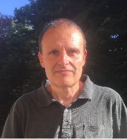 Antonio Pifferi合作论文数Dipartimento di Fisica, Politecnico di Milano12
 Alexander Flocke合作论文数11
 Andrea Giudice合作论文数Micro Photon Devices Srl11Elena Venturini合作论文数9
 Bogdan Rosinski合作论文数VERMON SA, F-37000 Tours, France9
 Peter Gordebeke合作论文数9
 Simon Arridge合作论文数Department of Computer Science, Faculty of Engineering Science, University College London9Pietro Panizza合作论文数9Mathieu Perriollat合作论文数8
 Carmelo Scarcella合作论文数8Jean-Marc Dinten合作论文数8
 Giulia Maffeis合作论文数7
 Bojan Markovic合作论文数Dipt. di Elettron. e Inf., Politec. di Milano;c;Dipt. di Elettron. e Inf., Politec. di Milano7
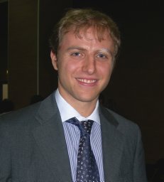 Andrea Farina合作论文数Laboratorio di Oncogenesi Molecolare, CRS-IRE7
 Daniel Durini合作论文数6
 Yu Zou合作论文数6Sascha Weyers合作论文数6
 Werner Brockherde合作论文数Fraunhofer IMS6
 Piotr Kolenderski合作论文数Institute of Physics;Nicolaus Copernicus University;Institute of Physics, Nicolaus Copernicus University6Pamela Zolda合作论文数5Giuseppe Di Sciacca合作论文数5Hélène Sportouche合作论文数5Simone Bellisai合作论文数5
 Sergio Cova合作论文数Department of Electronic Information and Bioengineering, Politecnico di Milano5Christophe Fraschini合作论文数4
 Vincenzo Sesta合作论文数Dipartimento di Elettronica, Informazione e Bioingegneria, Politecnico di Milano4Edoardo Ferocino合作论文数4
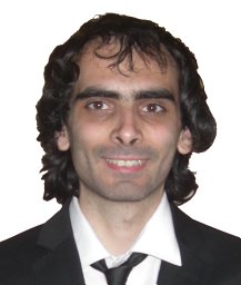 Rudi Lussana合作论文数4
 Kevin J Resch合作论文数Institute for Quantum Computing, University of Waterloo/Department of Physics and Astronomy, University of Waterloo4
 Thomas Jennewein合作论文数Department of Physics, Simon Fraser University;QEYNet;Universal Quantum Devices Inc.4
 Deny R. Hamel合作论文数Inst Quantum Comp, Univ Waterloo4
 Uwe Paschen合作论文数4
 Krister Shalm合作论文数National Institute of Standards and Technology4Johnsen, K.合作论文数3marco vitali合作论文数3s nikolic合作论文数3
 Bienvenu Ndagano合作论文数Sch Phys & Astron, Univ Glasgow3
 Ilya Starshynov合作论文数Sch Phys & Astron, Univ Glasgow3Ashley Lyons合作论文数3




