In the developing neocortex, a diverse array of neurons with defined types and abundances are systematically generated by a limited population of radial glial progenitors (RGPs) as they undergo successive fate changes. The molecular regulation behind this intricate temporal patterning remains elusive. We undertook in-depth single-cell multi-omics analyses, discovering a two-layered regulatory framework at the core of this process. Central to this are global temporal regulators positioned above a temporal network, consisting of series of transcriptional factor (TF) hub groups under sequential state transitions. This temporal network operates not by restricting TF expressions to discrete temporal windows, but through coordinated transcriptional and chromatin-accessibility dynamics that modulate transient TF regulatory activity. Moreover, global temporal regulators specify the duration of each cascading stage and, consequently, the number of progenies generated at each stage. Loss of global temporal regulators protracts RGP lineage progression, whereas their increased activity accelerates it. These findings suggest a two-layer temporal regulatory system controlling RGP lineage progression and neural progeny output duality in mammalian neocortical development. ### Competing Interest Statement The authors have declared no competing interest.
Mosaic analysis with double markers (MADM) is a powerful in vivo lineage tracing technique. It utilizes Cre recombinase-dependent interchromosomal recombination to restore the stable expression of two fluorescent proteins sparsely in individual dividing stem or progenitor cells and their progenies. Here, we describe the application of this technique for quantitative lineage analysis of radial glial progenitors in the developing mouse neocortex at the single-cell resolution.
Primary cilia are cellular antennae emanating from vertebrate cell surfaces to sense and transduce extracellular signals intracellularly to regulate cell behavior and function. However, their signal sensing and physiological functions in neocortical neurons remain largely unclear. Here, we show that, in response to various animal stressors, primary cilia in the mouse prefrontal cortex (PFC) exhibit consistent axonemal elongation. Selective removal of excitatory neuron primary cilia in the prefrontal but not sensory cortex leads to a reduction in animal stress sensing and response. Treatment with corticosterone, the major stress hormone, elicits an increase in primary ciliary cyclic adenosine 3',5'-monphosphate (cAMP) level in PFC excitatory neurons and a decrease in neuronal excitability dependent on primary cilia. Suppression of primary ciliary protein kinase A (PKA) activity in PFC excitatory neurons reduces animal stress. These results suggest that excitatory neurons in the PFC are involved in sensing and regulating animal stress via primary ciliary cAMP/PKA signaling.
Folding of the mammalian cerebral cortex into sulcal fissures and gyral peaks is the result of complex processes that are incompletely understood. Previously we showed that genetic deletion of Flrt1/3 adhesion molecules causes folding of the smooth mouse cortex into sulci resulting from increased lateral dispersion and faster neuron migration, without progenitor expansion. Here, we show in mice that combining the Flrt1/3 double knockout with an additional genetic deletion that causes progenitor expansion, greatly enhances cortex folding. Expansion of intermediate progenitors by deletion of Cep83 leads to a relative increase in Flrt-mutant neurons resulting in enhanced formation of sulci. Expansion of apical progenitors by deletion of Fgf10 leads to a relative reduction in Flrt-mutant neurons resulting in enhanced formation of gyri. These results together with computational modeling identify key developmental mechanisms, such as adhesive properties, cell densities and migration of cortical neurons, that cooperate to promote cortical gyrification.
Nuclear Factor I (NFI) misexpressions in humans are associated with severe brain malformations, yet the underlying mechanisms remain poorly understood. Here we show that NFIs regulate the broad lineage progression and lifespan of radial glial progenitors (RGPs), and consequently neocortical development in a dose-dependent manner. Human cerebral organoids carrying patient-mimicking NFI mutations exhibit expression level-dependent bidirectional impairments in RGP temporal development, coinciding with patient phenotypes. In mouse models, selective removal of NFIs leads to a dramatic protraction of RGP lineage progression and lifespan, excessive progeny output, and neocortical overgrowth and abnormal folding, whereas overexpression of NFIs accelerates RGP lineage progression, resulting in developmental stage-dependent precocious productions of diverse neural progenies. Moreover, NFIs exhibit a positive auto-regulation and progressive increase in expression, and regulate distinct temporal-specific targets underlying RGP lineage progression. These results suggest that NFIs act as evolutionarily conserved key global temporal regulators of RGP lineage progression and neocortical development.
The development of advanced neural modulation techniques is crucial to neuroscience research and neuroengineering applications. Recently, optical-based, nongenetic modulation approaches have been actively investigated to remotely interrogate the nervous system with high precision. Here, we show that a thin-film, silicon (Si)-based diode device is capable to bidirectionally regulate in vitro and in vivo neural activities upon adjusted illumination. When exposed to high-power and short-pulsed light, the Si diode generates photothermal effects, evoking neuron depolarization and enhancing intracellular calcium dynamics. Conversely, low-power and long-pulsed light on the Si diode hyperpolarizes neurons and reduces calcium activities. Furthermore, the Si diode film mounted on the brain of living mice can activate or suppress cortical activities under varied irradiation conditions. The presented material and device strategies reveal an innovated optoelectronic interface for precise neural modulations.
Cross-modal analysis of the same whole brain is an ideal strategy to uncover brain function and dysfunction. However, it remains challenging due to the slow speed and destructiveness of traditional whole-brain optical imaging techniques. Here we develop a new platform, termed Photoacoustic Tomography with Temporal Encoding Reconstruction (PATTERN), for non-destructive, high-speed, 3D imaging of ex vivo rodent, ferret, and non-human primate brains. Using an optimally designed image acquisition scheme and an accompanying machine-learning algorithm, PATTERN extracts signals of genetically-encoded probes from photobleaching-based temporal modulation and enables reliable visualization of neural projection in the whole central nervous system with 3D isotropic resolution. Without structural and biological perturbation to the sample, PATTERN can be combined with other whole-brain imaging modalities to acquire the whole-brain image with both high resolution and morphological fidelity. Furthermore, cross-modal transcriptome analysis of an individual brain is achieved by PATTERN imaging. Together, PATTERN provides a compatible and versatile strategy for brain-wide cross-modal analysis at the individual level.
The neocortex contains a vast collection of diverse neurons organized into distinct layers. While nearly all neocortical neurons are generated by radial glial progenitors (RGPs), it remains largely unclear how a complex yet organized neocortex is constructed reliably and robustly. Here, we show that the division behavior and neuronal output of RGPs are highly constrained with patterned variabilities to support the reliable and robust construction of the mouse neocortex. The neurogenic process of RGPs can be well-approximated by a consistent Poisson-like process unfolding over time, producing deep to superficial layer neurons progressively. The exact neuronal outputs regarding layer occupation are variable; yet, this variability is constrained systematically to support all layer formation, largely reflecting the variable intermediate progenitor generation and RGP neurogenic entry and exit timing differences. Together, these results define the fundamental features of neocortical neurogenesis with a balanced reliability and variability for the construction of the complex neocortex.
Sensory neurons in the neocortex exhibit distinct functional selectivity to constitute the neural map. While neocortical map of the visual cortex in higher mammals is clustered, it displays a striking "salt-and-pepper" pattern in rodents. However, little is known about the origin and basis of the interspersed neocortical map. Here we report that the intricate excitatory neuronal kinship-dependent synaptic connectivity influences precise functional map organization in the mouse primary visual cortex. While sister neurons originating from the same neurogenic radial glial progenitors (RGPs) preferentially develop synapses, cousin neurons derived from amplifying RGPs selectively antagonize horizontal synapse formation. Accordantly, cousin neurons in similar layers exhibit clear functional selectivity differences, contributing to a salt-and-pepper architecture. Removal of clustered protocadherins (cPCDHs), the largest subgroup of the diverse cadherin superfamily, eliminates functional selectivity differences between cousin neurons and alters neocortical map organization. These results suggest that developmental neuronal origin regulates neocortical map formation via cPCDHs.
In vivo detection of neurochemicals, including neurotransmitters and neuromodulators, is critical for both understanding brain mechanisms and diagnosing brain diseases. However, few sensors are competent in monitoring neurochemical dynamics in vivo at high specificity. Here, we propose the fiber-optic probes based on engineered cells (FOPECs) for plug-and-play, real-time detection of neurochemicals in freely moving animals. Taking advantages of life-evolved neurochemical receptors as key components, the chemical specificity of FOPECs is unprecedented. We demonstrate the applications of FOPECs in real-time monitoring of neurochemical dynamics under various physiology and pathology conditions. With no requirement of viral infection in advance and no dependence on animal species, FOPECs can be widely adopted in vertebrates, such as mice, rats, rabbits, and chickens. Moreover, FOPECs can be used to monitor drug metabolisms in vivo. We demonstrated the neurochemical monitoring in blood circulation systems in vivo. We expect that FOPECs will benefit not only neuroscience study but also drug discovery.
1H MRI maps brain structure and function non-invasively through versatile contrasts that exploit inhomogeneity in tissue micro-environments. Inferring histopathological information from magnetic resonance imaging (MRI) findings, however, remains challenging due to absence of direct links between MRI signals and cellular structures. Here, we show that deep convolutional neural networks, developed using co-registered multi-contrast MRI and histological data of the mouse brain, can estimate histological staining intensity directly from MRI signals at each voxel. The results provide three-dimensional maps of axons and myelin with tissue contrasts that closely mimic target histology and enhanced sensitivity and specificity compared to conventional MRI markers. Furthermore, the relative contribution of each MRI contrast within the networks can be used to optimize multi-contrast MRI acquisition. We anticipate our method to be a starting point for translation of MRI results into easy-to-understand virtual histology for neurobiologists and provide resources for validating novel MRI techniques.
The embryonic mouse brain undergoes drastic changes in establishing basic anatomical compartments and laying out major axonal connections of the developing brain. Correlating anatomical changes with gene-expression patterns is an essential step toward understanding the mechanisms regulating brain development. Traditionally, this is done in a cross-sectional manner, but the dynamic nature of development calls for probing gene-neuroanatomy interactions in a combined spatiotemporal domain. Here, we present a four-dimensional (4D) spatiotemporal continuum of the embryonic mouse brain from E10.5 to E15.5 reconstructed from diffusion magnetic resonance microscopy (dMRM) data. This study achieved unprecedented high-definition dMRM at 30- to 35-µm isotropic resolution, and together with computational neuroanatomy techniques, we revealed both morphological and microscopic changes in the developing brain. We transformed selected gene-expression data to this continuum and correlated them with the dMRM-based neuroanatomical changes in embryonic brains. Within the continuum, we identified distinct developmental modes comprising regional clusters that shared developmental trajectories and similar gene-expression profiles. Our results demonstrate how this 4D continuum can be used to examine spatiotemporal gene-neuroanatomical interactions by connecting upstream genetic events with anatomical changes that emerge later in development. This approach would be useful for large-scale analysis of the cooperative roles of key genes in shaping the developing brain.
The capability to selectively and precisely modulate neural activities represents a powerful tool for neuroscience research and clinical therapeutics. Traditional electrical stimulations associate with bulky and tethered implants, and optogenetic methods rely on genetic modification for cell targeting. Here, we report an optoelectronic, non-genetic strategy for exciting and inhibiting neural activities, accomplished by bioresorbable, thin-film silicon (Si) diodes. Under illumination, these devices establish polarity-dependent, positive or negative voltages at the semiconductor/solution interface. Such photovoltaic signals enable deterministic depolarization and hyperpolarization of cultured neurons, upregulating and downregulating intracellular calcium dynamics in vitro. Furthermore, flexible, thin-film Si based devices mounted on the nerve tissue selectively activate and silence in vivo activities, both in the peripheral nerve and the brain. Finally, these Si membranes naturally dissolve within the animal body. Such a Si-based material and device platform offers broad potential for biomedical applications.
The neocortex consists of a vast number of diverse neurons that form distinct layers and intricate circuits at the single-cell resolution to support complex brain functions(1). Diverse cell-surface molecules are thought to be key for defining neuronal identity, and they mediate interneuronal interactions for structural and functional organization2-6. However, the precise mechanisms that control the fine neuronal organization of the neocortex remain largely unclear. Here, by integrating in-depth single-cell RNA-sequencing analysis, progenitor lineage labelling and mosaic functional analysis, we report that the diverse yet patterned expression of clustered protocadherins (cPCDHs)-the largest subgroup of the cadherin superfamily of cell-adhesion molecules7-regulates the precise spatial arrangement and synaptic connectivity of excitatory neurons in the mouse neocortex. The expression of cPcdh genes in individual neocortical excitatory neurons is diverse yet exhibits distinct composition patterns linked to their developmental origin and spatial positioning. A reduction in functional cPCDH expression causes a lateral clustering of clonally related excitatory neurons originating from the same neural progenitor and a significant increase in synaptic connectivity. By contrast, overexpression of a single cPCDH isoform leads to a lateral dispersion of clonally related excitatory neurons and a considerable decrease in synaptic connectivity. These results suggest that patterned cPCDH expression biases fine spatial and functional organization of individual neocortical excitatory neurons in the mammalian brain.
Neural activities can be modulated by leveraging light-responsive nanomaterials as interfaces for exerting photothermal, photoelectrochemical or photocapacitive effects on neurons or neural tissues. Here we show that bioresorbable thin-film monocrystalline silicon pn diodes can be used to optoelectronically excite or inhibit neural activities by establishing polarity-dependent positive or negative photovoltages at the semiconductor/solution interface. Under laser illumination, the silicon-diode optoelectronic interfaces allowed for the deterministic depolarization or hyperpolarization of cultured neurons as well as the upregulated or downregulated intracellular calcium dynamics. The optoelectronic interfaces can also be mounted on nerve tissue to activate or silence neural activities in peripheral and central nervous tissues, as we show in mice with exposed sciatic nerves and somatosensory cortices. Bioresorbable silicon-based optoelectronic thin films that selectively excite or inhibit neural tissue may find advantageous biomedical applicability.
Achieving optimal behavior requires animals to flexibly retrieve prior knowledge. Here, we show that adult newborn granule cells (anbGCs) mediate emotional state-dependent adaptability of memory retrieval. We find that acute social reward (aSR) enhances memory retrieval by increasing the reactivation of engram cells, while acute social stress (aSS) weakens retrieval and reduces the reactivation. Such bidirectional regulation relies on the activation of distinct populations of anbGCs by aSR and aSS, triggering opposing modifications of dDG activity, which is sufficient to regulate and predict the performance of memory retrieval. Concordantly, in emotional disorder models, aSR-dependent memory adaptability is impaired, while the effect of aSS remains intact. Together, our data revealed that anbGCs mediate adaptability of memory retrieval, allowing animals to flexibly retrieve memory according to the current emotional state, and suggested the essential roles of anbGCs in translating emotional information to the regulation of memory expression.
Proper neural progenitor behavior in conjunction with orderly vasculature formation is fundamental to the development of the neocortex. However, the mechanisms coordinating neural progenitor behavior and vessel growth remain largely elusive. Here we show that robust metabolic production of lactate by radial glial progenitors (RGPs) co-regulates vascular development and RGP division behavior in the developing mouse neocortex. RGPs undergo a highly organized lineage progression program to produce diverse neural progeny. Systematic single-cell metabolic state analysis revealed that RGPs and their progeny exhibit distinct metabolic features associated with specific cell types and lineage progression statuses. Symmetrically dividing, proliferative RGPs preferentially express a cohort of genes that support glucose uptake and anaerobic glycolysis. Consequently, they consume glucose in anaerobic metabolism and produce a high level of lactate, which promotes vessel growth. Moreover, lactate production enhances RGP proliferation by maintaining mitochondrial length. Together, these results suggest that specific metabolic states and metabolites coordinately regulate vasculature formation and progenitor behavior in neocortical development.
As the primary microtubule-organizing center in animal cells, centrosomes regulate microtubule cytoskeleton to support various cellular behaviors. They also serve as the base for nucleating primary cilia, the hub of diverse signaling pathways. Cells typically possess one centrosome that contains two inequal centrioles and undergoes semi-conservative duplication during cell division, resulting in two centrosomes with an inherent asymmetry in age and properties. While the centrosome is ubiquitously present, mutations of centrosome proteins are strongly associated with human microcephaly characterized by a small cerebral cortex, underscoring the importance of an intact centrosome in supporting cortical neurogenesis. Here we review recent advances on centrosome regulation and function in mammalian cortical neural progenitors and discuss the implications for a better understanding of cortical neurogenesis and related disease mechanisms.
 Xinjun Zhang合作论文数Memorial Sloan-Kettering Cancer Center, Mem. Sloan-Kettering Cancer Center13Li Zhizhong合作论文数10Jian Ma合作论文数China Medical University Shenyang(China Medical University),Shenyang,China9
 Kun Huang合作论文数Department of Biostatistics & Health Data Science, Indiana University School of Medicine;Department of Biostatistics, School of Public Health, Indiana University;School of Computer Science, Indiana University9Zhongfu Shen合作论文数Tsinghua-Peking Center for Life Sciences, School of Life Sciences, Tsinghua University, Beijing, 100084, China.8Jiajun Yang合作论文数8
 Sultan Khadeejah T合作论文数Developmental Biology Program, Memorial Sloan-Kettering Cancer Center;Graduate Program in Neuroscience, Weill Cornell Medical College;Memorial Sloan-Kettering Cancer Center, Weill Cornell Medical College8
 Wei Shao合作论文数Developmental Biology Program, Memorial Sloan Kettering Cancer Center, 1275 York Avenue, New York, NY 10065, USA7Keith N. Brown合作论文数7
 She Chen (陈舌)合作论文数School of Basic Medical Sciences, Fudan University7
 Zhi Han (韩治)合作论文数MathWorks, Inc.7Ryan Insolera合作论文数Department of Neurology, University of Michigan7
 Xiaohui Lv (吕晓辉)合作论文数College of Life Sciences, Nankai University6
 Lily Jan (葉公杼)合作论文数Department of Biochemistry and Biophysics, University of California, San Francisco;Department of Physiology, University of California, San Francisco6
 Jiangyang Zhang合作论文数Department of Radiology, Grossman School of Medicine, New York University5
 Peng Gao合作论文数Alzheimers Dis & Brain Hlth, Eisai Inc5Lin Yang合作论文数4
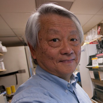 Yuh Nung Jan合作论文数Department of Physiology, University of California San Francisco;School of Medicine, University of California San Francisco;Howard Hughes Medical Institute4Xin Tan合作论文数4
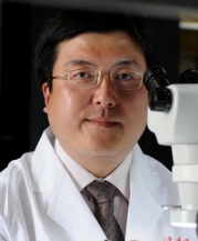 Yongchun Yu (禹永春)合作论文数Institues of Brain Science, Fudan University4
 Wei Shi (石玮)合作论文数Chinese Institute for Brain Research, Beijing4
 Roberto Malinow合作论文数Department of Neurobiology, School of Biological Sciences, University of California, San Diego4
 Lorenz Studer合作论文数Sloan Kettering Institute, Memorial Sloan-Kettering Cancer Center;Center for Stem Cell Biology, Memorial Sloan-Kettering Cancer Center4Luisirene Hernandez合作论文数3
 Shuijin He (何水金)合作论文数School of Life Science and Technology, ShanghaiTech University3Yunxiang Huang合作论文数3Hang Shi合作论文数3Xiuli Zhang合作论文数3
 Choong Heon Lee合作论文数Dept Radiol, Bernard & Irene Schwartz Ctr Biomed Imaging, NYU3Si-Qiang Ren合作论文数The State University of New Jersey3Wenying Angela Liu合作论文数3Xin Fu合作论文数3
 Lizhu Li合作论文数Tsinghua Univ, Beijing Natl Res Ctr Informat Sci & Technol, Dept Elect Engn, Beijing 100084, Peoples R China3
 Huachun Wang (王华春)合作论文数School of Integrated Circuits, Sun Yat-Sen University3
 Stewart A Anderson合作论文数Department of Psychiatry, Perelman School of Medicine, University of Pennsylvania3
 Xiaoqun Wang (王晓群)合作论文数State Key Laboratory of Cognitive Neuroscience and Leaning, Beijing Normal University3Xiaojian Li (李骁健)合作论文数3Lan Yin (尹斓)合作论文数3Ronald S. Bultje合作论文数3
 Jose A. Esteban合作论文数3
 Xing Sheng (盛兴)合作论文数Department of Electronic Engineering, Tsinghua University;IDG/McGovern Institute for Brain Research, Tsinghua University3
 Ben Simons合作论文数Department of Applied Mathematics and Theoretical Physics, Centre for Mathematical Sciences, University of Cambridge;Cambridge Stem Cell Institute, University of Cambridge3Rongqi Hong合作论文数2Oren Kodish合作论文数2Jia-Ling Teo合作论文数2Elizabeth Melendez合作论文数2David P Lin合作论文数2Yuwei Peng合作论文数2




