The first global analysis of the cerebral organoid (CO) response to 3D microvasculature indicates elaborate crosstalk between COs and ECs and points out potential vascularization factors for brain organoid vascularization on a chip.
Main function of glomerulus is to filter plasma and waste products from blood. The filtration function is known to be impaired and irreversibly damaged by a disease or a drug administration. Therefore, nephrotoxicity assessment is essential for a new drug development. However, conventional animal models for drug screening cannot appropriately assess toxicity of drugs to human glomerulus. In this study, we constructed a glomerulus-on-a-chip using human podocytes and evaluated the selectivity of inulin and albumin for filtration function.
Oxygen and nutrient supply provided via the vascular system is essential for organ development and tissue metabolism. Recent advances in stem cell technologies have led to the development of complex 3D tissue culture systems. However, direct supply of nutrients and oxygen by simple diffusion fails to reach inner areas resulting in impaired maturity of these complex tissues over time. In an attempt to alleviate this problem, 3D tissues are integrated with vascular networks (VNs) on a chip to ensure appropriate oxygen and nutrient supply. Such an in vitro vascular system must maintain a stable, robust, and perfusable network without degradation during entire culture period that may easily exceed several weeks. Current protocols for generation of VNs provide no information on the changes in these parameters in the long term. Here, using human umbilical vein endothelial cells, we generated a vascular network on a microfluidic chip and evaluated its morphology and function for 35 days. We demonstrate network stability, preservation of lumen structure and robust perfusability with negligible increase in permeability over time.
Development of the robust and functionally stable three-dimensional (3D) microvasculature remains challenging. One often-overlooked factor is the presence of potential anti-angiogenic agents in culture media. Sodium selenite, an antioxidant commonly used in serum-free media, demonstrates strong anti-angiogenic properties and has been proposed as an anticancer drug. However, its long-term effects on in vitro microvascular systems at the concentrations used in culture media have not been studied. In this study, we used a five-channel microfluidic device to investigate the concentration and temporal effects of sodium selenite on the morphology and functionality of on-chip preformed microvasculature. We found that high concentrations (∼3.0 μM) had adverse effects on microvasculature perfusion, permeability, and overall integrity within the first few days. Moreover, even at low concentrations (∼3.0 nM), a long-term culture effect was observed, resulting in an increase in vascular permeability without any noticeable changes in morphology. A further analysis suggested that vessel leakage may be due to vascular endothelial growth factor dysregulation, disruption of intracellular junctions, or both. This study provides important insight into the adverse effects caused by the routinely present sodium selenite on 3D microvasculature in long-term studies for its application in disease modeling and drug screening.
A large body of evidence points to the importance of cell adhesion molecules in cancer metastasis. Alterations in adhesion and attachment properties of neoplastic cells are important biomarkers of the metastatic potential of cancer. Loss of intracellular adhesion is correlated with more invasive phenotype by increasing the chances of malignant cells escaping from their site of origin, promoting metastasis. Therefore, there is great demand for rapid and accurate measurements of individual cell adhesion and attachment. Current technologies that measure adhesion properties in either suspension or bulk (microfluidics) remain very complex (e.g., atomic force microscopy [AFM], optical tweezers). Moreover, existing tools cannot provide measurements for fully attached individual adherent cells as they operate outside of such a force range. Even more importantly, none of the existing approaches permit concurrent and automated single-cell adhesion measurement and collection, which prohibits direct correlation between single-cell adhesion properties and molecular profile. Here, we report a fully automated and versatile platform, A-picK, that offers single-cell adhesion assay and isolation in parallel. We demonstrate the use of this approach for a time course analysis of human lung carcinoma A549 cells and substrate-specific adhesion potential using seven different substrates, including fibronectin, laminin, poly-l-lysine, carboxyl, amine, collagen, and gelatin.
We developed a MEMS based sensor, Nanopin, for mechanical characterization of individual adherent cells. Nanopin consists of sensing tip that forms a contact with a cell, a displacement sensor, and an actuator. The feasibility of sensing was evaluated using various concentrations of agarose gel, and then the stiffness measurements of human carcinoma cells were conducted on different surfaces. After the measurements of cells, we confirmed that cells grow normally in an incubator.
Single cell analysis (SCA) has become an essential part of basic and clinical research. A prerequisite to SCA studies is the isolation of individual cells from cell cultures. There are commercially available technologies including laser based systems and cell sorting instruments. However, they are typically complex and expensive making integration within standard lab/clinical sample processing workflows difficult. We have developed a universal platform for rapid acquisition of single cells from cultures and deposition into the single wells for further molecular analysis or clonal expansion. The instrument may also be used for tissue microdissection and measurements of individual cell adhesion. The main working principle is based on the capillary based vacuum pulse assisted technology (CTAS; UP8797644). Developed instrument collects individual cells from any adherent cultures grown in standard cell culture dishes in as small as 15 nl volume. It may be used with a wide range of inverted microscopes, so that the cells of interest can be identified based on morphology, location or fluorescent label. Automatic extraction and deposition algorithms were developed for acquisition of targeted cells and tissue regions of interest. Here, individual cells were collected from human neuroblastoma SH-SY5Y, CHO, and 3T3 cell lines. Collected cells were dispensed immediately into individual wells for clonal expansion. Clonal expansion and trypan blue assay revealed minimal effect on cellular viability (up to 99% when compared to controls). The benefits of the proposed technology include cost-efficiency, simple operation, complete workflow, and compatibility with a wide range of inverted microscopes and standard culture dishes.
This study combines the high-throughput capabilities of microfluidics with the sensitive measurements of microelectromechanical systems (MEMS) technology to perform biophysical characterization of circulating cells for diagnostic purposes. The proposed device includes a built-in microchannel that is probed by two opposing tips performing compression and sensing separately. Mechanical displacement of the compressing tip (up to a maximum of 14 µm) and the sensing tip (with a quality factor of 8.9) are provided by two separate comb-drive actuators, and sensing is performed with a capacitive displacement sensor. The device is designed and developed for simultaneous electrical and mechanical measurements. As the device is capable of exchanging the liquid inside the channel, different solutions were tested consecutively. The performance of the device was evaluated by introducing varying concentrations of glucose (from 0.55 mM (0.1%) to 55.5 mM (10%)) and NaCl (from 0.1 mM to 10 mM) solutions in the microchannel and by monitoring changes in the mechanical and electrical properties. Moreover, we demonstrated biological sample handling by capturing single cancer cells. These results show three important capabilities of the proposed device: mechanical measurements, electrical measurements, and biological sample handling. Combined in one device, these features allow for high-throughput multi-parameter characterization of single cells.
We present a mesodissection platform that retains the advantages of laser-based dissection instrumentation with the speed and ease of manual dissection. Tissue dissection in clinical laboratories is often performed by manually scraping a physician-selected region from standard glass slide mounts. In this manner, costs associated with dissection remain low, but spatial resolution is compromised. In contrast, laser microdissection methods maintain spatial resolution that matches the requirements for analysis of important tissue heterogeneity but remains costly and labor intensive. We demonstrate a microfluidic tool for rapid extraction of histological regions of interest from formalin-fixed paraffin-embedded tissue, which uses a simple and automated method that is compatible with most downstream enzymatic reactions, including protocols used for next-generation DNA sequencing.
Microtubule-associated protein (MAP) tau is a well-established hallmark of a large group of age related neurodegenerative diseases collectively called tauopathies. Under pathological conditions the equilibrium of tau binding to the MTs is perturbed, either by misregulation in the expression levels of specific tau isoforms or by MAPT gene mutations. Preclinical detection of such misregulated tau proteins in cerebrospinal fluid (CSF) is desirable for differential diagnosis and effective prognosis of neurodegeneration. Conventional tau protein detection methods utilize tau isoform-specific antibodies. Such immuno-based protocols, including enzyme-linked immunosorbent assay (ELISA) and Western blots have appropriate sensitivity and specificity, but often show high variability and are time consuming. Here, we established a non-immuno tau protein detection method utilizing microtubule (MT)-kinesin “gliding”assay. All the six tau isoforms expressed in the human brain (0N3R, 1N3R, 2N3R, 0N4R, 1N4R and 2N4R) and five MAPT gene mutants (V248L, G272V, P301L, V337M and R406W) were studied. The landing rate, binding density and gliding velocity of MTs with respect to each tau type were determined and are proposed as tau detection parameters. The detection parameters depicted the type of tau bound to the MTs. Furthermore, MT landing rate and density were found to be superior to gliding velocity in differentiating tau isoforms and mutants. The 3R vs. 4R isoforms, their admixtures, wild vs. mutant 2N4R and specific mutants were differentiated. Our data show that MT-kinesin gliding assay provides a convenient, lab-on-a-chip (LOC) compatible and antibody-free protocol for tau protein analysis.
Tau protein is a well-established biomarker for a group of neurodegenerative diseases collectively called tauopathies. So far, clinically relevant detection of tau species in cerebrospinal fluid (CSF) cannot be achieved without immunological methods. Recently, it was shown that different tau isoforms including the ones carrying various types of mutations affect microtubule (MT)-kinesin binding and velocity in an isoform specific manner. Here, based on these observations, we developed a microfluidic device to analyze tau mutations, isoforms and their ratios. The assay device consists of three regions: a MT reservoir which captures MTs from a solution to a kinesin-coated surface, a microchannel which guides gliding MTs, and an arrowhead-shaped collector which concentrates MTs. Tau-bound fluorescently labeled MTs (tau-MTs) were assayed, and the increase in fluorescence intensity (FI) corresponding to the total number of MTs accumulated was measured at the collector. We show that our device is capable of differentiating 3R and 4R tau isoform ratios and effects of point mutations within 5 minutes. Furthermore, radially oriented collector regions enable simultaneous FI measurements for six independent assays. Performing parallel assays in the proposed device with minimal image processing provides a cost-efficient, easy-to-use and fast tau detection platform.
Previously, we reported the application of micromachined silicon nanotweezers (SNT) integrated with a comb-drive actuator and capacitive sensors for capturing and mechanical characterization of DNA bundles. Here, we demonstrate direct DNA amplification on such a MEMS structure with subsequent electrical and mechanical characterization of a single stranded DNA (ssDNA) bundle generated between the tips of SNT via isothermal rolling circle amplification (RCA) and dielectrophoresis (DEP). An in situ generated ssDNA bundle was visualized and evaluated via electrical conductivity (I-V) and mechanical frequency response measurements. Colloidal gold nanoparticles significantly enhanced (P < 0.01) the electrical properties of thin ssDNA bundles. The proposed technology allows direct in situ synthesis of DNA with a predefined sequence on the tips of a MEMS sensor device, such as SNT, followed by direct DNA electrical and mechanical characterization. In addition, our data provides a "proof-of-principle" for the feasibility of the on-chip label free DNA detection device that can be used for a variety of biomedical applications focused on sequence specific DNA detection.
We report the successful demonstration of an on-chip tau detection system based on the difference in landing rate and binding density of microtubules (MTs) on a kinesin surface. Tau detection device comprises of a MT reservoir, channel and collector region with an overhang structure. We assayed MTs decorated with three tau types in the kinesin coated device. Since the increase in fluorescence intensity (FI) at the collector regions reflected the type of tau decorated on MTs, thus by measuring the FI we were able to distinguish wild 3R, 4R and P301L mutant tau.
 Lili Kudo合作论文数NeuroInDx, Inc.40
 Mehmet C Tarhan合作论文数16
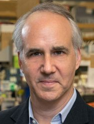 Daniel H. Geschwind合作论文数Center for Autism Research and Treatment, University of California, Los Angeles;Institute of Precision Health, University of California, Los Angeles;David Geffen School of Medicine, University of California, Los Angeles16
 Hiroyuki Fujita (藤田博之)合作论文数Tokyo City University14
 Ryuji Yokokawa合作论文数Ritsumeikan University, Kyoto University13Nancy Vi合作论文数13
 Anatol Bragin合作论文数School of, UCLA12Zhongcai Ma合作论文数9Koon-Sea Hui合作论文数8
 Liubov Parfenova合作论文数7Maria Hui合作论文数7Kimbley Lau合作论文数7Michelle Gray合作论文数6M-L Bondeson合作论文数6
 Momoko Kumemura合作论文数6
 Dominique Collard合作论文数6BM Carlberg合作论文数5
 X. William Yang合作论文数David Geffen School of Medicine, University of California, Los Angeles5
 Hidetoshi Kotera (小寺秀俊)合作论文数Osaka University;Kyoto University;Center for Research and Development Strategy, Japan Science and Technology Agency5Maneesha Shaji合作论文数4Hirofumi SHINTAKU合作论文数4Kazuya Fujimoto合作论文数Dept. Micro Eng., Kyoto Univ.;c;Dept. Micro Eng., Kyoto Univ.4
 Ulf Pettersson合作论文数Department of Immunology, Genetics and Pathology, Uppsala University4Orazov Yslam合作论文数4
 Michael Ross合作论文数The Lundquist Institute;David Geffen School of Medicine, University of California, Los Angeles;Fielding School of Public Health, University of California, Los Angeles4Martina Wiedau-Pazos合作论文数4Kristina Lagerstedt合作论文数4R. Yokokawa合作论文数3
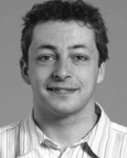 Laurent Jalabert合作论文数3
 Subhathirai Subramaniyan Parimalam合作论文数Concordia University3
 Jerome Engel合作论文数Brain Research Institute, University of California, Los Angeles;Epilepsy Telemetry Unit Seizure Disorder Center, University of California, Los Angeles;Neurology Psychiatry and Biobehavioral Sciences Jane and Terry Semel Institute for Neuroscience and Human Behavior Neurobiology, University of California, Los Angeles3Subramaniyan, S.P.合作论文数3
 Chiara Sabatti合作论文数Department of Biomedical Data Science, School of Medicine, Stanford University3Tony A. Fields合作论文数3Guijie Ren (任桂杰)合作论文数Department of Biochemistry & Molecular Biology, School of Basic Medical Sciences, Shandong University3
 Harley I. Kornblum合作论文数Department of Molecular and Medical Pharmacology, University of California Los Angeles3
 Nicolas Lafitte合作论文数2Daniel H Geshwind合作论文数2David Ma合作论文数2Wj Kleijer合作论文数2Atsuya Kitada合作论文数2
 Harry V. Vinters合作论文数Brain Research Institute, David Geffen School of Medicine, University of California, Los Angeles;Mental Retardation Research Center, David Geffen School of Medicine, University of California, Los Angeles;Mary S. Easton Center for Alzheimer’s Research and Care, David Geffen School of Medicine, University of California, Los Angeles2Berry R W合作论文数2Haykinson Michael J合作论文数2Avliyakulov Nuraly K合作论文数2Tippin Brigette L合作论文数2H. Fujita合作论文数2Lester I. Binder合作论文数2
 Mina Desai合作论文数Dept. of Ob/Gyn, LA BioMed),2




