Classically, the insula is considered an associative multisensory cortex where emotional awareness emerges through the integration of interoceptive and exteroceptive information, along with autonomic regulation. However, since early intracortical microstimulation (ICMS) studies, the insular cortex has also been conceived as a mosaic of anatomo-functional sectors processing various types of sensory information to generate specific overt behaviors. Based on this, the insula has been subdivided into distinct functional fields: an anterior field associated with oroalimentary behaviors, a middle field involved dorsally in hand movements and ventrally in emotional reactions, and a posterior field engaged in axial and proximal movements. Nevertheless, the anatomo-functional networks through which these fields produce motor behaviors remain largely unknown. To fill this gap in the present study, we investigated the connectivity of the macaque insula using a multimodal approach which combines resting-state fMRI with data from tract-tracing injections in insular functional fields defined by ICMS, as well as in brain areas known to be connected to the insula and characterized by specific somatotopic organization. The results revealed that each insular functional field takes part in distinct somatotopically organized network modulating specific motor or visceromotor behaviors, extending previous models that subdivide the insula primarily based on the types of interoceptive and exteroceptive information it receives. Our findings posit the various insular sectors as interfaces that synthesize diverse interoceptive and exteroceptive inputs into coherent subjective experiences and decision-making processes, within an embodied and enactive framework, that moves beyond the traditional dichotomy between sensory experience and motor behavior.
The lateral prefrontal cortex has been classically defined as an associative region involved in the so-called executive functions, such as guiding behavior based on abstract rules and mnemonic information. However, most neurophysiological studies on monkeys did not address the issue of whether distinct anatomical sectors of lateral prefrontal cortex play different functional roles. The main aim of this work is to study functional properties of neurons recorded from a large part of ventrolateral prefrontal cortex (VLPF) of two monkeys performing passive visual tasks and a visuo-motor task, and to map them on the anatomical areas defined on the basis of our recent parcellations. Our results show that some functional features are broadly distributed within VLPF, while others characterize specific areas. In particular, the temporal structuring of events and the general behavioral rule appear to be coded in all recorded areas, while each area differently contributes to the encoding of visual features and to the exploitation of contextual information for guiding behavior. Caudal VLPF areas, and especially caudal 12r, are characterized by a strong coding of visual information, both when passively presented or exploited for guiding behavior, while middle VLPF areas, and especially middle 46v, are rather more involved in the processing of contextual information for action organization. In this latter sector, visual stimuli/instructions appear to be encoded in a pragmatic format, that is, in terms of the associated behavioral outcome. Finally, area 45A and more anterior VLPF areas are characterized by a generally lower responsiveness to the employed tasks. Altogether, our findings indicate that caudal VLPF areas represent the first processing stage of visual input while middle VLPF areas primarily contribute to the selection and planning of contextually appropriate behaviors.
Previous studies indicated that the sense of body ownership (i.e., the feeling that our body parts belong to us; SBO) can be experimentally modulated in humans. Here, we focused on SBO from an across-species perspective, by investigating whether similar bottom-up and top-down constraints that consent to build SBO in humans also operate to build it in monkeys. To this aim, one monkey and a cohort of humans (N = 20) performed a paradigm combining the well-known rubber hand illusion (RHI), able to induce a fake hand embodiment, and a hand-identification reaching task, borrowed from the clinical evaluation of patients with SBO disorders. This task consisted of reaching one’s own hand with the other, while presenting a fake hand in different conditions controlling for bottom-up (synchronicity of the visuo-tactile stimulation) and top-down (congruency of the fake hand position relative to the monkey’s body) SBO constraints. Spatiotemporal kinematic features of such self-directed movements were measured. Our results show that, when the monkey aimed at the own hand, the trajectory of self-directed movements was attracted by the position of the hand believed to be one’s own (i.e., the fake hand), as in humans. Interestingly, such an effect was present only when both bottom-up and top-down constraints were met. Moreover, in the monkey, besides displacement of movement trajectory, also other kinematic parameters (velocity peak, deceleration phase) showed sensitivity to the embodiment effect. Overall, if replicated in a larger sample of monkeys, these results should support the view that human and non-human primates share similar body representation constraints and that they are able to modulate the motor behavior in both species.
The present study aimed to describe the cortical connectivity of a sector located in the ventral bank of the superior temporal sulcus in the macaque (intermediate area TEa and TEm [TEa/m]), which appears to represent the major source of output of the ventral visual stream outside the temporal lobe. The retrograde tracer wheat germ agglutinin was injected in the intermediate TEa/m in four macaque monkeys. The results showed that 58-78% of labeled cells were located within ventral visual stream areas other than the TE complex. Outside the ventral visual stream, there were connections with the memory-related medial temporal area 36 and the parahippocampal cortex, orbitofrontal areas involved in encoding subjective values of stimuli for action selection, and eye- or hand-movement related parietal (LIP, AIP, and SII), prefrontal (12r, 45A, and 45B) areas, and a hand-related dysgranular insula field. Altogether these data provide a solid substrate for the engagement of the ventral visual stream in large scale cortical networks for skeletomotor or oculomotor control. Accordingly, the role of the ventral visual stream could go beyond pure perceptual processes and could be also finalized to the neural mechanisms underlying the control of voluntary motor behavior.
In the past 2 decades, several attempts have been made to promote a correct diagnosis and possible restorative interventions in patients suffering from disorders of consciousness. Sensory stimulation has been proved to be useful in sustaining the level of arousal/awareness and to improve behavioural responsiveness with a significant effect on oro-motor functions. Recently, action observation has been proposed as a stimulation strategy in patients with disorders of consciousness, based on neurophysiological evidence that the motor cortex can be activated not only during action execution but also when actions are merely observed in the absence of motor output, or during listening to action sounds and speech. This mechanism is provided by the activity of mirror neurons. In the present study, a group of patients with disorders of consciousness (11 males, 4 females; median age: 55 years; age range: 19-74 years) underwent task-based functional MRI in which they had, in one condition, to observe and listen to the sound of mouth actions, and in another condition, to listen to verbs with motor or abstract content. In order to verify the presence of residual activation of the mirror neuron system, the brain activations of patients were compared with that of a group of healthy individuals (seven males, eight females; median age: 33.4 years; age range: 24-65 years) performing the same tasks. The results show that brain activations were lower in patients with disorders of consciousness compared with controls, except for primary auditory areas. During the audiovisual task, 5 out of 15 patients with disorders of consciousness showed only residual activation of low-level visual and auditory areas. Activation of high-level parieto-premotor areas was present in six patients. During the listening task, three patients showed only low-level activations, and six patients activated also high-level areas. Interestingly, in both tasks, one patient with a clinical diagnosis of vegetative state showed activations of high-level areas. Region of interest analysis on blood oxygen level dependent signal change in temporal, parietal and premotor cortex revealed a significant linear relation with the level of clinical functioning, assessed with coma recovery scale-revised. We propose a classification of the patient's response based on the presence of low-level and high-level activations, combined with the patient's functional level. These findings support the use of action observation and listening as possible stimulation strategies in patients with disorders of consciousness and highlight the relevance of combined methods based on functional assessment and brain imaging to provide more detailed neuroanatomical specificity about residual activated areas at both cortical and subcortical levels. Errante et al. have studied the activation of the mirror neuron system (MNS) in patients with disorders of consciousness. Their findings demonstrate a spared activation of the MNS, thus suggesting the potential use of observation of actions and listening to their sound as stimulation programme in this challenging condition. Graphical Abstract
The view of the functions of the motor system has radically changed in past years. It is now clear that the motor system not only is a "producer" of movements but also is involved in cognitive functions. In this chapter we review the anatomical and functional organization of the cortical motor system in monkeys and humans with particular emphasis on those areas that are endowed of the mirror mechanism and involved in communication. We discuss the possible evolutionary link between basic unintentional communication and voluntary communication. We conclude examining the hypothesis that purports that motor activation is involved in phonemes and understanding of action words.
The prefrontal cortex plays an important role in coding rules and producing context-appropriate behaviors. These processes necessarily require the generation of goals based on current context. Indeed, instructing stimuli are prospectively encoded in prefrontal cortex in relation to behavioral demands, but the coding format of this neural representation is, to date, largely unknown. In order to study how instructions and behaviors are encoded in prefrontal cortex, we recorded the activity of monkeys (Macaca mulatta) ventrolateral prefrontal neurons in a task requiring to perform (Action condition) or withhold (Inaction condition) grasping actions on real objects. Our data show that there are neurons responding in different task phases, and that the neuronal population discharge is stronger in the Inaction condition when the instructing cue is presented, and in the Action condition in the subsequent phases, from object presentation to action execution. Decoding analyses performed on neuronal populations showed that the neural activity recorded during the initial phases of the task shares the same type of format with that recorded during the final phases. We propose that this format has a pragmatic nature, that is instructions and goals are encoded by prefrontal neurons as predictions of the behavioral outcome.
The brain mechanisms underlying the emergence of a normal sense of body ownership can be investigated starting from pathological conditions in which body awareness is selectively impaired. Here, we focused on pathological embodiment, a body ownership disturbance observed in brain-damaged patients who misidentify other people's limbs as their own. We investigated whether such body ownership disturbance can be classified as a disconnection syndrome, using three different approaches based on diffusion tensor imaging: (i) reconstruction of disconnectome maps in a large sample (N = 70) of stroke patients with and without pathological embodiment; (ii) probabilistic tractography, performed on the age-matched healthy controls (N = 16), to trace cortical connections potentially interrupted in patients with pathological embodiment and spared in patients without this pathological condition; (iii) probabilistic 'in vivo' tractography on two patients without and one patient with pathological embodiment. The converging results revealed the arcuate fasciculus and the third branch of the superior longitudinal fasciculus as mainly involved fibre tracts in patients showing pathological embodiment, suggesting that this condition could be related to the disconnection between frontal, parietal and temporal areas. This evidence raises the possibility of a ventral self-body recognition route including regions where visual (computed in occipito-temporal areas) and sensorimotor (stored in premotor and parietal areas) body representations are integrated, giving rise to a normal sense of body ownership. Errante et al.'s study demonstrates that pathological embodiment, a body ownership disturbance observed in stroke patients who misidentify other people's limbs as their own, emerges from the disconnection of multiple temporal, parietal and premotor areas, which play a key role in integrating visual and sensorimotor body representations.
Objective:Psychopathology (and depression in particular) is a cardiovascular risk factor independent from any co-occurring pathology. This link is traced back to the mind-heart-body connection, whose underlying mechanisms are still not completely known. To study psychopathology in relation to the heart, it is necessary to observe the autonomic nervous system, which mediates among the parts of that connection. Its gold standard of evaluation is the study of heart rate variability (HRV). To investigate whether any association exists between the HRV parameters and sub-threshold depressive symptoms in a sample of healthy subjects. Method:In this cross-sectional study, two short-term HRV recordings (5 min - supine and sitting) have been analyzed in 77 healthy subjects. Here we adopted a three-fold approach to evaluate HRV: a set of scores belonging to the time domain; to the frequency domain (high, low, and very low frequencies) and a set of 'nonlinear' parameters. The PHQ-9 (Patient Health Questionnaire-9) scale was used to detect depressive symptoms. Results:Depressive symptoms were associated only with a parameter from the non-linear approach and specifically the long-term fluctuations of fractal dimensions (DFA-α2). This association remained significant even after controlling for age, gender, BMI (Body-Mass-Index), arterial hypertension, anti-hypertensive drugs, dyslipidemia, and smoking habits. Moreover, the DFA-α2 was not affected by the baroreflex (postural change), unlike other autonomic markers. Conclusions:Fractal analysis of HRV (DFA-α2) allows then to predict depressive symptoms below the diagnostic threshold in healthy subjects regardless of their health status. DFA-α2 may be considered as an imprint of subclinical depression on the heart rhythm.
The ventral part of lateral prefrontal cortex (VLPF) of the monkey receives strong visual input, mainly from inferotemporal cortex. It has been shown that VLPF neurons can show visual responses during paradigms requiring to associate arbitrary visual cues to behavioral reactions. Further studies showed that there are also VLPF neurons responding to the presentation of specific visual stimuli, such as objects and faces. However, it is largely unknown whether VLPF neurons respond and differentiate between stimuli belonging to different categories, also in absence of a specific requirement to actively categorize or to exploit these stimuli for choosing a given behavior. The first aim of the present study is to evaluate and map the responses of neurons of a large sector of VLPF to a wide set of visual stimuli when monkeys simply observe them. Recent studies showed that visual responses to objects are also present in VLPF neurons coding action execution, when they are the target of the action. Thus, the second aim of the present study is to compare the visual responses of VLPF neurons when the same objects are simply observed or when they become the target of a grasping action. Our results indicate that: (1) part of VLPF visually responsive neurons respond specifically to one stimulus or to a small set of stimuli, but there is no indication of a "passive" categorical coding; (2) VLPF neuronal visual responses to objects are often modulated by the task conditions in which the object is observed, with the strongest response when the object is target of an action. These data indicate that VLPF performs an early passive description of several types of visual stimuli, that can then be used for organizing and planning behavior. This could explain the modulation of visual response both in associative learning and in natural behavior.
Psychopathology, and in particular depression, is a cardiovascular risk factor independent from co-occurring pathology. This link is traced back to the mind-heart-body connection, whose underlying mechanisms are, to date, not completely known. It is clear, however, that the autonomic nervous system plays a leading role in the mediation between the parts of that connection. Therefore, to study psychopathology in relation to the heart, it is necessary to observe the autonomic nervous system, whose gold standard of evaluation is the study of heart rate variability (HRV). Two short-term HRV recordings (5 min - supine and sitting) were analysed in 77 healthy subjects. Here we adopted a three-fold approach to evaluate HRV: a set of scores belonging to the time domain (SDNN, pNN50, RMSSD); the frequency analyses that gauges three main components (high, low, and very low frequencies) and a new set of complexity nonlinear parameters. The PHQ-9 scale was used to detect depressive symptoms. Depressive symptoms were associated only with a parameter from the non-linear approach and specifically the long-term fluctuations of fractal dimensions (DFA-2). This association remained significant even after controlling for age, gender, BMI, arterial hypertension, anti-hypertensive drugs, dyslipidaemia, and smoking habit. Moreover, the DFA-2 was not affected by the baroreflex (postural change), unlike other autonomic markers. In conclusion, fractal analysis of HRV (DFA-2) allows to predict depressive symptoms below diagnostic threshold in healthy subjects regardless of their health status. DFA-2 may be then considered as an imprint of subclinical depression on the heart rhythm.
In the macaque brain, projections from distant, interconnected cortical areas converge in specific zones of the striatum. For example, specific zones of the motor putamen are targets of projections from frontal motor, inferior parietal, and ventrolateral prefrontal hand-related areas and thus are integral part of the so-called “lateral grasping network.” In the present study, we analyzed the laminar distribution of corticostriatal neurons projecting to different parts of the motor putamen. Retrograde neural tracers were injected in different parts of the putamen in 3 Macaca mulatta (one male) and the laminar distribution of the labeled corticostriatal neurons was analyzed quantitatively. In frontal motor areas and frontal operculum, where most labeled cells were located, almost everywhere the proportion of corticostriatal labeled neurons in layers III and/or VI was comparable or even stronger than in layer V. Furthermore, within these regions, the laminar distribution pattern of corticostriatal labeled neurons largely varied independently from their density and from the projecting area/sector, but likely according to the target striatal zone. Accordingly, the present data show that cortical areas may project in different ways to different striatal zones, which can be targets of specific combinations of signals originating from the various cortical layers of the areas of a given network. These observations extend current models of corticostriatal interactions, suggesting more complex modes of information processing in the basal ganglia for different motor and nonmotor functions and opening new questions on the architecture of the corticostriatal circuitry. SIGNIFICANCE STATEMENT Projections from the ipsilateral cerebral cortex are the major source of input to the striatum. Previous studies have provided evidence for distinct zones of the putamen specified by converging projections from specific sets of interconnected cortical areas. The present study shows that the distribution of corticostriatal neurons in the various layers of the primary motor and premotor areas varies depending on the target striatal zone. Accordingly, different striatal zones collect specific combinations of signals from the various cortical layers of their input areas, possibly differing in terms of coding, timing, and direction of information flow (e.g., feed-forward, or feed-back).
In this chapter we describe the anatomical and functional characteristics of the Posterior Parietal Cortex (PPC) of macaque monkeys and humans. We critically discuss the classical notion that the posterior parietal lobule is an association region, and contrast it with the more recent view that PPC is endowed with motor properties. We conclude that PPC motor organization represents the basic scaffold of this region on the top of which sensory information is organized.
Based on neural tracer injections we found evidence for 3 connectionally distinct sectors of the dorsal part of the macaque prefrontal area 46 (46d), located at different rostro-caudal levels. Specifically, a rostral sector displayed an almost exclusive and extensive intraprefrontal connectivity and extraprefrontal connections limited to superior temporal areas and the caudal cingulate area 31. Conversely, both a middle and a caudal sector were characterized by robust, topographically organized connections with parietal and frontal sensorimotor areas. Both these sectors shared connections with caudal and medial superior parietal areas (V6A and PGm) where visuospatial information is combined with gaze- and arm-related signals for visuomotor control of arm reaching and/or eye movements. However, the caudal sector was preferentially connected to parietal and frontal oculomotor areas, whereas the middle one was preferentially connected to skeletomotor, mostly arm-related, parietal and premotor areas. The present study provides evidence for a rostro-caudal organization of area 46d similar to that described for the ventrolateral prefrontal cortex, in which more caudal areas are relatively more directly involved in controlling different aspects of motor behavior and more rostral areas are most likely involved in higher order, possibly more abstract, cognitive functions.
Continuing investigations of corticostriatal connections in rodents emphasize an intricate architecture where striatal projections originate from different combinations of cortical layers, include an inhibitory component, and form terminal arborizations which are cell-type dependent, extensive, or compact. Here, we report that in macaque monkeys, deep and superficial cortical white matter neurons (WMNs), peri-claustral WMNs, and the claustrum proper project to the putamen. WMNs retrogradely labeled by injections in the putamen (four injections in three macaques) were widely distributed, up to 10 mm antero-posterior from the injection site, mainly dorsal to the putamen in the external capsule, and below the premotor cortex. Striatally projecting labeled WMNs (WMNsST) were heterogeneous in size and shape, including a small GABAergic component. We compared the number of WMNsST with labeled claustral and cortical neurons and also estimated their proportion in relation to total WMNs. Since some WMNsST were located adjoining the claustrum, we wanted to compare results for density and distribution of striatally projecting claustral neurons (ClaST). ClaST neurons were morphologically heterogeneous and mainly located in the dorsal and anterior claustrum, in regions known to project to frontal, motor, and cingulate cortical areas. The ratio of ClaST to WMNsST was about 4:1 averaged across the four injections. These results provide new specifics on the connectional networks of WMNs in nonhuman primates, and delineate additional loops in the corticostriatal architecture, consisting of interconnections across cortex, claustralstriatal and striatally projecting WMNs.
 Marzio Gerbella合作论文数Istituto Italiano di Tecnologia (IIT),, Università di Parma and Rete Multidisciplinare Tecnologica35
 Giuseppe Luppino合作论文数Istituto di Fisiologia;Universitadi Parma;Istituto di Fisiologia, Universitadi Parma35
 Elena Borra合作论文数Sezione di Fisiologia, Università di Parma30Leonardo Fogassi合作论文数22
 Pier Francesco Ferrari合作论文数Istituto di Fisiologia Umana, Università di Parma14Luciano Simone合作论文数Center for Human Technologies, Istituto Italiano di Tecnologia12
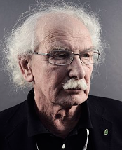 Giacomo Rizzolatti合作论文数University of Parma11Abdelouahed Belmalih合作论文数7
 Francesca Roda合作论文数Dept Med & Surg, Univ Parma6
 Luca Bonini合作论文数6Gino Coude合作论文数6Roberta Calzavara合作论文数Department of Pharmacology and Physiology, University of Rochester School of Medicine6
 Marco Bimbi合作论文数Institut des Sciences Cognitives Marc Jeannerod, CNRS;Universite Claude Bernard Lyon 1, Universite de Lyon;Universite Claude Bernard Lyon 1, Universite de Lyon5
 Monica Maranesi合作论文数5Massimo Matelli合作论文数5Francesca Ugolotti Serventi合作论文数4matteo baccarini合作论文数3
 Alfonso Gravante合作论文数3
 Massimo Contini合作论文数3
 Fausto Caruana合作论文数Brain Center for Social and Motor Cognition, Italian Institute of technology (IIT),3
 Ahmad Jezzini合作论文数Department of Neuroscience, University of Parma3Pierluca Marazzi合作论文数2Maria Carsillo合作论文数2
 Piergiorgio Mandarano合作论文数Univ Parma2
 Alice Rossi Sebastiano合作论文数2
 Antonino Errante合作论文数Dept Med & Surg, Univ Parma2
 Davide Lazzeroni合作论文数IRCCS, Fondazione Don Carlo Gnocchi2
 Francesca Garbarini合作论文数2
 Paolo Ossola合作论文数2Mangiaracina Chiara合作论文数2
 Bruni Stefania合作论文数2Tonelli Simone合作论文数2L. Bonini合作论文数1A Errante合作论文数1
 Matilde Leonardi合作论文数1Akiko Ikkai合作论文数1valentine l silakov合作论文数1Marianna Rizzo合作论文数1Wh Goldstein合作论文数1
 Niping Wang合作论文数1Giorgio Cappellaro合作论文数1
 Valentina Bruno合作论文数1
 Nicolo Castellani合作论文数1
 Carolina Giulia Ferroni合作论文数Dept Med & Surg, Univ Parma1
 Settimio Ziccarelli合作论文数Dept Med & Surg, Univ Parma1Greta Demichelis合作论文数1R. Massini合作论文数1Valentina Giorgetti合作论文数1Lan Zhou合作论文数1




