Background:Nuclear protein in testis (NUT) carcinomas are aggressive, poorly or undifferentiated cancers, generally arising from midline structures. This subtype of squamous cell carcinoma is rare and has a poor prognosis. NUT cancers are defined by NUTM1 fusions. Rearrangements of the NUTM1 gene have rarely been described in primary thyroid cancer and are mainly reported in patients ≤45 years old. NSD3::NUTM1 translocation is the most common NUTM1 fusion transcript reported in thyroid cancer. As they are very infrequent, NUTM1 fusions are not routinely sought in poorly differentiated thyroid cancer (PDTC) or anaplastic thyroid cancer (ATC). Case presentation:We report two PDTC patients >65 years old with locally advanced disease and 18FDG-avid distant metastases. NSD3::NUTM1 translocation was evidenced in both patients by RNA sequencing using the next-generation sequencing panel of our institution. Conclusion:We suggest including the search for NUTM1 fusions in the RNA sequencing panel for advanced and refractory thyroid cancers.
Background ESTIMABL2, a multicentre randomised phase 3 trial in patients with low-risk differentiated thyroid cancer (ie, pT1am or pT1b, N0 [no evidence of regional nodal involvement] or Nx [involvement of regional lymph nodes that cannot be assessed in the absence of neck dissection]), showed the non-inferiority of a follow-up strategy without radioactive iodine (131I) administration compared with a postoperative 131I administration at 3 years post-randomisation. Here, we report a pre-specified analysis after 5 years of follow-up. Methods Patients treated with total thyroidectomy with or without prophylactic neck lymph node dissection, without postoperative suspicious findings on neck ultrasonography, were randomly assigned to the no-radioiodine group or to the radioiodine group (11 GBq-30 mCi after recombinant human thyrotropin-stimulating hormone). Follow-up consisted of annual thyroglobulin and thyroglobulin antibody determinations during levothyroxine treatment and neck ultrasonography in odd-numbered years. An event was defined as abnormal foci of 131I uptake on the post-treatment whole-body-scan requiring subsequent treatment, abnormal neck ultrasonography, elevated thyroglobulin levels, increasing titres or appearance of thyroglobulin antibody (using the same laboratory assay), or a combination of these definitions. Non-inferiority of the proportion of patients without an event in one group compared with the other at 5 years after randomisation was shown if this proportion and its CI did not differ by more than -5%. This study was registered on ClinicalTrials.gov (NCT01837745) and is completed. Findings Of the 776 patients (n=642 [827%] female and n=134 [173%] male, median age 529 years [IQR 426-631]) enrolled, 698 were evaluable at 5 years. The proportions of patients without events were 932% in the no-radioiodine group and 948% in the radioiodine group, for a difference of -16% (90% CI -45 to 14). Events consisted of structural or functional abnormalities (n=11) and biological abnormalities (n=31). Interpretation The non-inferiority of a follow-up strategy compared with postoperative 131I administration in low risk differentiated thyroid cancer was confirmed at 5 years. There is no loss of opportunity in following these patients without postoperative ablation. Copyright (c) 2024 The Author(s). Published by Elsevier Ltd. This is an Open Access article under the CC BY 4.0 license.
Anaplastic thyroid cancer is a rare and rapidly deadly disease. In case of clinical suspicion (rapid growth, stony neck mass), diagnostic work-up should be carried out as a matter of urgency to enable prompt treatment. Multidisciplinary assessment involving the patient's referring specialists, the support care team, and if necessary, a geriatric oncology specialist should be performed and must take account of disease extent, comorbidities, general health status and the patient's wishes. Patients and their families should receive realistic information about the prognosis; either active treatment in parallel to support care or exclusive palliative care can be recommended from the outset. Despite the dismal prognosis, recent advances in tumor molecular profiling and treatment with the advent of targeted treatment and immunotherapy hold out great promise for the future. This article summarizes the consensus recommendations on management of anaplastic thyroid cancers by the ENDOCAN TUTHYREF network, a rare-cancer network of the French National Institute for Cancer (INCa).
Radioactive-iodine-refractory differentiated thyroid cancer (RAIR DTC) represents 3-5% of follicular-derived DTCs, with approximately 200-300 new cases diagnosed annually in France. Median overall survival in the French RAIR DTC database is 9.5years, underscoring the importance of long-term support for caregivers and patients. To guide treatment decision-making, the French ENDOCAN TUTHYREF network has provided algorithms for RAIR DTC management, available at the TUTHYREF website. The present article summarizes these recent practical recommendations, focusing on 5 points. (1) RAIR DTC has long been defined by locally advanced disease not amenable to surgery or metastatic disease not fully responding to radioactive iodine (RAI) therapy, a definition that can be further refined considering prognostic factors. (2) Treatment should be tailored according to tumor burden and progression, with local treatments prioritized for non-progressive or slowly progressive disease. (3) Early tumor molecular testing should be performed to identify driver oncogenes such as BRAF mutation or RET/NTRK/ALK fusion, to optimize access to existing selective targeted therapies. (4) For symptomatic or progressive RAIR DTC, tyrosine multikinase inhibitors, such as sorafenib, lenvatinib or cabozantinib, are the standard therapies, but alternative and 2nd-line kinase inhibitors are also available. (5) Since most therapies are associated with common side-effects such as fatigue and cardiovascular, digestive and skin issues, preparing and monitoring patients for systemic therapy should include careful assessment of comorbidities, toxicity prevention and individual dose adjustment. Overall, management of RAIR DTC requires a multidisciplinary approach, with an emphasis on personalized treatment strategies and proactive therapeutic education.
PurposeMRI is essential in the management of brain tumours. However, long waiting times reduce patient accessibility. Reducing acquisition time could improve access but at the cost of spatial resolution and diagnostic quality. A commercially available artificial intelligence (AI) solution, SubtleMR™, can increase the resolution of acquired images. The objective of this prospective study was to evaluate the impact of this algorithm that halves the acquisition time on the detectability of brain lesions in radiology and radiotherapy.Material and methodsThe T1/T2 MRI of 33 patients with brain metastases or meningiomas were analysed. Images acquired quickly have a matrix divided by two which halves the acquisition time. The visual quality and lesion detectability of the AI images were evaluated by radiologists and radiation oncologist as well as pixel intensity and lesions size.ResultsThe subjective quality of the image is lower for the AI images compared to the reference images. However, the analysis of lesion detectability shows a specificity of 1 and a sensitivity of 0.92 and 0.77 for radiology and radiotherapy respectively. Undetected lesions on the IA image are lesions with a diameter less than 4mm and statistically low average gadolinium-enhancement contrast.ConclusionIt is possible to reduce MRI acquisition times by half using the commercial algorithm to restore the characteristics of the image and obtain good specificity and sensitivity for lesions with a diameter greater than 4mm.
Les cancers thyroïdiens de souche folliculaire (CT) sont rares chez les enfants et les adolescents nécessitant une prise en charge experte multidisciplinaire conformément aux recommandations ETA 2022. Cette étude rétrospective multicentrique du réseau national TUTHYREF a pour objectif de décrire le parcours de soins actuel et le devenir de ces patients. 52 patients mineurs pris en charge pour un CT entre 2010 et 2022 ont été inclus ce jour avec un âge médian de 14 ans. 63 % des patients avaient un nodule Bethesda V ou IV ; 11 % Bethesda IV et 4 % Bethesda III. La chirurgie thyroïdienne était validée en RCP dans 31 % des cas, impliquant un pédiatre dans 35 % des cas et une consultation génétique dans 26 % des cas. 16 % des patients étaient opérés par un chirurgien pédiatrique. 88 % et 12 % des cancers étaient respectivement papillaires ou folliculaires. 40 % des patients présentaient une hypoparathyroïdie transitoire et 10 % une hypoparathyroïdie définitive. Après une thyroïdectomie totale dont 77 % avec un curage ganglionnaire, un traitement par iode radioactif était proposé à 98 % des patients, dont 58 % en sevrage et 42 % sous Thyrogen®, avec une Tg médiane au moment de l’iode à 4,8ng/mL et plusieurs cures pour 38 % des patients. Des métastases ganglionnaires et à distance étaient présentes chez 50 % et 9 % enfants. Après le traitement initial, 65 % des patients étaient d’emblée en rémission, 26 % présentaient une récidive locorégionale. Après un suivi médian de 3 ans, 93 % des patients étaient en rémission et 7 % en réponse biologique incomplète sans aucun décès.
Introduction Le vandetanib est indiqué en première ligne (L1) dans les carcinomes médullaires thyroïdiens (CMT) requérant un traitement systémique. Dans la littérature, la durée médiane de traitement est d’environ 20 mois, un quart des patients peuvent présenter une réponse prolongée. Objectifs Évaluer la proportion et les caractéristiques des patients avec CMT du réseau ENDOCAN-TUTHYREF en traitement prolongé par vandétanib. Méthode Étude rétrospective multicentrique incluant les CMT traités par vandetanib L1 plus de 3 mois, comparant les patients traités plus de 24 mois (LgTT) aux autres patients. La survie globale (SG) a été évaluée par modèle de Kaplan-Meier. Résultats Au total, 126 patients ont été suivis 38 mois en médiane (Q1=18 ;Q3=81), dont 53/126 LgTT (durée médiane de traitement=49 mois). En analyse univariée, les facteurs associés au statut LgTTT (a), tendant à y être associés (b) et non associés (c) étaient (a) un meilleur ECOG (p-value=0,04), un délai plus long diagnostic-traitement (p-value=0,02), (b) des métastases à distance métachrones (p-value=0,05), un âge plus jeune au moment du traitement (p-value=0,06), (c) statut mutationnel RET (NA=8), sexe, posologie du vandétanib et localisation métastatique. Les causes d’arrêt du traitement n’étaient pas différentes dans les deux groupes.La SG depuis le début du traitement était supérieure dans le groupe LgTT (90,7[71,5–176,4]) vs 26(18,7–42,7) mois, p-value=410-7) mais la SG après l’arrêt du traitement était comparable (17,7[10,4–38,6]) vs 12,9(8,85–27), p-value=0,7). Conclusion 40 % des CMT traités par vandetanib L1 ont présenté un contrôle à long terme et une survie significativement prolongée indépendamment de la mutation RET.
ImportanceWhether F18-choline (FCH) positron emission tomographic (PET)/computed tomographic (CT) scan can replace Tc99m-sestaMIBI (MIBI) single-photon emission (SPE)CT/CT as a first-line imaging technique for preoperative localization of parathyroid adenomas (PTA) in patients with primary hyperparathyroidism (PHPT) is unclear.ObjectiveTo compare first-line FCH PET/CT vs MIBI SPECT/CT for optimal care in patients with PHPT needing parathyroidectomy and to compare the proportions of patients in whom the first-line imaging method resulted in successful minimally invasive parathyroidectomy (MIP) and normalization of calcemia 1 month after surgery.Design, Setting, and ParticipantsA French multicenter randomized open diagnostic intervention phase 3 trial was conducted. Patients were enrolled from November 2019 to May 2022 and participated up to 6 months after surgery. The study included adults with PHPT and an indication for surgical treatment. Patients with previous parathyroid surgery or multiple endocrine neoplasia type 1 (MEN1) were ineligible.InterventionsPatients were assigned in a 1:1 ratio to receive first-line FCH PET/CT (FCH1) or MIBI SPECT/CT (MIBI1). In the event of negative or inconclusive first-line imaging, they received second-line FCH PET/CT (FCH2) after MIBI1 or MIBI SPECT/CT (MIBI2) after FCH1. All patients underwent surgery under general anesthesia within 12 weeks following the last imaging. Clinical and biologic (serum calcemia and parathyroid hormone levels) assessments were performed 1 and 6 months after surgery.Main Outcomes and MeasuresThe primary outcome was a true-positive first-line imaging-guided MIP combined with uncorrected serum calcium levels of 2.55 mmol/l or less 1 month after surgery, corresponding to the local upper limit of normality.ResultsOverall, 57 patients received FCH1 (n = 29) or MIBI1 (n = 28). The mean (SD) age of patients was 62.8 (12.5) years with 15 male (26%) and 42 female (74%) patients. Baseline patient characteristics were similar between groups. Normocalcemia at 1 month after positive first-line imaging-guided MIP was observed in 23 of 27 patients (85%) in the FCH1 group and 14 of 25 patients (56%) in the MIBI1 group. Sensitivity was 82% (95% CI, 62%-93%) and 63% (95% CI, 42%-80%) for FCH1 and MIBI1, respectively. Follow-up at 6 months with biochemical measures was available in 43 patients, confirming that all patients with normocalcemia at 1 month after surgery still had it at 6 months. No adverse events related to imaging and 4 adverse events related to surgery were reported.ConclusionsThis randomized clinical trial found that first-line FCH PET/CT is a suitable and safe replacement for MIBI SPECT/CT. FCH PET/CT leads more patients with PHPT to correct imaging-guided MIP and normocalcemia than MIBI SPECT/CT thanks to its superior sensitivity.Trial RegistrationClinicalTrials.gov Identifier: NCT04040946
Nuclear medicine has long been a mainstay in the management of thyroid cancers. In patients with differentiated thyroid cancer (DTC), the most common histotype, radioiodine (RAI, 131I) has been for years a cornerstone for the treatment of RAI-avid metastases. Post-therapeutic 131I scintigraphy helps guide these treatments and contributes to the definition of refractory cancers. In these refractory patients, who represent fewer than 5% of CTDs, 18FDG PET plays a central diagnostic and prognostic role. From a therapeutic perspective, RAI uptake can be re-induced in some of these patients with the BRAF mutation by using redifferentiation protocols. In anaplastic thyroid cancer (ATC) that is rare, aggressive and undifferentiated, 18FDG PET remains the metabolic imaging of choice. In medullary thyroid cancer (MTC), PET imaging is mainly based on the use of 18F-DOPA, even if 18FDG also provides prognostic data and 68Ga-DOTATOC could allow a theranostic approach. Other radiopharmaceuticals offering new theranostic avenues in thyroid cancers are also discussed, such as prostate-specific membrane antigen (PSMA) and fibroblast activation protein (FAP). After decades of a "one-size fits all" approach in thyroid cancer management, molecular imaging is paving the way towards personalized medicine.
Background Nintedanib is a triple-angiokinase inhibitor with potential activity in patients with advanced thyroid cancers, as radioiodine refractory differentiated thyroid cancer (RAIR DTC) and medullary thyroid cancer (MTC).Design EORTC-1209 (NCT01788982) was a double-blind randomized (2:1 ratio) placebo-controlled phase II, multi-cohort study exploring the efficacy and safety of nintedanib in patients with progressive, locally advanced, and/or metastatic RAIR DTC and MTC. The primary endpoint was progression-free survival (PFS) in the per-protocol (PP) population for both cohorts. Secondary endpoints included response rate, duration of response, overall survival (OS), and safety.Results RAIR DTC cohort: Seventy out of the 75 planned patients with RAIR DTC (median age, 66 years; 39 women) who had progressed after one (76%) or two lines (24%) of previous systemic therapy were randomized to receive either nintedanib (N = 45) or placebo (N = 25). Of these, 69 patients started treatment and 56 met all inclusion criteria (PP). At data cutoff, the median duration of follow-up was 26.3 months in the nintedanib arm and 19.8 months in the placebo arm. In the PP population, the median PFS was 3.7 months [80% confidence interval (CI), 1.9-6.5] in the nintedanib arm and 2.9 months (80% CI, 2.0-5.6) in the placebo arm (HR = 0.65; 80% CI, 0.42-0.99; one-sided log-rank test P = 0.0947). No objective response was observed. The median OS was 29.6 months [80% CI, 15.2-not reached (NR)] in the nintedanib arm and not reached in the placebo arm. Grade 3-4 adverse events of any attribution occurred in 50% of patients receiving nintedanib and in 36% of patients receiving placebo. MTC cohort: Thirty-one out of the 67 planned patients with MTC (median age, 57 years; eight women) who had progressed after one (68%) or two (32%) lines of previous systemic therapy were randomized to receive either nintedanib (N = 22) or placebo (N = 9). Of these, 20 patients (15 in the nintedanib arm and five in the placebo arm) started treatment and met all inclusion criteria (PP). The median PFS was 7.0 months (80% CI, 1.9-8.7) in the nintedanib arm and 3.9 months (80% CI, 3.0-5.5) in the placebo arm (HR = 0.49; 95% CI, 0.16-1.53). No objective response was reported. The median OS was 16.4 months (80% CI, 12.1-24.9) in the nintedanib arm and 12.3 months (80% CI, 7.1-NR) in the placebo arm. Grade 3-4 adverse events of any attribution during the blinded period occurred in 59.1% of patients receiving nintedanib and in 33.3% of patients receiving placebo.Conclusion This study did not suggest a clinically significant improvement of PFS with nintedanib over placebo in patients with pretreated RAIR DTC and MTC.
L'indication en première intention de la TEP/TDM à la F18-choline (FCH TEP) ou de la TEMP/TDM au Tc99m-sestaMIBI (MIBI TEMP) pour la localisation préopératoire d'adénome parathyroïdien dans l'hyperparathyroïdie primaire (pHPT) est source de débat. Nous avons mené une étude randomisée multicentrique de phase III chez des adultes atteints de pHPT avec indication chirurgicale. Les patients ont eu en première intention soit une FCH TEP/TDM (FCH1), soit un MIBI TEMP/TDM (MIBI1). En cas d'imagerie négative ou non concluante, les patients ont eu un examen de deuxième ligne, FCH TEP/TDM (FCH2) après MIBI1 ou un MIBI TEMP/TDM (MIBI2) après FCH1. L'objectif principal était de comparer les proportions de patients pour lesquels l'imagerie de première intention a permis une parathyroïdectomie mini-invasive (MIP) efficace associée à une guérison (normalisation des taux sériques de calcium et d'hormone parathyroïdienne à 1 mois). Nous avons formulé l'hypothèse d'une supériorité de 30 % de la FCH1 sur la MIBI1 pour déterminer l'échantillon (PMID : 33413316). Entre novembre 2019 et mai 2022, 58 patients ont été randomisés, 30 dans le bras FCH1 et 28 dans MIBI1. Les caractéristiques des patients à l'inclusion étaient similaires entre les groupes. La FCH1, positive chez 24/29 (83 %) patients, a permis une MIP efficace chez 23/24 patients. Le MIBI1, positif chez 18/26 patients (69 %), a conduit à une MIP pour 16/17 patients opérés avec guérison chez 14/16 patients et à une thoracoscopie vidéo-assistée (VATS) chez 1 patient (guéri). La proportion de patients pour lesquels l'imagerie de première intention a conduit à une MIP efficace avec guérison était de 79% (n = 23) pour FCH1 et 54 % (n = 14) pour MIBI1 (p = 0,043). La sensibilité de l'examen à l'échelle de la lésion était supérieure pour FCH1 que pour MIBI1 : 83 % vs 61 %. Quatre événements indésirables transitoires liés à la chirurgie ont été rapportés, aucun lié à l'imagerie. Cette étude montre que la FCH TEP en première intention est supérieure au MIBI TEMP pour orienter les patients atteints de pHPT vers une MIP efficace. Une analyse coût-bénéfices est en cours pour valider cette stratégie sur le plan médico-économique.
Searchable abstracts of presentations at key conferences in endocrinology ISSN 1470-3947 (print) | ISSN 1479-6848 (online)
La médecine nucléaire occupe depuis longtemps une place importante dans la prise en charge des cancers thyroïdiens. Dans les cancers thyroïdiens différenciés (CTD), les plus fréquents, l’iode radioactif (131I) garde un rôle fondamental dans le traitement des métastases iodofixantes. La scintigraphie post-thérapeutique à l’131I permet de guider ces traitements et contribue à la définition des cancers réfractaires. Chez ces patients réfractaires, qui représentent moins de 5 % des CTD, la TEP/TDM 18FDG joue un rôle diagnostique et pronostique central. Sur le plan thérapeutique, la redifférenciation, c’est-à-dire la restauration de la fixation de l’131I, est possible chez des CTD mutés BRAF. Dans les cancers anaplasiques de la thyroïde (CAT), rares et agressifs, et par définition indifférenciés, la TEP au 18FDG reste l’imagerie métabolique de choix. Dans les cancers médullaires (CMT), l’imagerie TEP repose principalement sur la 18F-DOPA mais le 18FDG et le 68Ga-DOTATOC peuvent être utiles, en raison des données pronostiques et théranostiques qu’ils peuvent fournir. D’autres radiopharmaceutiques TEP qui sont encore du domaine de la recherche et dont certains offrent des perspectives théranostiques, tels l’antigène membranaire spécifique de la prostate (PSMA) et la protéine d’activation des fibroblastes (FAP), seront également discutés. Après des décennies où seul l’131I était disponible et administré systématiquement dans les CTD, quel que soit le risque de maladie résiduelle, l’arrivée de nouveaux traceurs TEP vient enrichir les outils d’imagerie et l’arsenal thérapeutique. Les développements rapides et récents de l’imagerie moléculaire permettent une évolution vers une prise en charge encore plus personnalisée des cancers thyroïdiens.
Searchable abstracts of presentations at key conferences in endocrinology ISSN 1470-3947 (print) | ISSN 1479-6848 (online)
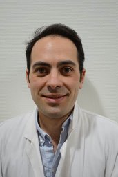 Ciappuccini Renaud合作论文数Departments of Nuclear Medicine and Thyroid Unit, Centre François Baclesse53
 Zerdoud Slimane合作论文数32
 Sophie Leboulleux合作论文数Hôpitaux Universitaires De Genève31Godbert Yann合作论文数29
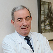 Martin Schlumberger合作论文数Université Paris-Saclay28
 Isabelle Borget合作论文数Faculté de Pharmacie de Chatenay-Malabry, Université Paris-Sud;Gustave Roussy26
 Nicolas Aide合作论文数Medecine Nucleaire;Hopital Tenon AP-HP;Hopital Beaujon;Hopital Tenon AP-HP, Hopital Beaujon AP-HP25
 Elske Quak合作论文数afdeling Medische Oncologie, Universitair Medisch Centrum St Radboud23Claire Bournaud合作论文数22Vincent Rohmer合作论文数21
 Pierre Vera合作论文数Centre Henri-Becquerel;Université de Rouen Normandie21
 Natacha Heutte合作论文数18Christine Do Cao合作论文数Service d’Endocrinologie, CHRU de Lille Lille18
 Francoise Borson-Chazot合作论文数Fac Med, Univ Lyon 117Clarisse Bénédicte合作论文数17Livia Lamartina合作论文数Department of Internal and Specialized Medicine, Sapienza University of Rome16
 Bernard Charbonnel合作论文数Faculté de Médecine, University of Nantes16P Sai合作论文数15
 Charline Lasnon合作论文数UNICANCER, François Baclesse Cancer Centre15Laurence Leenhardt合作论文数Departement Maladies Chroniques et Traumatismes;Institut de Veille Sanitaire;Departement Maladies Chroniques et Traumatismes, Institut de Veille Sanitaire15
 Michel Henry-Amar合作论文数Centre François Baclesse15
 Christelle De la Fouchardiere合作论文数Institut Paoli-Calmettes15
 Michel Meignan合作论文数Assistance Publique – Hôpitaux de Paris14Abir Al Ghuzlan合作论文数14
 Cyril Jaudet合作论文数Laboratoire de Physique Theorique;Universite Paul Sabatier;Laboratoire de Physique Theorique, Universite Paul Sabatier14Jean-Pierre Rame合作论文数13
 Jean-François CHATAL合作论文数GIP Arronax;University of Nantes12Buffet Camille合作论文数12Benisvy Danielle合作论文数12Emmanuel Itti合作论文数12D. Maugendre合作论文数12Jean-Philippe Vuillez合作论文数12
 Weyts Kathleen合作论文数Institut Claudius Regaud, 20-24 Rue du Pont Saint-Pierre, Toulouse 31052, France.11
 David Blanchard合作论文数11Antoine Tabarin合作论文数11
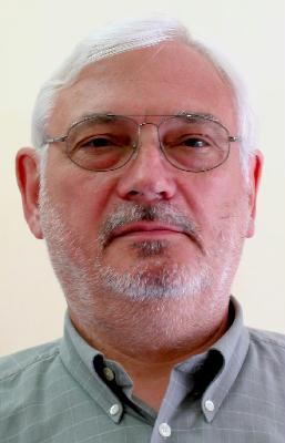 Jacques Barbet合作论文数Centre de Recherche en Cancérologie de Nantes-Angers;Nantes Université11Pierre-Yves Salaun合作论文数Centre Hospitalier Universitaire de Brest;University Hospital of Brest;Hôpital Morvan Brest11
 Yves Reznik合作论文数CHU Caen, Transplantation and Liver Surgery, CHU cote de nacre11
 Delphine Drui合作论文数10Marcel Ricard合作论文数10Jean-Jacques Michels合作论文数9Bogdan Catargi合作论文数9Hubert Allannic合作论文数9Eric Mirallie合作论文数Department of Endocrine Surgery, University Hospital La Timone9N. Aide合作论文数8Audrey Lasne-Cardon合作论文数8Claire Schvartz合作论文数8
 Marie-Elisabeth Toubert合作论文数8Olivier Schneegans合作论文数8




