Background & objectives: Acute myocardial infarction (AMI) is characterized by irreparable and irreversible loss of cardiac myocytes. Despite major advances in the management of AMI, a large number of patients are left with reduced left ventricular ejection fraction (LVEF), which is a major determinant of short and long term morbidity and mortality. A review of 33 randomized control trials has shown varying improvement in left ventricular (LV) function in patients receiving stem cells compared to standard medical therapy. Most trials had small sample size and were underpowered. This phase III prospective, open labelled, randomized multicenteric trial was undertaken to evaluate the efficacy in improving the LVEF over a period of six months, after injecting a predefined dose of 5-10 x 10(8) autologous mononuclearcells (MNC) by intra-coronary route, in patients, one to three weeks post ST elevation AMI, in addition to the standard medical therapy.Methods: In this phase III prospective, multicentric trial 250 patients with AMI were included and randomized into stem cell therapy (SCT) and non SCT groups. All patients were followed up for six months. Patients with AMI having left ventricular ejection fraction (LVEF) of 20-50 per cent were included and were randomized to receive intracoronary stem cell infusion after successfully completing percutaneous coronary intervention (PCI).Results: On intention-to-treat analysis the infusion of MNCs had no positive impact on LVEF improvement of >= 5 per cent. The improvement in LVEF after six months was 5.17 +/- 8.90 per cent in non SCT group and 4.82 +/- 10.32 per cent in SCT group. The adverse effects were comparable in both the groups. On post hoc analysis it was noted that the cell dose had a positive impact when infused in the dose of >= 5 X 10(8) (n=71). This benefit was noted upto three weeks post AMI. There were 38 trial deviates in the SCT group which was a limitation of the study.Interpretation & conclusions: Infusion of stem cells was found to have no benefit in ST elevation AMI. However, the procedure was safe. A possible benefit was seen when the predefined cell dose was administered which was noted upto three weeks post AMI, but this was not significant and needs confirmation by larger trials.
Lt Col Navreet Singh *, Gp Capt Hemant Madan , Brig Y.K. Arora, VSM, Brig Rajat Dutta , Col Sunil Sofat , Col Prashant Bhardwaj, VSM, Rahul Sharma , Gp Capt D.S. Chadha , Col A.K. Ghosh , Lt Col Sanjeev Sengupta f Classified Specialist (Cardiology), CH (SC), Pune-40, India b Senior Advisor (Cardiology), Army Hosp (R&R), New Delhi, India Consultant (Cardiology), Base Hosp, Delhi Cantt, India Consultant (Cardiology), Army Hosp (R&R), New Delhi, India Resident (Cardiology), Army Hosp (R&R), New Delhi, India Classified Specialist (Cardiology), MH (CTC), Pune-40, India
BACKGROUND:Radiofrequency catheter ablation is an emerging curative modality of treatment for several types of cardiac arrhythmias. The aim of our study was to evaluate our experience of electrophysiology procedures over a 10-year-period and compare it with the published literature.METHODS AND RESULT:All patients undergoing cardiac electrophysiology and radiofrequency ablation procedures during the period from 01 January 2003 to 30 April 2012 were included in this study. The study analyzed 892 patients of which atrioventricular nodal reentrant tachycardia (AVNRT) was n = 513 (57.5%), accessory pathways n = 230 (25.8%), atrial tachycardia n = 26 (2.9%), atrial flutter n = 15 (1.7%), atrial fibrillation = 9 (1.0%), ventricular tachycardia n = 37 (4.2%) and diagnostic studies n = 62 (6.9%). Three-dimensional electro-anatomic mapping was performed in n = 17 (1.9%). The success rate was 99% with AVNRT, 94% with accessory pathway, 81% with atrial tachycardia, 64% with ventricular tachycardia and 34% with atrial fibrillation. The major and serious complications rate was 0.45%.CONCLUSIONS:The procedural success rate and the complication rate in this real-world study of the Armed Forces were comparable/superior to the reported literature.
 Hemant Madan合作论文数4
 Prashant Bharadwaj合作论文数4Rajat Datta合作论文数3
 Sunil Chandy合作论文数2Alka Sharma合作论文数2Neelam Marwah合作论文数2Jacob Jose合作论文数2
 Sujata Mohanty合作论文数Stem Cell Facility, All India Institute of Medical Sciences2Ganguli Prosenjit合作论文数2
 Balram Airan合作论文数2Balram Bhargava合作论文数2
 Naveen Garg合作论文数2
 Soniya Nityanand合作论文数Department of Hematology, Sanjay Gandhi Post-Graduate Institute of Medical Sciences (SGPGIMS)2
 Shinjini Bhatnagar合作论文数Department of Pediatrics, All India Institute of Medical Sciences2Ajay Bahl合作论文数Department of Cardiology, Post Graduate Institute of Medical Education and Research2
 Pravin K Goel合作论文数2
 Lt Gen Dr Velu Nair合作论文数Department of Haematology & Bone Marrow Transplantation, Army Hospital (Research & Referral),2Sanjay Gambhir合作论文数Department of Nuclear Medicine, Sanjay Gandhi Postgraduate Institute of Medical Sciences2Oomen K George合作论文数1Prashant Bhardwaj合作论文数1Rahul Sharma合作论文数1V. Mathews合作论文数1A. Ghosh合作论文数1K K Talwar合作论文数1
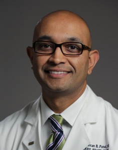 Chetan B. Patel合作论文数Department of Medicine, School of Medicine, Duke University1Sengupta Sanjeev合作论文数1Davinder Chadha合作论文数Classified Specialist (Cardiology),, MH (CTC),1Dutta Rajat合作论文数1Arora Y K合作论文数1
 Navreet Singh合作论文数1Kabde Ajit合作论文数1
 Oommen K George合作论文数1
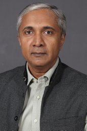 Vikram Mathews合作论文数Department of Haematology, Christian Medical College1Paulv George合作论文数1




