Periodontitis is traditionally regarded as an oral biofilm-driven inflammatory disease that leads to progressive loss of the tooth-supporting alveolar bone. However, accumulating evidence indicates that periodontal bone loss is more accurately understood as a state of pathological uncoupling of bone remodeling, in which exaggerated bone resorption coexists with inadequate bone formation response. In this review, we reposition periodontitis within the broader context of inflammatory skeletal diseases and synthesize current mechanistic insights from osteoimmunology, bone biology, and mechanobiology. We discuss how excessive osteoclastogenesis in periodontitis is sustained by receptor activator of nuclear factor kappa-B ligand (RANKL) dominance derived from osteocytes, osteoblast-lineage cells, stromal cells, monocytes/macrophages, B and T lymphocytes, and neutrophils within a cytokine-rich microenvironment characterized by tumor necrosis factor (TNF)-α, interleukin (IL)-1β, IL-6, and IL-17A signaling. Persistent activation of nuclear factor kappa-B (NF-κB) and mitogen-activated protein kinase (MAPK) pathways further enhance osteoclast differentiation, survival, and resorptive activity. At the same time, inflammatory mediators actively suppress osteoblast-lineage commitment by inhibiting Runx2 and Osterix, antagonizing canonical Wnt/β-catenin signaling through the upregulation of sclerostin and Dickkopf-1 (DKK1), and impairing bone matrix production and mineralization. We further examine how disruption of key osteoclast-osteoblast coupling mechanisms, including ephrinB2/EphB4 and semaphorin signaling, prevents the effective transition from resorption to formation, while osteocyte dysfunction amplifies the uncoupled phenotype by integrating inflammatory and mechanical signals. Comparisons with rheumatoid arthritis, inflammatory bowel disease-associated bone loss, and peri-implantitis reveal shared immune-driven mechanisms of remodeling imbalance, whereas the unique features of alveolar bone, including high turnover, continuous mechanical loading, and chronic microbial exposure, make it particularly susceptible to inflammatory uncoupling. Together, these concepts support a therapeutic shift toward restoring physiological coupling instead of solely inhibiting resorption and position periodontitis as a clinically accessible model for understanding and targeting inflammatory bone loss across skeletal diseases.
Myeloid-derived suppressor cells (MDSCs) are immature, immunosuppressive myeloid cells whose role in periodontal disease (PD) is not well defined. Here, we show that gingiva from PD patients exhibit increased infiltration of MDSCs, including granulocytic (G-MDSC) and monocytic (M-MDSC) subsets, alongside elevated CD4+ T cells and CD19+ B cells. Functionally, these MDSC subsets suppress autologous CD4+ T cell proliferation in co-culture, confirming their immunoregulatory capacity. In a murine ligature-induced periodontitis (LIP) model, both gingival MDSC subsets increased over time and correlated with CD4+ T cell and CD19+ B cell infiltration. Adoptive transfer of G-MDSCs and M-MDSCs into LIP mice reduced gingival CD4+ T cell infiltration and pro-inflammatory cytokines, while increasing arg1, Il-10, and CD4+CD25+FoxP3+ Tregs. In contrast, anti-Gr-1 depletion exacerbated periodontal inflammation, reduced Treg counts, and increased alveolar bone loss. Collectively, these findings identify anti-inflammatory and osteoprotective roles for MDSCs in PD and support their therapeutic potential for periodontal inflammation.
Periodontitis is a chronic, host-mediated inflammatory disease in which microbial dysbiosis and dysregulated immune responses drive the destruction of tooth-supporting tissues. Although conventional therapy remains centered on mechanical biofilm control, persistent inflammation and alveolar bone loss in susceptible individuals underscore the need for adjunctive strategies. Translating preclinical discoveries into predictable clinical outcomes, however, remains a major challenge in periodontal research. This narrative review integrates two interrelated themes, translational research methodology and adjunctive therapeutic innovation, to critically examine how preclinical findings can be more effectively bridged to clinical practice in periodontitis management. Evidence was synthesized from experimental, translational, and clinical studies retrieved from PubMed, Scopus, and Web of Science up to September 2025. Emphasis was placed on mechanistic insights, model validity, and translational feasibility across host-modulatory, natural, probiotic, and device-based adjuncts. Animal models remain indispensable for mechanistic understanding and therapeutic testing but face biological and methodological limitations that hinder direct extrapolation to humans. Interspecies differences, short disease kinetics, and non-standardized endpoints constrain translational predictability. Addressing these gaps requires refined modeling, standardized outcomes, and integration of systemic risk factors. Within this methodological framework, several adjunctive modalities, including specialized pro-resolving mediators, probiotics, natural compounds such as curcumin and resveratrol, and device-based therapies like antimicrobial photodynamic therapy demonstrate promising anti-inflammatory, osteoimmunomodulatory, and regenerative effects. Emerging translational tools such as bioresponsive drug delivery systems, nanocarriers, 3D-printed scaffolds, and AI-driven precision periodontics may further enhance clinical relevance and patient-specific targeting. Advancing adjunctive periodontal therapy demands harmonized translational models, bioresponsive delivery platforms, and precision diagnostic tools that bridge preclinical efficacy with real-world outcomes. By aligning methodological rigor with therapeutic innovation, translational research can accelerate the safe and effective clinical integration of next-generation adjunctive treatments in periodontitis.
This study aimed to investigate the effects of soluble epoxide hydrolase (sEH) inhibition on osteoclast differentiation and activity in vitro and in vivo, as well as to elucidate the signaling pathways associated with osteoclastogenesis. Primary murine bone marrow monocytes were stimulated with macrophage colony-stimulating factor and receptor activator of nuclear factor kappa B ligand to induce osteoclastogenesis and treated with the sEH inhibitor 1-(1-propanoylpiperidin-4-yl)-3-[4-(trifluoromethoxy)phenyl]urea (TPPU) (0.1-10 μM). Tartrate-resistant acid phosphatase staining, gene expression analyses, and immunofluorescence were used to evaluate osteoclast formation, transcriptional regulation, and cell fusion. A murine model of ligature-induced periodontitis was used to assess in vivo effects of sEH inhibition (TPPU 10 mg/kg). Alveolar bone loss was quantified by histomorphometry, and gingival gene expression was analyzed. In vitro, sEH inhibition significantly reduced tartrate-resistant acid phosphatase-positive multinucleated osteoclast formation, downregulated the expression of key transcription factors and osteoclast activity-related genes. Immunofluorescence analysis revealed attenuation of mitogen-activated protein kinase signaling and reduced dendritic cell-specific transmembrane protein expression, indicating impaired cell fusion. In vivo, TPPU treatment preserved alveolar bone structure, reduced osteoclast-like cell numbers, and decreased the expression of osteoclastic markers in gingival tissues during experimental periodontitis. sEH acts as a crucial regulator of osteoclast differentiation and function. Pharmacological inhibition of sEH suppresses osteoclastogenesis and protects against inflammatory bone loss. Therefore, targeting sEH may represent a novel therapeutic approach to modulate osteoclast activity and prevent bone destruction in periodontitis and other bone-resorptive diseases. SIGNIFICANCE STATEMENT: This study provides direct evidence that soluble epoxide hydrolase inhibition modulates osteoclast differentiation and fusion, contributing to reduced inflammatory bone loss. By demonstrating effects on osteoclast-intrinsic pathways while also influencing the inflammatory microenvironment, our findings support soluble epoxide hydrolase as a pharmacological target for chronic inflammatory bone-resorptive diseases.
Diabetes is a major risk factor for severe periodontitis. This is partly due to impaired wound healing associated with concomitant failure to resolve inflammation and chronic infection. Studies from the Joslin Medalist Cohort, people with >50 years of type 1 diabetes, have reported that protective factors exist to delay the onset of severe retinopathy, nephropathy, and periodontitis, even with persistent hyperglycemia. Proteomic analysis of healthy gingival tissues from individuals with hyperglycemia versus those with periodontitis and good glycemic control showed substantial upregulation of oxidative phosphorylation, particularly mitochondrial complex I and II, as validated by immunoblot analysis. Diabetes reduced mitochondrial enzyme expression and function, measured by oxygen consumption rate in the gingiva, compared with nondiabetic mice, in parallel, with exacerbation of ligature-induced periodontal bone loss, elevation of inflammatory cytokines (Il-1b, Il-6, and Il-17a) and activation of osteoclasts. However, activation of pyruvate kinase M2 reversed diabetes and ligature-induced mitochondrial dysfunction, reduced levels of inflammatory cytokines, and mitigated periodontal bone loss, without affecting glycolytic function. These results suggested that enhancing mitochondrial protein expression and function can decrease periodontal inflammation and osteoclast activation in diabetes, thereby protecting against the onset of periodontitis and bone loss, suggesting a potential therapy for chronic wounds, even with persistent hyperglycemia. ARTICLE HIGHLIGHTS:Characterization of protective factors against periodontal inflammation with long durations of diabetes. Proteomic analysis of gingival tissue from people with type 1 diabetes >50 years and poor glycemic control without periodontitis showed elevated mitochondrial enzymes and functions. These mitochondrial dysfunctions, as well as inflammatory and osteoclast activation, were validated in diabetic mouse models of periodontitis. Elevation of mitochondrial functions by pyruvate kinase 2 activation reversed diabetes-induced gingival inflammation, osteoclast activation, and periodontitis, even in the presence of hyperglycemia. Mitochondrial activation could be a potential therapy for periodontitis and, perhaps, chronic wound healing in diabetes.
Objective To assess expression of specialized pro-resolving lipid mediators (SPMs) and inflammation resolution receptors (IRRs) on oral neutrophils (oPMNs) in oral lichen planus: FFAR2 binds short chain fatty acids, FFAR4 binds long chain fatty acids, ERV1 binds resolvin E1, and ALX binds lipoxin A4. The hypothesis is that there is increased expression of IRRs, but decreased levels of SPMs, reflecting failure of the distinct process of inflammation resolution, and hence progression from acute to chronic inflammation in OLP pathogenesis. SPMs activate the inflammation resolution pathway and are potential therapeutic options for steroid-refractory OLP. Methods OLP cases and healthy controls were recruited from the oral medicine clinic at Brigham and Women's Hospital. Oral rinse samples were collected for oPMN isolation and lipidomics analysis using liquid chromatography-mass spectrometry. oPMNs were labeled and analyzed by flow cytometry. The number/percentage of cells positive for specific receptors, quantities of receptors per cell, and levels of SPMs were compared using 2-sample t tests. Principle component analysis was performed to identify clustering trends in lipid profiles. Results A total of 27 controls and 17 cases were included. Cases had at least double the number of mean total oPMNs (14 × 103, P < .01) and ERV1+ oPMNs (11 × 103, P < .01). The mean percentage of ERV1+ oPMNs was greater in cases than controls (84% vs 67%, P < .01). Mean fluorescent intensity of receptors in cases vs controls were 63 × 103 vs 90 × 103 for ERV1 (P < .01), 83 × 103 vs 66 × 103 for FFAR2 (P = .02), 53 × 103 vs 36 × 103 for FFAR4 (P = .02), and 604 × 103 vs 690 × 103 for ALX (P = .45). Concentration of SPM biosynthetic intermediates, 8-hydroxydocosahexaenoic acid (8-HDoHE) and 5S,12S-dihydroxyeicosatetraenoic acid (5S,12S-DiHETE) were significantly lower in cases (P = .01). Principle component analysis demonstrated distinct clusters for cases and controls with loading plots showing 8-HDoHE to contribute the most variance. Conclusion In OLP, there were more ERV1+ oPMNs, but less expression of ERV1. There is variation in lipid profiles between groups, with downregulation of pro-resolution mediators in OLP. These findings possibly indicate failure of inflammation resolution in OLP and potentially explain acute/symptomatic exacerbations of this chronic condition. Further research is required to support translational studies using oral rinses containing SPMs for OLP management.
The central question addressed in this review revisits the historical chicken-and-egg debate: "In periodontitis, does microbial dysbiosis drive inflammation, or does inflammation shape the subgingival microbiome?" This question is reframed through the lens of inflammation resolution. Specialized pro-resolving mediators (SPMs) provide a mechanistic framework for understanding how inflammation intersects with microbial dysbiosis. Derived from omega-3 and omega-6 fatty acids, SPMs actively promote the resolution of inflammation through binding of specific cell surface receptors rather than nonspecifically suppressing it, highlighting their therapeutic potential as side-effect-free host modulators, with implications beyond periodontitis to other chronic inflammatory diseases. The evidence reviewed shows how SPMs can: (1) control inflammation by resolution rather than inhibition, (2) reverse microbial dysbiosis as a consequence of inflammation control, and (3) promote tissue regeneration through diverse biological pathways. Whether the primary dysregulation in periodontitis lies solely in resolution failure or involves additional-possibly still unidentified-mechanisms, remains unclear. All individuals harbor periodontal pathobionts, yet only a subset develop severe disease. Why do some individuals with significant biofilm accumulation maintain attachment levels, while others with reasonable plaque control become edentulous? This remains one of the most significant unanswered questions in periodontology. What is evident, however, is the need for a paradigm shift. While bacteria initiate the inflammatory process in all individuals, it is the host response that ultimately determines the progression to periodontitis.
Background: All-cause mortality consisting of several heterogeneous subgroups does not have a well-defined set of risk factors. Despite the well-described role of oral hygiene on mortality, the association between the condition of the existing dentition and mortality remains unclear. Therefore, we embarked on the current study to assess the association of oral hygiene self-care (OHS) with all-cause mortality. Methods: We assessed whether edentulism and the levels of OHS are associated with all-cause mortality in 476 subjects without missing values participating in the KOHH study using proportional hazard models. We designated the edentulous group as OHS0, and poor, fair, and good OHS groups as OHS1, OHS2, and OHS3, respectively. The self-reported OHS was validated against clinical measures of oral inflammation and dental cleanliness, i.e., gingival bleeding and plaque indices. We, then, compared all-cause mortality at three levels of OHS (poor, fair, good) to that of the edentulous group. To test whether the association of OHS to all-cause mortality was mediated by inflammation, we adjusted for CRP. Results: The validity of self-reported OHS was good demonstrating an inverse association with gingival inflammation and plaque index in a dose-response manner. The group with good OHS lived significantly longer, with a 50% lower risk of all-cause mortality. The Hazard ratio (HR) = 0.50 (95% confidence limit: 0.25-0.99), p = 0.045, in a model adjusted for age, smoking, body mass index, and education. Adjusting for CRP attenuated the association of OHS to all-cause mortality slightly, suggesting that this association was mediated, at least in part, by inflammation. In the final model, the poor OHS group exhibited HR = 0.98 (0.51-1.89), p = 0.95. The HR and p-value so close to 1 suggested poor OHS has a similar risk to edentulism. Conclusions: OHS was associated with reduced risk for all-cause mortality: the better OHS, the lower the risk for all-cause mortality. Poor oral hygiene showed a similar risk for all-cause mortality to edentulism.
Peri-implant inflammation and periodontitis share a common etiology rooted in periodontopathic bacterial invasion, with periodontitis notably linked to systemic inflammatory comorbidities involving T cells. However, the intricate processes within the peri-implant microenvironment and systemic repercussions of implants, particularly related to implant materials, remain inadequately understood. We aim to elucidate the impact of contact with titanium materials, widely employed in dental implants for their high biocompatibility and excellent corrosion resistance, on diverse T cell subpopulations. This study adopts a comprehensive approach, encompassing (1) transcriptomic profiling of peri-implant epithelium in a rat model, (2) examination of phenotypic and functional changes in T cell immunity in human blood cells cultured on titanium discs, and (3) in vivo validation of T cells in implanted mice. Transcriptomic evidence and functional in vitro results revealed that exposure to titanium materials promoted T cell activation and differentiation towards inflammatory subsets, and escalated the secretion of corresponding cytokines. In vivo results showed that most of the gingiva-extracted T cells were activated in both healthy and implanted mice, the latter exhibiting significant lymphadenitis. High-dimensional flow cytometric findings in the in vivo lymphadenitis model indicated titanium-induced T cell immunity, involving preferential activation of Th1, Th17, and Tc1 cells over Tregs in adjacent lymph nodes within three days after implant placement. These findings highlight the pivotal role of T cells in the initiation of peri-implant inflammation, emphasizing the need to understand extra-periodontal inflammatory complications associated with implant surgeries. Our study provides a foundation for future therapeutic strategies targeting T cell responses to enhance the success and longevity of dental implant treatments.
The regeneration of periodontal, periapical, and pulpal tissues is a complex process requiring the direct involvement of cells derived from pluripotent stem cells in the periodontal ligament and dental pulp. Dental pulp stem cells (DPSCs) and periodontal ligament stem cells (PDLSCs) are spatially distinct with the potential to differentiate into similar functional and phenotypic cells. We aimed to identify the cell heterogeneity of DPSCs and PDLSCs and explore the differentiation potentials of their specialized organ-specific functions using single-cell transcriptomic analysis. Our results revealed 7 distinct clusters, with cluster 3 showing the highest potential for differentiation. Clusters 0 to 2 displayed features similar to fibroblasts. The trajectory route of the cell state transition from cluster 3 to clusters 0, 1, and 2 indicated the distinct nature of cell differentiation. PDLSCs had a higher proportion of cells (78.6%) at the G1 phase, while DPSCs had a higher proportion of cells at the S and G2/M phases (36.1%), mirroring the lower cell proliferation capacity of PDLSCs than DPSCs. Our study suggested the heterogeneity of stemness across PDLSCs and DPSCs, the similarities of these 2 stem cell compartments to be potentially integrated for regenerative strategies, and the distinct features between them potentially particularized for organ-specific functions of the dental pulp and periodontal ligament for a targeted regenerative dental tissue repair and other regeneration therapies.
Poor oral health conditions in adults are associated to chronic pain. A nationwide cross-sectional study was conducted to investigate the link between tooth loss and chronic pain. The study involved 8,662 participants from the National Health and Nutrition Examination Survey (NHANES). Tooth count was categorized into four groups and chronic pain was defined as persistent pain lasting over three months despite treatment. Location of the chronic pain, demographics, comorbidities, lifestyle determinants, and dietary intake were retrieved. Univariate and multivariate logistic regression were used to explore cross-sectional associations between tooth count and chronic pain. Compared to participants with more than 20 teeth, those with severe tooth loss presented with greater odds of chronic pain (adjusted odds ratio (aOR)=2.111, 95%CI=1.213-3.676 f for patients with 1-8 teeth). Edentulous participants presented with significant higher odds of chronic pain in lower extremities (78.4%) and buttocks (49.5%). In the multivariate model, apart from rheumatic arthritis (aOR=4.004, 95%CI=2.766-5.798), variables of higher chronic pain included smoking (aOR=1.518, 95%CI=1.228-1.878), and hypertension (aOR=1.463, 95%CI=1.013-2.112). On the contrary, being Mexican American (aOR=0.603, 95%CI=0.414-0.880) was associated with lower odds of chronic pain. The findings suggested a significant link between chronic pain and tooth loss, independent of ethnicity, lifestyle determinants, and immune-mediated inflammatory diseases including rheumatoid arthritis.PerspectiveA US nationwide study examined tooth loss and chronic pain. Those with severe tooth loss had increased odds of chronic pain. Edentulous individuals presented higher odds of pain in lower extremities and buttocks. This study highlighted the link between tooth loss and chronic pain, independent of comorbidities and lifestyle factors.
Chronic inflammation poses challenges to effective cancer treatment. Although anti-inflammatory therapies have shown short-term benefits, their long-term implications may be unfavorable because they fail to initiate the necessary inflammatory responses. Recent research underscores the promise of specialized pro-resolving mediators, which play a role in modulating the cancer microenvironment by promoting the resolution of initiated inflammatory processes and restoring tissue hemostasis. This review addresses current insights into how inflammation contributes to cancer pathogenesis and explores recent strategies to resolve inflammation associated with cancer.
Epoxyeicosatrienoic acids (EETs) and other epoxy fatty acids are short -acting lipids involved in resolution of inflammation. Their short half-life, due to its metabolism by soluble epoxide hydrolase (sEH), limits their effects. Specialized proresolving mediators (SPMs) are endogenous regulatory lipids insufficiently synthesized in uncontrolled and chronic inflammation. Using an experimental periodontitis model, we pharmacologically inhibited sEH, examining its impact on T cell activation and systemic SPM production. In humans, we analyzed sEH in the gingival tissue of periodontitis patients. Mice were treated with sEH inhibitor (sEHi) and/or EETs before ligature placement and treated for 14 d. Bone parameters were assessed by microcomputed tomography and methylene blue staining. Blood plasma metabololipidomics were carried out to quantify SPM levels. We also determined T cell activation by reverse transcription -quantitative PCR and flow cytometry in cervical lymph nodes. Human gingival samples were collected to analyze sEH using ELISA and electrophoresis. Data reveal that pharmacological sEHi abrogated bone resorption and preserved bone architecture. Metabololipidomics revealed that sEHi enhances lipoxin A4, lipoxin B4, resolvin E2, and resolvin D6. An increased percentage of regulatory T cells over Th17 was noted in sEHi-treated mice. Lastly, inflamed human gingival tissues presented higher levels and expression of sEH than did healthy gingivae, being positively correlated with periodontitis severity. Our findings indicate that sEHi preserves bone architecture and stimulates SPM production, associated with regulatory actions on T cells favoring resolution of inflammation. Because sEH is enhanced in human gingivae from patients with periodontitis and connected with disease severity, inhibition may prove to be an attractive target for managing osteolytic inflammatory diseases. The Journal of Immunology, 2024, 212: 433-445.
Craniofacial bone defects caused by tumors, trauma, long-term tooth loss, or periodontal disease are a major challenge in the field of tissue engineering. In periodontitis and peri-implantitis, reconstructive therapy is also a major challenge for the dental surgeon. Lipoxins, resolvins, protectins, and maresins, known as specialized pro-resolving lipid mediators (SPMs), have been widely studied in the field of dental, oral, and craniofacial research for bone regeneration for their actions in restoring tissue homeostasis and promoting tissue healing and regeneration. Therefore, this study focuses on a survey of the use of SPMs for craniofacial and alveolar bone regeneration. Thus, electronic searches of five databases were performed to identify pre-clinical studies that evaluated the actions of SMPs on craniofacial and alveolar bone regeneration. Of the 523 articles retrieved from the electronic databases, 19 were included in the analysis. Resolvin (Rv) E1 was the mostly assessed SPM (n=8), followed by maresins (Ma) R1 (n=3), lipoxins (Lx) A4 (n=3), RvD1 (n=3), RvD2 (n=1), LxB4 (n=1), and maresin (M)-CTR3 (n=1). Meta-analysis showed that SPMs increased the newly formed bone by 14.85% compared to the control group (p<0.00001), decreased the area of the remaining defect by 0.35 mm2 (p<0.00001), and decreased the linear distance between the defect to the bone crest by 0.53 mm (p<0.00001). RvE1 reduced inflammatory bone resorption in periodontal defects and calvarial osteolysis and enhanced bone regeneration when RvE1 was combined with a bovine bone graft. RvD2 induced active resolution of inflammation and tissue regeneration in periapical lesions, while RvD1 controlled the inflammatory microenvironment in calvarial defects in rats, promoting bone healing and angiogenesis. MaR1 induced the proliferation and migration of mesenchymal stem cells, osteogenesis, and angiogenesis in calvarial defects, and benzo (b)-LxA4 and LxA4 promoted bone regeneration calvarial and alveolar bone defects in rats, inducing regeneration under inflammatory conditions. In summary, SPMs have emerged as pivotal contributors to the resolution of inflammation and the facilitation of bone neoformation within craniofacial and alveolar bone defects. These results are based on pre-clinical studies, in vivo and in vitro, and provide an updated review regarding the impact of SPMs in tissue engineering.
There is a strong association between vitamin D levels and periodontal disease based on numerous epidemiological studies. We have previously shown that experimental deficiency of serum vitamin D in mice leads to gingival inflammation and alveolar bone loss. Treatment of cultured oral epithelial cells with the active form of vitamin D, 1,25(OH)2 vitamin D3 (1,25(OH)2D3), inhibits the extracellular growth and intracellular invasion of bacteria associated with periodontal disease. Maintenance of periodontal health may be due in part to the anti-inflammatory activities of vitamin D. Furthermore, this hormone can induce the expression of an antimicrobial peptide in cultured oral epithelial cells. We have shown that oral epithelial cells are capable of converting inactive vitamin D to the active form, suggesting that topical treatment of the oral epithelium with inactive vitamin D could prevent the development of periodontitis. We subjected mice to ligature-induced periodontitis (LIP), followed by daily treatment with inactive vitamin D or 1,25(OH)2D3. Treatment with both forms led to a reduction in ligature-induced bone loss and inflammation. Gingival tissues obtained from vitamin D-treated LIP showed production of specialized proresolving mediators (SPM) of inflammation. To examine the mechanism, we demonstrated that apical treatment of 3-dimensional cultures of primary gingival epithelial cells with vitamin D prevented lipopolysaccharide-induced secretion of proinflammatory cytokines and led to a similar production of SPM. Analysis of the oral microbiome of the mice treated with vitamin D showed significant changes in resident bacteria, which reflects a shift toward health-associated species. Together, our results show that topical treatment of oral tissues with inactive vitamin D can lead to the maintenance of periodontal health through the regulation of a healthy microbiome and the stimulation of resolution of inflammation. This strongly supports the development of a safe and effective vitamin D-based topical treatment or preventive agent for periodontal inflammation and disease.
AimsPeriodontitis is a prevalent inflammatory disorder affecting the oral cavity, driven by dysbiotic oral biofilm and host immune response interactions. While the major clinical focus of periodontitis treatment is currently controlling oral biofilm, understanding the immune response is crucial to prevent disease progression. Soluble epoxide hydrolase (sEH) inhibition has shown promise in preventing alveolar bone resorption. Triggering receptors expressed on myeloid cells (TREMs) play pivotal roles in regulating inflammation and bone homeostasis, and dysregulation of TREM signaling is implicated in periodontitis. Here, we investigated the impact of sEH inhibition on TREM 1 and 2 expression, associated with inflammatory cytokines, and histologically assessed the inflammatory infiltrate in periodontal tissue.MethodsThe experimental periodontitis model was induced by placing a ligature around the upper second molar. For 14 days, animals were treated daily with a sEH inhibitor (TPPU) or vehicle. The alveolar bone loss was examined using a methylene blue stain. Gingival tissues were used to measure the mRNA expression of TREM-1, TREM-2, IKK beta, NF-kappa B, IL-1 beta, IL-6, IL-8, and TNF-alpha by RT-qPCR. Another set of experiments was performed to determine the histological inflammatory scores.ResultsIn a ligature-induced periodontitis model, sEH inhibition prevented alveolar bone loss and reduced TREM1 expression, albeit with a slight elevation compared to the disease-free group. In contrast, TREM2 expression remained elevated, suggesting sustained immunomodulation favoring resolution. The inhibition of sEH reduced the expression of NF-kappa B, IL-1 beta, and TNF-alpha, while no differences were found in the expression of IL-6, IL-8, and IKK beta. In histological analysis, sEH inhibition reduced the inflammatory leukocyte infiltrate in periodontal tissues close to the ligature.ConclusionThese findings underscore the potential of sEH inhibition to modulate periodontal inflammation by regulating TREM-1 alongside decreased IL-1 beta and TNF-alpha expression, highlighting a promising therapeutic approach for periodontitis management. The pharmacological inhibition of soluble epoxide hydrolase prevented the alveolar bone loss induced by experimental periodontitis. Additionally, these effects were associated with a reduction of leukocyte infiltrate, low levels of inflammatory markers, and decreased gene expression of TREM1.image
Background: Gingipains are important virulence factors present in Porphyromonas gingivalis. Arginine-specific gingipains (RgpA and RgpB) are critically associated with increased proteolytic activity and immune system dysfunction, including neutrophilic activity. In this study, we assessed the impact of gingipains (RgpA and RgpB) on neutrophil function. Methods: Peripheral blood samples were obtained; neutrophils were isolated and incubated with P. gingivalis A7436, W50, and the double RgpA/RgpB double knockout mutant E8 at MOI 20 for 2 hours. Neutrophil viability was assessed by Sytox staining. Phagocytic capacity and apoptosis were measured by flow cytometry. Superoxide release was measured by superoxide dismutase and cytochrome c reduction assay. Gene expression of TLR2, p47-phox, p67-phox, and P2 x 7was measured by qPCR. Inflammatory cytokine and chemokine production was measured by IL-1 beta, IL-8, RANTES, and TNF-alpha in cell supernatants. Results: Neutrophil TLR2 gene expression was reduced in the absence of RgpA/RgpB (p < 0.05), while superoxide production was not significantly impacted. RgpA/RgpB(-/-) significantly impaired neutrophil phagocytic function (p < 0.05) and increased TNF-alpha production when compared with the wild-type control (p < 0.05). Neutrophil apoptosis was not altered when exposed to RgpA/RgpB(-/-) E8 (p > 0.05). Conclusion: These data suggest that arginine-specific gingipains (RgpA/RgpB) can modulate neutrophil responses against P. gingivalis infection.
Immune checkpoint inhibitors (ICIs) cause immune-related adverse events (irAEs) across various organ systems including oral health complications such as dry mouth and stomatitis. In this study, we aimed to determine the risk of periodontitis among patients on immune checkpoint inhibitors (ICIs) and to test the associations between ICI-associated periodontitis and other immune-related adverse events (irAEs). We performed a retrospective cohort study involving adult cancer patients between January 2010 and November 2021. Patients on an ICI were propensity score-matched to patients not on an ICI. The primary outcome was the occurrence of periodontitis. ICIs included programmed cell death 1 (PD-1) inhibitors programmed cell death ligand 1 (PD-L1) inhibitors, and cytotoxic T-lymphocyte-associated protein 4 (CTLA-4) inhibitors. The risk of periodontitis following ICI use was derived through a Cox proportional hazard model and Kaplan-Meier survival analysis. Overall, 868 patients on an ICI were matched to patients not on an ICI. Among the ICI cohort, 41 (4.7%) patients developed periodontitis. The incidence rate of periodontitis was significantly higher in patients on an ICI than in patients not on an ICI (55.3 vs 25.8 per 100 patient-years, incidence rate ratio=2.14, 95% CI=1.38-3.33). Both the use of PD-L1 inhibitors (multivariate HR=2.5, 95%CI=1.3-4.7) and PD-1 inhibitors (multivariate HR=2.0, 95%CI=1.2-3.2) were associated with the risk of periodontitis. The presence of immune-related periodontitis was associated with better overall survival (not reached vs 17 months, log-rank p-value<0.001), progression-free survival (14.9 vs 5.6 months, log-rank p-value=0.01), and other concomitant immune-related cutaneous adverse events. In conclusion, ICI was associated with an increased risk of periodontitis. Immune-related periodontitis as an irAE was associated with better cancer survival and concomitant cutaneous irAEs.
Periodontal inflammation is largely governed by infiltration of myeloid cells, in particular macrophages. Polarization of Mφ within the gingival tissues is a well-controlled axis and has considerable consequences for how Mφ participate in inflammatory and resolution (tissue repair) phases. We hypothesize that periodontal therapy may instigate a pro-resolution environment favoring M2 Mφ polarization and contribute towards resolution of inflammation post-therapy. We aimed to evaluate the markers of macrophage polarization before and after periodontal therapy. Gingival biopsies were excised from human subjects with generalized severe periodontitis, undergoing routine non-surgical therapy. A second set of biopsies were excised after 4-6 weeks to assess the impact of therapeutic resolution at the molecular level. As controls, gingival biopsies were excised from periodontally healthy subjects, undergoing crown lengthening. Total RNA was isolated from gingival biopsies to evaluate pro- and anti-inflammatory markers associated with macrophage polarization by RT-qPCR. Mean periodontal probing depths, CAL and BOP reduced significantly after therapy and corroborated with the reduced levels of periopathic bacterial transcripts after therapy. Compared to heathy and treated biopsies, higher load of Aa and Pg transcripts were observed in disease. Lower expression of M1Mφ markers (TNF-α, STAT1) were observed after therapy as compared to diseased samples. Conversely, M2Mφ markers (STAT6, IL-10) were highly expressed in post-therapy as opposed to pre-therapy, which correlated with clinical improvement. These findings corroborated with murine ligature-induced periodontitis and resolution model, comparing the respective murine Mφ polarization markers (M1 Mφ: cox2 , iNOS2 and M2 Mφ: tgm2 and arg1 ). Our findings suggest that imbalance in M1 and M2 polarized macrophages by assessment of their markers can provide relevant clinical information on the successful response of periodontal therapy and can be used to target non-responders with exaggerated immune responses.
BACKGROUND:This study aimed to investigate the contribution of myeloid differentiation primary-response gene 88 (MyD88) on the differentiation of T helper type 17 (Th17) and regulatory T (Treg) cells and the emerging subgingival microbiota dysbiosis in Porphyromonas gingivalis-induced experimental periodontitis. METHODS:Alveolar bone loss, infiltrated inflammatory cells, immunostained cells for tartrate-resistant acid phosphatase (TRAP), the receptor activator of nuclear factor-kB ligand (RANKL), and osteoprotegerin (OPG) were quantified by microcomputerized tomography and histological staining between age- and sex-matched homozygous littermates (wild-type [WT, Myd88+/+] and Myd88-/- on C57BL/6 background). The frequencies of Th17 and Treg cells in cervical lymph nodes (CLNs) and spleen were determined by flow cytometry. Cytokine expression in gingival tissues, CLNs, and spleens were studied by quantitative polymerase chain reaction (qPCR). Analysis of the composition of the subgingival microbiome and functional annotation of prokaryotic taxa (FAPROTAX) analysis were performed. RESULTS:P. gingivalis-infected Myd88-/- mice showed alleviated bone loss, TRAP+ osteoclasts, and RANKL/OPG ratio compared to WT mice. A significantly higher percentage of Foxp3+CD4+ T cells in infected Myd88-/- CLNs and a higher frequency of RORγt+CD4+ T cells in infected WT mice was noted. Increased IL-10 and IL-17a expressions in gingival tissue at D14-D28 then declined in WT mice, whereas an opposite pattern was observed in Myd88-/- mice. The Myd88-/- mice exhibited characteristic increases in gram-positive species and species having probiotic properties, while gram-negative, anaerobic species were noted in WT mice. FAPROTAX analysis revealed increased aerobic chemoheterotrophy in Myd88-/- mice, whereas anaerobic chemoheterotrophy was noted in WT mice after P. gingivalis infection. CONCLUSIONS:MyD88 plays an important role in inflammation-induced bone loss by modulating the dynamic equilibrium between Th17/Treg cells and dysbiosis in P. gingivalis-induced experimental periodontitis.
 Alpdoğan Kantarci合作论文数Division of Periodontology, Department of Developmental and Surgical Sciences, School of Dentistry, University of Minnesota;Forsyth Institute90
 Hasturk Hatice合作论文数Dept Periodontol, Forsyth Inst82
 Charles Nicholas Serhan合作论文数Center for Experimental Therapeutics and Reperfusion Injury, Harvard Institutes of Medicine, Brigham and Women's Hospital, Harvard Medical School24
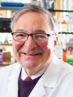 R Genco合作论文数Periodontal Disease Clinical Research Center, University at Buffalo;Department of Oral Biology, School of Dental Medicine, University at Buffalo18
 Steven Offenbacher合作论文数Department of Periodontology, Adams School of Dentistry, The University of North Carolina at Chapel Hill15
 Jukka H. Meurman合作论文数Institute of Dentistry, University of Helsinki;Department of Oral and Maxillofacial Diseases, Helsinki University Central Hospital13
 J. Max Goodson合作论文数Forsyth Institute;Department of Oral Medicine, Infection, and Immunity, Harvard School of Dental Medicine, Harvard University12
 Robert Gyurko合作论文数12
 Salomon Amar合作论文数Touro College of Dental Medicine, New York Medical College;Touro College and University System11
 Bruno S. Herrera合作论文数Department of Physiology and Pathology, UNESP-University Estadual Paulista11Sok-Ja Janket合作论文数School of Dental Medicine, Boston University11Batista Eraldo L合作论文数Goldman School of Dental Medicine, Boston University9
 Daniel Nguyen合作论文数9
 Marcelo Freire合作论文数Genomic Medicine and Infectious Disease Department, J. Craig Venter Institute9
 Danielle Stephens合作论文数9
 Corneliu Sima合作论文数Department of Oral Medicine, Infection and Immunity, Harvard School of Dental Medicine, Harvard Medical School9
 Kenneth Kornman合作论文数Department of Periodontics & Oral Medicine, School of Dentistry, University of Michigan-Ann Arbor8
 Bruce J. Paster合作论文数Harvard School of Dental Medicine;The Forsyth Institute8Peter Mark Bartold合作论文数8Qisheng Tu合作论文数8
 Kevin Sheng-Kai Ma合作论文数National Taiwan University7Luis Carlos Spolidorio合作论文数7Anne D Haffajee合作论文数7
 Jake Jinkun Chen合作论文数Division of Oral Biology, School of Dental Medicine, Tufts University;Department of Genetics, Molecular and Cellular Biology, Graduate School of Biomedical Sciences, Tufts University7
 James Beck合作论文数Adams School of Dentistry, The University of North Carolina at Chapel Hill7
 Gabrielle Fredman合作论文数Harvard Institutes of Medicine, and Harvard Medical School7
 Henrique Ballassini Abdalla合作论文数Laboratoy of Neuroimmune Interface of Pain Research, Instituto de Pesquisas São Leopoldo Mandic6Ayilavarapu Srinivas合作论文数Goldman School of Dental Medicine, Boston University6
 Kholy Karim El合作论文数Forsyth Institute;Harvard University;Forsyth Institute, Harvard University6
 Martha E Nunn合作论文数6
 Judith Jones合作论文数Center for Clinical Research, Henry M. Goldman School of Dental Medicine, Boston University6
 Barbara S Nikolajczyk合作论文数Department of Biology, Rosenstiel Basic Medical Sciences Research Center;Brandeis University;Rosenstiel Basic Medical Sciences Research Center, Brandeis University6Wei Qiu合作论文数5Chia-Dan Cheng合作论文数5Wan-Chien Cheng合作论文数5
 Nidia Castro dos Santos合作论文数Department of Periodontology and Oral Implantology, Guarulhos University5Takanori Shinjo合作论文数5Cheng-En Sung合作论文数5
 Marcelo H Napimoga合作论文数Lab Immunol & Mol Biol, Sao Leopoldo Mandic Inst & Res Ctr5Baird Alison E合作论文数5
 Atsushi Ishikado合作论文数5
 Lee Chun-Teh合作论文数Department of Oral Medicine, Infection and Immunity, Harvard School of Dental Medicine5
 Evangelos Papathanasiou合作论文数Department of Periodontology, Tufts University School of Dental Medicine5
 George L. King合作论文数Department of Ophthalmology, Harvard Medical School;Joslin Diabetes Center5
 Maurizio S. Tonetti合作论文数Department of Periodontology and Implant Dentistry, Faculty of Dentistry, The University of Hong Kong;Shanghai Perio Implant Innovation Center, School of Medicine, Shanghai Jiao Tong University;European Research Group on Periodontology5
 Jan Lindhe合作论文数Department of Periodontology, Sahlgrenska Academy, University of Gothenburg5
 Joao Paulo Steffens合作论文数5
 Ren-Yeong Huang合作论文数5
 Ahmed Tawakol合作论文数Massachusetts General Hospital, Harvard Medical School5




