Pulmonary bone marrow embolism is most strongly associated with trauma or chest compressions during cardiopulmonary resuscitation. Embolism associated with robot-assisted lung resection has not been previously reported. A female patient underwent left upper lobectomy with robot-assisted thoracic surgery for lung cancer. On postoperative Day 3, chest X-ray revealed a fracture on the dorsal side of the eighth rib. Her postoperative course was uneventful. Pathology of the resected lung revealed pulmonary bone marrow embolism. Owing to anatomical limitation in the mobility of the dorsal intercostal rib space, there is a risk of rib fracture by the metallic port of the robot. The path from the skin incision to the intercostal space should be directed straight towards the target anatomy, and attention should be given to the direction of movement of the most dorsal port during surgery to avoid rib fractures.
Background/objectives: The efficacy of lung metastasectomy in patients with urothelial carcinoma remains inconclusive, as there is only limited evidence from small studies. In this study, we aimed to assess the prognostic outcomes of excising pulmonary metastases from urothelial carcinoma. Methods: In this study, we utilized data from the Metastatic Lung Tumor Study Group of Japan database, a multi-institutional prospective database of pulmonary metastasectomies. We examined the data of patients who had undergone pulmonary metastasectomy for urothelial carcinoma between 1985 and 2021. Exclusion criteria included insufficient clinical information and follow-up of <3 months. Results: The study cohort comprised 100 patients (63 bladder cancer, 37 renal pelvic and ureteral cancer), with a median follow-up of 34 months. There were 70 male and 30 female patients of average age 66.5 ± 10.4 years at lung metastasectomy. The median interval from treatment of the primary lesion to metastasectomy was 19 months and the maximum tumor diameter was 21 ± 15 mm. Three- and five-year overall survival rates were 69% and 59%, respectively. Three- and five-year disease-free survival rates were 56% and 46%, respectively. Multivariate analysis identified larger tumor diameter (hazard ratio: 1.62, 95% confidence interval: 1.21–2.17) and distant metastases at the time of treatment of the primary cancer (hazard ratio: 4.23; 95% confidence interval: 1.54–11.6) as significant adverse prognostic factors for overall survival. Conclusions: To our knowledge, this is the largest published case series of pulmonary resection for metastatic urothelial carcinoma, providing benchmark data for the assessment of long-term outcomes of this rare entity.
BACKGROUND/AIM:The dynamic interplay between cancer cells and the microenvironment involves a wide range of intricate relationships that evolve during different stages of tumor progression. Recent attention has focused on high endothelial venules (HEVs), specialized endothelial cells in tumors with a unique cuboidal shape similar to those in lymph nodes. Previous animal studies have shown that normalization of tumor angiogenesis through anti-VEGFR2 therapy promotes HEV formation. However, few reports exist regarding the relationship between HEVs and preexisting blood vessels or interstitial fibers. In this study, we histologically examined whether tumor vascular structure correlates with HEV neogenesis. PATIENTS AND METHODS:A total of 109 patients with pathological stage I lung adenocarcinoma who had undergone curative lung resection at our Institute between 2012 and 2016 were included. HEVs were identified by anti-peripheral node addressin (PNAd) staining. Immunostaining and Elastica-Masson-Goldner staining were performed on tumor sections and quantified. RESULTS:PNAd-positive cells were identified in 102 (93.6%) patients. Nearly all PNAd-positive cells were located within or near immune cell clusters. We investigated the correlation between microvessel structures or interstitial fibers and the number/density of PNAd-positive vessels, but no significant correlation was found. Since PNAd-positive cells were concentrated in immune cell aggregates, we focused our analysis specifically on these regions. Immune cell aggregates with abundant PNAd-positive vessels had a greater microvessel density along with by rich collagen fiber production, and displayed a more mature morphological phenotype of HEVs. CONCLUSION:The generation of PNAd-positive cells in tumors is governed by an angiogenetic mechanism distinct from that of broader tumor microenvironment. Furthermore, the accumulation of immune cells is associated with increased HEV maturation.
Hemoglobin vesicles (HbVs), considered as red blood cell substitutes, are liposomes encapsulating purified hemoglobin, with a phospholipid bilayer membrane (diameter: 250 nm; P50, 28 Torr). In this study, we aimed to investigate HbV function during hemorrhagic shock in lung resection and analyze the details of oxygen delivery. Left pneumonectomy was performed in dogs under mechanical ventilation, followed by rapid exsanguination of approximately 30% of the total circulating blood volume, which led to shock, reducing the mean arterial pressure (MAP) by approximately 60% of baseline. Subsequently, either 5% human serum albumin (HSA) or HbVs suspended in 5% HSA were infused for resuscitation. The MAP only recovered to 75% of baseline after HSA administration, but fully recovered (100%) after HbV administration, with significant differences between the groups (P < 0.005). Oxygen delivery was restored in the HbV group and was significantly higher than that in the HSA group (P < 0.0001). The infusion of HbVs dispersed in a 5% HSA solution compensated for the rapid loss of approximately 30% of the total circulating blood volume in a dog pneumonectomy model, even with impaired lung function. Thus, HbVs can be used for resuscitation from hemorrhagic shock during thoracic surgery.
Supplementary Figure 3 - Expression of VEGFR-1 and IL-1ß in TAMs in PAN02 tumors.
Effects of VEGFR-1 signaling ablation on glucose/insulin tolerance and insulin production by the pancreas during obesity.
Figure S1 is a schematic of lymphocytes shown as CD3+ cells against side scatter (SSC). Figure S2 is a Kaplan-Meier time-to-progression graph.
56歳女性.呼吸困難と体重減少を主訴に受診した.CTで前縦隔から両側胸腔の大半を占拠する30 cm大の腫瘤を認めた.画像所見から脂肪肉腫と診断し,手術の方針とした.巨大な腫瘍であるため麻酔や手術中の換気不全,循環虚脱のリスクを考慮し,膜型人工肺をスタンバイした.Clamshellアプローチで手術を行い,腫瘍を切除した.腫瘍の最大径は38 cm,重量は2855 gであり,病理学的診断は脱分化型脂肪肉腫であった.術後2年経過し再発なく経過観察中である.
Supplementary Figure 5 - Effects of VEGFR-1 signaling ablation on PAN02 tumor progression in body-weight matched mice.
Supplementary Table 1: Protein quantification of cytokines in tumors and plasma of WT and Flt1TK-/- obese using ELISA; Supplementary Table 2: Quantification of the expression of IL-1ÃŽÃ,² and VEGFR-1 in TAMs from PAN02 tumors by mmunofluorescence.
Supplementary Figure 1 - A) Vessel density in PAN02 tumors from lean and obese mice, and effect of VEGFR-1-TK-deletion on tumor vessel density and hypoxia markers in obese mice. Supplementary Figure 2 - Gene expression of M1 / M2 markers in TAMs isolated from PAN02 tumors in lean and obese mice.
Table S1 displays second line therapies that patients pursued. Table S2 displays baseline and change (%) in plasma and cellular biomarkers.
Supplementary Figure 7 - Effects of VEGFR-1 signaling ablation on IGF-1R/IR signaling pathways in PAN02 and E0771 tumors implanted in obese mice; Supplementary Figure 8 - Additional effects of VEGFR-1 signaling ablation and metformin on tumor metabolism and vessel density in obese mice.
Supplementary Figure 10 - Additional measurements of PlGF and VEGF-B; Supplementary Figure 11 - Additional effects of PlGF deletion on obesity-induced tumor progression and systemic metabolism.
Effects of VEGFR-1 signaling ablation on E0771 tumor growth, vascular and immune environment in lean and obese mice.
 Rakesh K. Jain合作论文数Harvard Medical School, Harvard University;Edwin L. Steele Laboratories for Tumor Biology, Department of Radiation Oncology, Massachusetts General Hospital30
 Dan G. Duda合作论文数Houston Methodist Research Institute28
 Kohno Mitsutomo合作论文数19Masatoshi Yamaguchi合作论文数17
 Dai Fukumura合作论文数Massachusetts General Hospital;Mass General Research Institute;Harvard Medical School, Harvard University17Trupti Vardam合作论文数Edwin L. Steele Laboratory, Massachusetts General Hospital;Edwin L. Steele Laboratory, Massachusetts General Hospital17Hirotoshi Horio合作论文数17
 Babykutty Suboj合作论文数Department of Biochemistry, Sree Chitra Tirunal Institute for Medial Sciences and Technology16Suboj Priya合作论文数16
 Joao Incio合作论文数Edwin L. Steele Laboratory for Tumor Biology, Department of Radiation Oncology,16Masahiko Harada合作论文数16
 Andrew X. Zhu (朱秀轩)合作论文数Jiahui International Cancer Center, Jiahui International Hospital (Shanghai)15Mitsuo Nakayama合作论文数14Aoki Kohei合作论文数13Shan M. Chin合作论文数12Fukuda Hiroki合作论文数12Ato Sugiyama合作论文数11Inoue Yoshiaki合作论文数11Dan T. McManus合作论文数10Masatoshi Gika合作论文数10Udo Hoffmman合作论文数10Anna Khachatryan合作论文数10
 Ivy Chen合作论文数10Masabumi Shibuya (澁谷正史)合作论文数Graduate School of Medical and Dental Sciences, Tokyo Medical and Dental University;The University of Tokyo;Nagoya University;Institute of Physiology and Medicine, Jobu University10
 Christopher Schlett合作论文数Massachusetts General Hospital, Harvard Medical School10
 Ian Krop合作论文数Yale Cancer Center;School of Medicine, Yale Univeristy;Harvard Medical School10
 Nuh Rahbari合作论文数Department of General and Visceral Surgery, University Hospital Ulm10
 Gregory Wojtkiewicz合作论文数Massachusetts General Hospital10
 Peter Carmeliet合作论文数Department of Oncology, Faculty of Medicine, KU Leuven;Laboratory of Angiogenesis and Vascular Metabolism, VIB-KU Leuven Center for Cancer Biology10
 Stefan B. Puchner合作论文数10Jennifer A. Ligibel合作论文数10
 Raquel Soares合作论文数Faculty of Medicine, University of Porto10
 Marek Ancukiewicz合作论文数10Keehoon Jung合作论文数Graduate School of Biomedical Science and Engineering, Korea Advanced Institute of Science and Technology (KAIST),10Ramjiawan Rakesh R合作论文数9
 Takashi Ohtsuka合作论文数Jikei University, School of Medicine, Division of Thoracic Surgery, Department of Surgery9Kamiyama Ikuo合作论文数8
 Kohei Shigeta合作论文数8
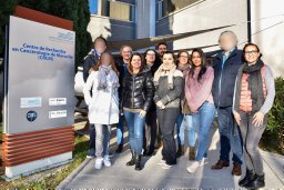 Emilie Mamessier合作论文数INSERM, Univ Aix Marseille 27Hishima Tsunekazu合作论文数7Peigen Huang合作论文数7
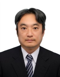 Taichiro Goto合作论文数Lung Cancer and Respiratory Disease Center, Yamanashi Central Hospital6
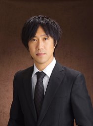 Kitahara Shuji合作论文数6
 Vikram Deshpande合作论文数Department of Pathology, Beth Israel Deaconess Medical Center, Harvard Medical School6
 Shigeki Suzuki合作论文数6
 Lipika Goyal合作论文数Division of Oncology, Department of Medicine, School of Medicine, Stanford University;Stanford Cancer Institute6
 Nabeel M. Bardeesy合作论文数Center for Cancer Research, Massachusetts General Hospital;Harvard Medical School;KFCCR Faculty, Mass General Research Institute;Harvard Stem Cell Institute;OncoRev, LLC6
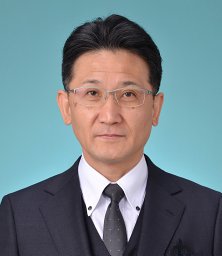 Yuichi Oike (尾池雄一)合作论文数Department of Molecular Genetics, Kumamoto University6
 Yunching Chen合作论文数Edwin L. Steele Laboratory, Massachusetts General Hospital and Harvard Medical School5




