Supplementary Fig. S9: Expression levels of Cxcr3 and its ligands are increased in ICC tissues after GC/dual ICB treatment in murine 425-ICC.
Supplementary Fig. S8: Bulk tissue RNA sequencing analysis of ICC after GC/dual ICB combination therapy in orthotopic murine 425-ICC model.
Supplementary Fig. S12: Effect of ICB treatment scheduling on efficacy and toxicity.
Supplementary Fig. S2: Standard chemotherapy converts ICB-resistant ICCs to ICB-responsive tumors, significantly delays tumor progression and increases survival in mice.
Supplementary Fig. S6: CTLA-4 blockade mediates the efficacy of GC/ICB therapy in ICC and increases CD8+CTL frequency in murine ICC.
Abstract Introduction: Pancreatic ductal adenocarcinoma (PDAC) is the third leading cause of cancer-related death in the United States, with a 5-year overall survival of ∼13%. Delayed diagnosis, limited response to current treatments, and the predominance of locally advanced or metastatic disease contribute to poor outcomes. Leveraging our preclinical findings in PDAC murine models, we showed that adding losartan—an angiotensin II type-1 receptor (AT1) blocker—to FOLFIRINOX followed by chemoradiation doubled R0 resection rates to ∼70% in locally advanced PDAC in a phase II trial (NCT01821729) (PMID: 31145418). However, the patient-response was variable. Here we recapitulated this variability in orthotopic PDAC mouse models and revealed the underlying mechanism. Methods and Results: To investigate potential causes of the variable response, we mapped AT1 expression using light-sheet microscopy in AT1 reporter mice bearing orthotopic PDAC. We observed abundant AT1 throughout the tumor microenvironment. To define its functional relevance, we generated Agtr1a knockout (KO) PDAC cell lines and inducible KO mouse models. AT1 deletion in either cancer cells or stromal cells (but not α-SMA+ myCAFs or pericytes) significantly reduced tumor growth. Because losartan is a pro-drug that needs activation by liver enzymes CYP3A4 and CYP2C9, we next examined its metabolism. We subcutaneously administered two major losartan metabolites, EXP3179 and EXP3174, to orthotopic PDAC-bearing mice and found that EXP3174 mediated the anti-tumor effects of losartan. Mass spectrometry of plasma samples revealed that while some tumor-bearing mice efficiently converted losartan to its active metabolite, others showed limited or no conversion. To determine the cause, we assessed CYP2C9 and CYP3A4 activity in liver microsomes from non-metastatic PDAC-bearing mice. CYP2C9 activity was markedly reduced and inversely correlated with tumor burden, providing a mechanistic basis for differential losartan activation. Ongoing work aims to determine whether PDAC patients likewise display variable plasma levels of losartan and EXP3174. Conclusion: These findings identify impaired hepatic metabolism as a key driver of variable losartan responses in locally advanced PDAC. Mass-spectrometry-based assessment of losartan and EXP3174 levels may help determine which patients can effectively activate the drug. Given losartan’s safety and low cost, its oral administration remains appropriate for most patients; however, in individuals with compromised CYP2C9 activity, administration of the active metabolite — EXP3174 may represent a more effective therapeutic option. Citation Format: Heena Kumra, Ryo Morisue, Benjamin E. Wolf, Vasiliki Salameti, Sonu Subudhi, Nilesh P. Talele, Eric F. Zaniewski, Robert Morris, Tsion H. Tale, Karim El-Marouk, Cora Schueller, Mariagiovanna Barresi, Jennifer Schulz, Halil I. Corbali, Rieke Schleinhege, Peigen Huang, Pascal Bernatchez, Wilhelm Haas, Yves Boucher, Dai Fukumura, Rakesh K. Jain. Improving response of pancreatic cancer to losartan: Mechanistic insights and implications for personalized therapy [abstract]. In: Proceedings of the American Association for Cancer Research Annual Meeting 2026; Part 1 (Regular Abstracts); 2026 Apr 17-22; San Diego, CA. Philadelphia (PA): AACR; Cancer Res 2026;86(7 Suppl):Abstract nr 1781.
Abstract Group 3 medulloblastomas (G3MB) carry the worst prognosis among medulloblastoma subtypes, yet molecularly targeted therapies remain elusive. Standard treatments cause severe long-term morbidity in survivors. Here, we identify tumor-derived sphingosine kinase 2 (SPHK2) as an essential driver of G3MB initiation and progression. SPHK2 exacerbates local immunosuppression by suppressing cytotoxic T-cell and NK-cell activity while promoting regulatory T-cell infiltration. Genetic or pharmacologic SPHK2 inhibition using Opaganib attenuates pro-survival tumor signaling and restores anti-tumor immunity, significantly improving survival in syngeneic G3MB mouse models. Combining Opaganib with fractionated low-dose radiation (f-LDRT) further enhances antigen presentation and reprograms tumor-associated myeloid cells toward an anti-tumor phenotype. This combination therapy markedly prolongs survival without inducing significant toxicity. Overall, our study establishes SPHK2 as a previously unrecognized therapeutic target and presents a safe, effective, microenvironment-reprogramming regimen for G3MB. One Sentence Summary Direct inhibition of tumor-derived SPHK2 overcomes local immunosuppression and downregulates pro-survival signaling in Group 3 medulloblastoma, while combination with fractionated low-dose radiation further enhances anti-tumor immunity and significantly improves survival.
Supplementary Fig. S11: Cxcr3 in CD8 T cells mediates the benefit of GC/ICB combination therapy in orthotopic murine 425-ICC model.
Abstract Introduction: Chronic arterial hypertension (CAH) is one of the most prevalent medical conditions worldwide and plays a causal role in cardiovascular diseases. Cancer patients with CAH have inferior outcomes compared to non-hypertensive cancer patients, especially in pancreatic ductal adenocarcinoma (PDAC). Furthermore, retrospective analyses have shown that antihypertensive therapy targeting angiotensin signaling is associated with improved oncological outcomes compared to other anti-hypertensive treatments in cancer patients. Methods and Results: To investigate this relationship, we employed a syngeneic, orthotopic PDAC model in mice with chronic hypertension induced by subcutaneously implanted osmotic minipumps delivering angiotensin II. Using highly multiplexed spectral flow cytometry, we found that chronic angiotensin II administration re-wires the anti-tumor immune response, leading to an increased presence of myeloid cells, particularly Ly6C+ monocytes, and CD206+ macrophages. Blockage of the angiotensin II receptor type I (AT1) with losartan leads to a marked decrease in tumor growth and a reduction in MDSCs. This effect was observed only with the synchronous administration of angiotensin II and losartan, not with losartan alone, and was consistent across different PDAC treatment regimens. In addition, we observed a trend toward improved tumor vessel perfusion in hypertensive mice treated with losartan, indicating vascular repair. Additional findings suggest that inhibition of the angiotensin II receptor type 2 (AT2) and the Mas receptor (which physiologically counterbalance AT1 signaling) abrogated the beneficial effect of AT1 blockade with losartan, suggesting a role for AT2 and the Mas receptor in shaping the immune microenvironment. Conclusion: Overall, our findings show that CAH reprograms the tumor microenvironment in PDAC and that inhibition of angiotensin signaling reverses these changes. This may indicate that inhibition of angiotensin signaling in cancer patients with CAH may exert its beneficial effect by reprogramming the anti-tumor immune response. Citation Format: Benjamin Wolf, Heena Kumra, Ryo Morisue, Karim El-marouk, Igor L. Gomes-Santos, Rieke Schleinhege, Tsion H. Tale, Marc Charabati, Sonu Subudhi, Dai Fukumura, Rakesh K. Jain. Angiotensin-II-induced chronic hypertension reprograms the antitumor-immune-response in pancreatic ductal adenocarcinoma [abstract]. In: Proceedings of the American Association for Cancer Research Annual Meeting 2026; Part 1 (Regular Abstracts); 2026 Apr 17-22; San Diego, CA. Philadelphia (PA): AACR; Cancer Res 2026;86(7 Suppl):Abstract nr 2084.
Supplementary Fig. S4: IMC analysis of ICC after GC-based therapies in orthotopic murine 425-ICC model.
Supplementary Fig. S5: CTLA-4 blockade is critical for efficacy of combined chemotherapy with ICB in ICC by grafting 425 murine cells in C57Bl/6/FVB F1 mice.
Supplementary Fig. S3: ScRNAseq analysis of ICC TME after GC-based therapies in orthotopic murine 425-ICC model.
Relationship between pathological response and circulating biomarker levels in the plasma of treated patients.
 Rakesh K. Jain合作论文数Harvard Medical School, Harvard University;Edwin L. Steele Laboratories for Tumor Biology, Department of Radiation Oncology, Massachusetts General Hospital330
 Dan G. Duda合作论文数Houston Methodist Research Institute123
 Lance Munn合作论文数Department of Radiation Oncology, Mass General Research Institute, Massachusetts General Hospital;Harvard Medical School;Université de Technologie de Compiègne82
 Soichiro Miura合作论文数Graduate School of Medicine, International University of Health and Welfare Graduate School;Graduate School of Public Health, International University of Health and Welfare Graduate School65
 I Kurose合作论文数50Peigen Huang合作论文数46Masaharu Tsuchiya合作论文数44
 Zohreh Amoozgar合作论文数Department of Radiation Oncology, Massachusetts General Hospital, Harvard Medical School41
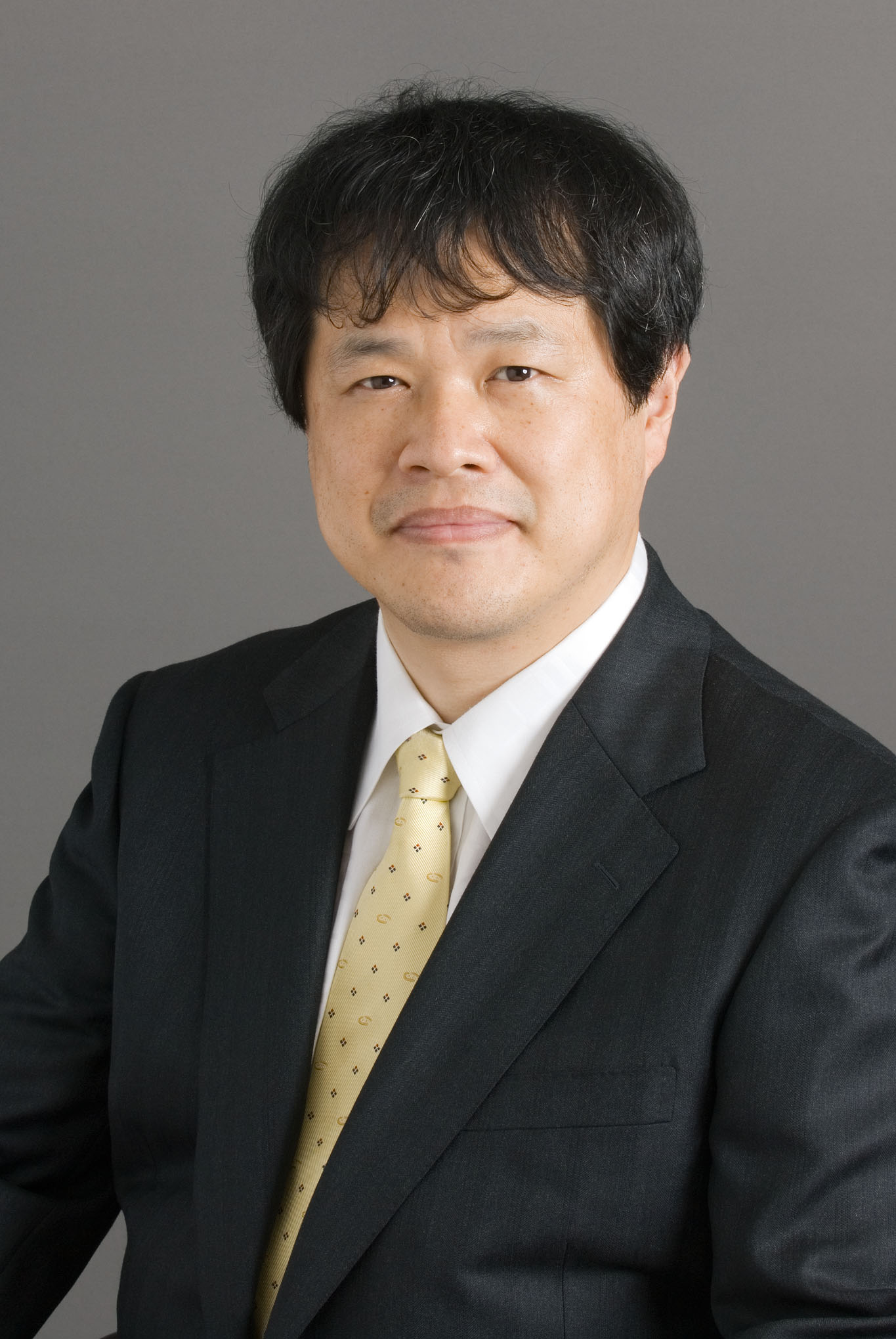 Makoto Suematsu (末松誠)合作论文数School of Medicine, Keio University39
 Meenal Datta合作论文数Edwin L. Steele Laboratory for Tumor Biology, Massachusetts General Hospital and Harvard Medical School;Department of Chemical and Biological Engineering, Tufts University;Harvard Medical School, Tufts University38
 Joao Incio合作论文数Edwin L. Steele Laboratory for Tumor Biology, Department of Radiation Oncology,32
 Yves Boucher合作论文数31Lei Xu合作论文数30Keehoon Jung合作论文数Graduate School of Biomedical Science and Engineering, Korea Advanced Institute of Science and Technology (KAIST),30
 Peter Carmeliet合作论文数Department of Oncology, Faculty of Medicine, KU Leuven;Laboratory of Angiogenesis and Vascular Metabolism, VIB-KU Leuven Center for Cancer Biology27
 Timothy P. Padera合作论文数Edwin L. Steele Laboratories, Massachusetts General Hospital;Harvard Medical School27
 Ashwin S Kumar合作论文数Harvard-Massachusetts Institute of Technology Division of Health Sciences and Technology, Massachusetts Institute of Technology25
 Moungi Bawendi (莫吉·巴旺迪)合作论文数Department of Chemistry, Massachusetts Institute of Technology25Posada, Jessica M.合作论文数24
 Sonu Subudhi合作论文数Massachusetts General Hospital, Harvard Medical School24
 Nuh Rahbari合作论文数Department of General and Visceral Surgery, University Hospital Ulm23Suboj Priya合作论文数22Trupti Vardam合作论文数Edwin L. Steele Laboratory, Massachusetts General Hospital;Edwin L. Steele Laboratory, Massachusetts General Hospital22
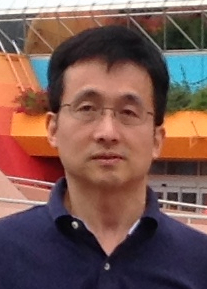 Yuhui Huang (黄玉辉)合作论文数Hematology Center, Cyrus Tang Medical Institute, Soochow University22H Ishii合作论文数21
 Jeffrey W Clark合作论文数Harvard Cancer Center, Harvard Medical School;Dana-Farber/Partners Cancer Care, Massachusetts General Hospital;Cancer Center Protocol Office, Massachusetts General Hospital21Pinji Lei合作论文数College of Life Sciences, Wuhan University21
 Talele Nilesh合作论文数21
 Walid S. Kamoun合作论文数Merrimack, Cambridge, MA;Merrimack, Cambridge, MA;21Sylvie Roberge合作论文数20
 Heena Kumra合作论文数Faculty of MedicineDepartment of Anatomy and Cell BiologyMcGill UniversityMontrealQC H3A 0C7Canada20
 Babykutty Suboj合作论文数Department of Biochemistry, Sree Chitra Tirunal Institute for Medial Sciences and Technology20
 Gregory Wojtkiewicz合作论文数Massachusetts General Hospital20Vasileios Askoxylakis合作论文数Merrimack Pharmaceuticals, Inc.20
 Mikael Pittet合作论文数Department of Pathology and Immunology, Faculty of Medicine, University of Geneva19H Higuchi合作论文数18Shan M. Chin合作论文数18
 John D. Martin合作论文数Edwin L. Steele Laboratory, Massachusetts General Hospital and Harvard Medical School;Department of Chemical Engineering, Massachusetts Institute of Technology;Massachusetts General Hospital and Harvard Medical School, Massachusetts Institute of Technology18
 Batista Ana合作论文数18
 Shuichi Aoki合作论文数Dept. of Surgery, Tohoku University Graduate School of Medicine18
 Ivy Chen合作论文数17
 Gino Ferraro合作论文数17Kirkpatrick Nathaniel D合作论文数17Yoshikazu Tsuzuki合作论文数17
 Tai Hato合作论文数School of Medicine, Keio University17
 Mari Mino-Kenudson合作论文数Harvard Medical School;Massachusetts General Hospital16
 Carlos Fernandez-Del Castillo合作论文数Mass General Research Institute;Harvard Medical School;GI Cancer Center, Massachusetts General Hospital16
 Theodore Hong合作论文数Tucker Gosnell Center for Gastrointestinal Cancers, Massachusetts General Hospital;Harvard Medical School16
 J Kloepper合作论文数16




