Objectives Inclusion body myositis (IBM) is an idiopathic inflammatory myopathy characterised by type 1 inflammation, driven by muscle-infiltrating KLRG1+ and TBX21+ cytotoxic T cells. However, the cellular sources of the stimuli that trigger this effector response in the muscle of patients with IBM remain unclear. Given their role as antigen-presenting cells, we hypothesised that these may be myeloid dendritic cells (mDCs), which have previously been reported in skeletal muscle of patients with IBM. We used immunohistochemistry, immunofluorescence, single-nucleus RNA-sequencing (snRNA-seq) and bulk RNA-sequencing (RNA-seq) data from skeletal muscle of patients with IBM, other myositis, and controls to identify mDCs and characterise their contribution to IBM inflammation. Methods We first analysed snRNA-seq data from 3 datasets to identify, quantify, and characterise 3 mDC subsets: type 1 conventional dendritic (cDC1) cells, type 2 conventional dendritic (cDC2) cells, and mature immunoregulatory dendritic (mregDC) cells. We then analysed RNA-seq data from 2 datasets to correlate specific markers of these subsets with markers of IBM disease activity. We used immunohistochemistry and immunofluorescence to confirm mDC presence. Results All 3 mDC subsets, and especially cDC1 cells, are relatively increased in the muscle of patients with IBM and correlate strongly with IBM-specific inflammatory markers, including KLRG1 and TBX21. In particular, cDC1 cells specifically express the KLRG1 ligands, CDH1 and CDH2, and, along with mregDC cells, IL12B, representing possible juxtacrine and paracrine signals for effector T cells in IBM. Conclusions Skeletal muscle of patients with IBM is specifically characterised by mDC subsets that correlate with markers of cytotoxic T cells and type 1 inflammation.
OBJECTIVE:Sporadic inclusion body myositis (IBM) is the most common adult idiopathic inflammatory myopathy. IBM etiology has been elusive, due to both degenerative and autoimmune disease features found on muscle biopsy, and significant disease heterogeneity. Investigating the role of antibodies in the muscle of IBM patients may improve our understanding of disease pathogenesis. METHODS:We used an IBM xenograft mouse model in which muscle biopsy tissue from IBM patients (n = 98) and controls (n = 131; including 54 from other types of myopathy) were implanted into immunodeficient mice (NOD-Rag1null-IL2rγnull). We quantified the amount of human IgG, IgA, and IgM in xenografted mouse sera using MesoScale Diagnostics (MSD) assay. We detected donor-derived antibody reactivities targeting autoantigens and infectious agents using Phage ImmunoPrecipitation Sequencing (PhIP-Seq). Finally, we used FR3 AmplifiKation Sequencing (FR3AK-Seq) to sequence the antibody mRNAs from a separate cohort of 146 patient biopsies (14 IBM, 22 healthy controls, 110 other myositis subtypes). RESULTS:With the MSD assay we found human IgG, IgA, and IgM in a larger percentage of IBM xenografted mice versus controls. Using PhIP-Seq, we found anti-microbial reactivities secreted from IBM muscle are prevalent amongst a healthy control population but autoantigen reactivities in IBM are more unique at the peptide and protein level. Additionally, NT5C1A (IgG/IgA and IgM) and TIF1γ (IgG/A) autoantibodies are secreted from muscle tissues of 4/18 and 10/18 IBM xenograft donors, respectively. CONCLUSION:Our characterization of antibody responses within the muscle of IBM patients reveals that muscle-infiltrating B cells produce both disease-associated autoantibodies and a broad spectrum of antibodies targeting non-self antigens.
Background and Objectives:Valosin Containing Protein-associated multisystem proteinopathy (VCP-MSP) is a progressive, autosomal dominant disorder caused by pathogenic variants in the VCP gene, resulting in a heterogeneous clinical presentation. Muscle biopsy findings are characteristic but not pathognomonic. This study aimed to comprehensively analyse VCP-related myopathology and explore correlations with clinical phenotypes, genetic variants, and disease progression. Methods:Muscle biopsy images and data were collected retrospectively from adults (≥18 years) with pathogenic or likely pathogenic VCP variants enrolled in the VCP Multicentre International Study. Biopsy data were standardized using the "Common Data Elements for Muscle Biopsy Reporting." Variations in biopsy findings were analysed by biopsy site, time from disease onset, the four most common VCP variants, and clinical phenotypes. Result:A total of 112 muscle biopsies were included. Most individuals were male (66.0%). The mean age at biopsy was 53.3 years (SD 10.0), with a mean disease duration of 6.5 years (SD 4.5). The most frequent VCP variant was c.464G>A (p.Arg155His) (18.8%). The top clinical phenotypes were isolated myopathy (37.5%), myopathy with Paget disease of bone (17.9%), and myopathy with motor neuron involvement (13.4%). The vastus lateralis was the most common biopsy site (34.8%), and 91% were open biopsies. Histopathologic findings included atrophic fibres (87.5%), rimmed vacuoles (72.3%), endomysial fibrosis (58.0%), and protein aggregates (51.8%), primarily p62 (60.3%) and VCP (36.2%). Degeneration niches with fibrofatty replacement and atrophic fibres were seen in 33.3% of biopsies without frequency differences by clinical phenotypes. There were no differences in biopsy findings among the 4 most common VCP gene variants, except for the absence of degeneration niches in muscle biopsies of 12 patients with c.277C>T (p.Arg93Cys). MRI data from 30 patients showed fat pockets corresponding to these niches and STIR hyperintensity correlated with inflammatory infiltrates in 42.9%. Concordance between clinical phenotype, biopsy, and neurophysiology was observed in only 49.4% of cases, indicating significant heterogeneity. Discussion:VCP-MSP muscle biopsies consistently show myopathic or mixed patterns with rimmed vacuoles and p62/VCP-positive inclusions, regardless of clinical phenotype, age, or progression. Some lack vacuoles, challenging diagnosis. Discrepancies between clinical, neurophysiology, and biopsy findings should prompt consideration of VCP-MSP to improve detection and management.
Defective nucleocytoplasmic transport (NCT) has emerged as a contributing factor in the pathogenesis of neurodegenerative diseases and aging. Valosin-containing protein (VCP) is an AAA+ATPase required for disassembly of protein complexes, and mutations in VCP cause neurodegenerative and neuromuscular diseases. We find that VCP is required for quality control of nuclear pore complexes (NPCs) by extracting selected nucleoporins from NPCs for proteasome-mediated degradation. Pathogenic VCP variants cause a reduction in nucleoporins in Drosophila, induced pluripotent stem cell (iPSC)-derived motor neurons, and muscle biopsies from patients, indicating a dominant gain-of-function mechanism. Mechanistically, disease-associated mutations in VCP result in increased recruitment to NPCs through interactions with Ufd1-Npl4, leading to the removal of a subset of nucleoporins from NPCs and disrupting NCT. These findings show that the VCP-Ufd1-Npl4 pathway regulates NPC quality control and that disease-associated variants aberrantly activate the VCP-Ufd1-Npl4 complex to degrade NPCs and disrupt NCT.
Inclusion body myositis (IBM) is an idiopathic inflammatory myopathy characterized by muscle-infiltrating KLRG1+ and TBX21+ cytotoxic T cells and type 1 inflammation. Myeloid dendritic cells (mDCs), including type 1 conventional dendritic (cDC1) cells, type 2 conventional dendritic (cDC2) cells, and mature immunoregulatory dendritic (mregDC) cells, have previously been reported in skeletal muscle of IBM patients and may activate these cytotoxic T cells. Here, we analyzed single-nucleus RNA-sequencing (snRNA-seq) and bulk RNA-sequencing (RNA-seq) data from skeletal muscle of IBM, other myositis, and control patients to identify and quantify these mDC subsets and characterize their contribution to IBM inflammation. Our findings reveal that all three mDC subsets are relatively increased and activated in muscle of IBM patients and correlate with IBM-specific inflammatory markers. Our data specifically implicates cDC1 cells in CD8+ T cell activation via specific expression of both KLRG1 ligands, CDH1 and CDH2, as well as IL12B in IBM muscle.
OBJECTIVE:Inclusion body myositis (IBM) is an idiopathic inflammatory myopathy with muscle pathology characterized by endomysial inflammation, rimmed vacuoles, and cytoplasmic mislocalization of transactive response DNA-binding protein 43 (TDP-43). We aimed to determine whether loss of TDP-43 splicing repression led to the production of "cryptic peptides" that could be detected in muscle biopsies as a useful biomarker for IBM. METHODS:We used an antisera against a neoepitope encoded by a TDP-43-dependent cryptic exon within hepatoma-derived growth factor-like protein 2 (HDGFL2) for immunohistochemical analysis on muscle biopsy samples of 122 patients with IBM, 181 disease controls, and 16 healthy controls without abnormal muscle pathology. In situ hybridization was also utilized to detect the localization of cryptic HDGFL2 transcripts. RESULTS:We found cryptic HDGFL2 peptides localized within myonuclei from muscle biopsies in 79 of 122 patients with IBM (65%), and this staining correlated with TDP-43 depletion. In contrast, cryptic HDGFL2 immunoreactivity was absent in 197 muscle biopsies from a variety of disease controls, except for 2 patients with vacuolar myopathies. Notably, we show that cryptic HDGFL2 transcripts are accompanied by the detection of cryptic HDGFL2 in muscle fibers of IBM without rimmed vacuoles and TDP-43 aggregates. INTERPRETATION:Together, our findings establish that loss of TDP-43 splicing repression occurs in myonuclei of IBM skeletal muscle and suggest that detection of cryptic peptides in muscle biopsies may be a useful biomarker. We suggest that a therapeutic strategy designed to restore TDP-43 function should be considered to attenuate the degeneration of skeletal muscle in this devastating disease. ANN NEUROL 2025;97:629-641.
Idiopathic inflammatory myopathies (IIMs), or myositis, are rare diseases marked by immune-driven muscle damage and complications like skin lesions and interstitial lung disease (ILD). Despite advances, challenges in diagnosis and treatment persist, particularly in inclusion body myositis (IBM), where no effective therapy exists. Recent breakthroughs, including transcriptomics and insights into antibody-mediated immunity and interferon (IFN) signaling, have clarified IIM pathophysiology and spurred the development of new therapies, such as chimeric antigen receptor (CAR) T cells and Janus kinase (JAK) inhibitors. We explore the latest findings on the mechanisms underlying adult-onset IIMs, emphasizing IBM pathobiology and its unique immune and degenerative pathways, such as a selective type 2 myofiber damage and severe cell stress. Finally, we highlight the recent advances in transcriptomics, single-cell analysis, and machine learning in transforming IIM research by improving diagnostic accuracy, uncovering therapeutic targets, and supporting the development of personalized treatment strategies.
Anti-NT5c1A antibodies, directed against cytosolic 5’-nucleotidase that is abundant in skeletal muscle, were identified as the first serological biomarker for IBM. Prior research suggested that NT5c1A seropositivity prognosticated a more severe motor phenotype with more severe motor weakness and bulbar involvement. Subsequent studies produced conflicting data, either confirming previous observations or not showing any relationship. The debate remains whether serological status may provide insight into functional severity and disease behavior. INSPIRE-IBM is a prospective NIH-funded observational study including patients ages 40 years or older with clinically defined IBM fulfilled by the ENMC 2011 criteria, and disease onset within the past 10 years of the Baseline visit. Serology for NT5c1A was collected at Baseline. Functional assessments to evaluate disease severity included Manual Muscle Testing (MMT), Timed get up-and-go (TUG), Sydney Swallow Questionnaire (SSQ), and EAT-10. Serological status was available for 140 out of 150 participants with IBM who were enrolled. Sixty-nine of the 140 IBM patients (49%) were seropositive for NT5c1A antibodies at Baseline. Patients were divided into two groups (Group A with disease duration between 0-5 years and Group B with disease duration between 6-10 years). Seropositive group A showed significantly greater difficulty swallowing (EAT-10 and SSQ) than seronegative group A. Seropositive group B showed a trend towards more difficulty swallowing (EAT-10 and SSQ) and motor function weakness (MMT) compared to the seronegative group but did not reach statistical significance. Seropositive IBM patients appear to have more swallowing difficulties than seronegative patients, and this difference appears early on in the disease course.
Inclusion body myositis (IBM) is a common muscular disorder in individuals over the age of 40 years, characterized by atrophy and progressive muscle weakness. Patient-reported outcomes such as the IBMFRS or the sIFA questionnaire provide valuable insights into disease impact from the patient's perspective on their symptoms, functional limitations, and quality of life. However, it remains a topic of further investigation to determine which of these questionnaires exhibits stronger correlations with disease progression. The INSPIRE-IBM is a natural history study involving 150 IBM patients across 13 different US sites. Evaluations are conducted biannually over two years and patients complete IBMFRS, sIFA, EAT-10, Sydney Swallow Questionnaire, PROMIS, along with manual muscle testing and pulmonary functions tests. This abstract analyzes correlations between IBMFRS and sIFA with the other assessments by regression analysis to identify which is a stronger correlator with disease progression. Preliminary analysis, involving 87 patients who completed three time points, revealed a strong correlation between IBMFRS and sIFA (R2=0.7, p=3.21E-96). Both outcomes show moderate correlation with PFTs (R2 between 0.5-0.7), with no significant difference in strength of correlation. IBMFRS and sIFA exhibit similar correlation with MMTs (R2=0.43, p=0.93). As the study is ongoing, more timepoints will be available per patient closer to the conference date and will be included in the analysis.
OBJECTIVES:Autoantibodies targeting intracellular proteins are common in various autoimmune diseases. In the context of myositis, the pathologic significance of these autoantibodies has been questioned due to the assumption that autoantibodies cannot enter living muscle cells. This study aims to investigate the validity of this assumption. METHODS:Confocal immunofluorescence microscopy was employed to localise antibodies and other proteins of interest in myositis muscle biopsies. Bulk RNA sequencing was used to examine the transcriptomic profiles of 669 samples, including those from patients with myositis, disease controls and healthy controls. Additionally, antibodies from myositis patients were introduced into cultured myoblasts through electroporation, and their transcriptomic profiles were analysed using RNA sequencing. RESULTS:In patients with myositis autoantibodies, antibodies accumulated inside myofibres in the same subcellular compartment as the autoantigen. Bulk RNA sequencing revealed that muscle biopsies from patients with autoantibodies targeting transcriptional regulators exhibited transcriptomic patterns consistent with dysfunction of the autoantigen. For instance, in muscle biopsies from patients with anti-PM/Scl autoantibodies recognising components of the nuclear RNA exosome complex, an accumulation of divergent transcripts and long non-coding RNAs was observed; these RNA forms are typically degraded by the nuclear RNA exosome complex. Introducing patient antibodies into cultured muscle cells recapitulated the transcriptomic effects observed in human disease. Further supporting evidence suggested that myositis autoantibodies recognising other autoantigens may also disrupt the function of their targets. CONCLUSIONS:This study demonstrates that, in myositis, autoantibodies are internalised into living cells, causing biological effects consistent with the disrupted function of their autoantigen.
TAR DNA binding protein 43 (TDP-43) is an RNA binding protein that accumulates as aggregates in the central nervous systems of some patients with neurodegenerative diseases. However, TDP-43 aggregation is also a sensitive and specific pathologic feature found in a family of degenerative muscle diseases termed inclusion body myopathy. TDP-43 aggregates from amyotrophic lateral sclerosis (ALS) and frontotemporal dementia brain lysates may serve as self-templating aggregate seeds in vitro and in vivo, supporting a prion-like spread from cell to cell. Whether a similar process occurs in patient muscle is not clear. We developed a mouse model of inducible, muscle-specific cytoplasmic localized TDP-43. These mice develop muscle weakness with robust accumulation of insoluble and phosphorylated sarcoplasmic TDP-43, leading to eosinophilic inclusions, altered proteostasis, and changes in TDP-43-related RNA processing that resolve with the removal of doxycycline. Skeletal muscle lysates from these mice also have seeding-competent TDP-43, as determined by a FRET-based biosensor, that persists for weeks upon resolution of TDP-43 aggregate pathology. Human muscle biopsies with TDP-43 pathology also contain TDP-43 aggregate seeds. Using lysates from muscle biopsies of patients with sporadic inclusion body myositis (IBM), immune-mediated necrotizing myopathy (IMNM), and ALS, we found that TDP-43 seeding capacity was specific to IBM. TDP-43 seeding capacity anticorrelated with TDP-43 aggregate and vacuole abundance. These data support that TDP-43 aggregate seeds are present in IBM skeletal muscle and represent a unique TDP-43 pathogenic species not previously appreciated in human muscle disease.
Inclusion body myositis is an enigmatic slowly progressive acquired myopathy. There is strong evidence favoring autoimmune origin, including the invasion of non-necrotic myofibers by clonally expanded cytotoxic CD8+ T cells, which have the hallmark of highly and possibly terminally differentiated T cells, expressing the killer cell lectin-like receptor G1 (KLRG1) cell surface ligand, and have features of senescence. Previous research suggests there is greater T cell differentiation with longer disease severity, though there is a paucity of information surrounding how muscle-invading T cells may influence disease behavior. Understanding the frequency of highly differentiated lymphocytes and their relationship to disease severity and behavior may influence novel therapeutic solutions. INSPIRE-IBM is a longitudinal NIH-funded multicenter study including patients ages 40 years or older with clinically defined IBM fulfilled by the ENMC 2011 criteria, and disease onset within the past 10 years of the Baseline visit. Complete dataset for the baseline cross-sectional visit was available from 60 participants. Flow cytometry was used on PBMCs to analyze percentage of immunosenescent lymphocytes staining for CD8+, KRLG1+, TEMRAs, and Tregs. Functional assessments to evaluate disease severity included Manual Muscle Testing (MMT), Timed get up-and-go (TUG), Sydney Swallow Questionnaire (SSQ), and EAT-10. Data will be analyzed in May 2024 and results from Baseline will be presented.
Macroautophagy/autophagy is a major pathway for the clearance of protein aggregates and damaged organelles, and multiple intracellular organelles participate in the process of autophagy, from autophagosome formation to maturation and degradation. Dysregulation of the autophagy pathway has been implicated in the pathogenesis of neurodegenerative diseases including amyotrophic lateral sclerosis (ALS) and frontotemporal dementia (FTD), however the mechanisms underlying autophagy impairment in these diseases are incompletely understood. Since the expansion of GGGGCC (G4C2) repeats in the first intron of the C9orf72 gene is the most common inherited cause of both ALS and FTD (C9-ALS-FTD), we investigated autophagosome dynamics in Drosophila motor neurons expressing 30 G4C2 repeats (30 R). In vivo imaging demonstrates that expression of expanded G4C2 repeats markedly impairs biogenesis of autophagosomes at synaptic termini, whereas trafficking and maturation of axonal autophagosomes are unaffected. Motor neurons expressing 30 R display marked disruption in endoplasmic reticulum (ER) structure and dynamics in the soma, axons, and synapses. Disruption of ER morphology with mutations in Rtnl1 (Reticulon-like 1) or atl (atlastin) also impairs autophagosome formation in motor neurons, suggesting that ER integrity is critical for autophagosome formation. Furthermore, live imaging demonstrates that autophagosomes are generated from dynamic ER tubules at synaptic boutons, and this process fails to occur in a C9-ALS-FTD model. Together, these findings suggest that dynamic ER tubules are required for formation of autophagosomes at the neuromuscular junction, and that this process is disrupted by expanded G4C2 repeats that cause ALS-FTD.
Background The diagnosis of patients with mutations in the VCP gene can be complicated due to their broad phenotypic spectrum including myopathy, motor neuron disease and peripheral neuropathy. Muscle MRI guides the diagnosis in neuromuscular diseases (NMDs); however, comprehensive muscle MRI features for VCP patients have not been reported so far. Methods We collected muscle MRIs of 80 of the 255 patients who participated in the “VCP International Study” and reviewed the T1-weighted (T1w) and short tau inversion recovery (STIR) sequences. We identified a series of potential diagnostic MRI based characteristics useful for the diagnosis of VCP disease and validated them in 1089 MRIs from patients with other genetically confirmed NMDs. Results Fat replacement of at least one muscle was identified in all symptomatic patients. The most common finding was the existence of patchy areas of fat replacement. Although there was a wide variability of muscles affected, we observed a common pattern characterized by the involvement of periscapular, paraspinal, gluteal and quadriceps muscles. STIR signal was enhanced in 67% of the patients, either in the muscle itself or in the surrounding fascia. We identified 10 diagnostic characteristics based on the pattern identified that allowed us to distinguish VCP disease from other neuromuscular diseases with high accuracy. Conclusions Patients with mutations in the VCP gene had common features on muscle MRI that are helpful for diagnosis purposes, including the presence of patchy fat replacement and a prominent involvement of the periscapular, paraspinal, abdominal and thigh muscles.
Inclusion body myositis (IBM) is the most prevalent inflammatory muscle disease in older adults with no effective therapy available. In contrast to other inflammatory myopathies such as subacute, immune-mediated necrotizing myopathy (IMNM), IBM follows a chronic disease course with both inflammatory and degenerative features of pathology. Moreover, causal factors and molecular drivers of IBM progression are largely unknown. Therefore, we paired single-nucleus RNA sequencing with spatial transcriptomics from patient muscle biopsies to map cell-type-specific drivers underlying IBM pathogenesis compared with IMNM muscles and noninflammatory skeletal muscle samples. In IBM muscles, we observed a selective loss of type 2 myonuclei paralleled by increased levels of cytotoxic T and conventional type 1 dendritic cells. IBM myofibers were characterized by either upregulation of cell stress markers featuring GADD45A and NORAD or protein degradation markers including RNF7 associated with p62 aggregates. GADD45A upregulation was preferentially seen in type 2A myofibers associated with severe tissue inflammation. We also noted IBM-specific upregulation of ACHE encoding acetylcholinesterase, which can be regulated by NORAD activity and result in functional denervation of myofibers. Our results provide promising insights into possible mechanisms of myofiber degeneration in IBM and suggest a selective type 2 fiber vulnerability linked to genomic stress and denervation pathways. Inclusion body myositis (IBM) is a progressive inflammatory muscle disease of unknown cause, prevalent in older adults. Through spatial and single nuclear profiling, the authors identify a selective type 2 myofiber pathology in IBM, linked to genomic stress and denervation.
 Mammen Andrew L合作论文数52
 Iago Pinal-Fernandez合作论文数National Institute of Arthritis and Musculoskeletal and Skin Disease40
 Lisa Christopher Stine合作论文数Division of Rheumatology, Department of Medicine, Johns Hopkins University36Charlotte J. Sumner合作论文数60029
 Jemima Albayda合作论文数Division of Rheumatology, Department of Medicine, Johns Hopkins University26
 Paik Julie J合作论文数Johns Hopkins University School of Medicine26
 Maria Casal Dominguez合作论文数Muscle Dis Unit, NIH21Pak Katherine合作论文数20Selva-O'Callaghan Albert合作论文数18Chiseko Ikenaga合作论文数17
 Hugo J Bellen合作论文数Department of Molecular and Human Genetics, Baylor College of Medicine;Jan and Dan Duncan Neurological Research Institute, Texas Children Hospital17
 Jeremy M. Sullivan合作论文数Wellesley College Neuroscience Program17
 Sonye K Danoff合作论文数Department of Pharmacology, University of Cambridge Hills Road17
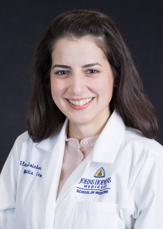 Eleni Tiniakou合作论文数Departments of Neurology and Neurosurgery, School of Medicine, Biomedical Engineering, Johns Hopkins University16
 Milisenda José C合作论文数16
 Jeffrey Rothstein合作论文数The Solomon H. Snyder Department of Neurology, School of Medicine, Johns Hopkins University;Institute for Clinical and Translational Research, Johns Hopkins University16
 Ke Zhang (张轲)合作论文数Institute of Neurological Diseases, Shenzhen Bay Laboratory15
 Tahseen Mozaffar合作论文数College of Medicine, University of California13
 Josep Maria Grau合作论文数Medical School, Fundació d'Investigació de la Santa Creu i Sant Pau Hospital de Sant Pau13Mazen M. Dimachkie合作论文数University of Texas–Houston Medical School13
 Michael Shy合作论文数Department of Neurology, Roy J. and Lucille A. Carver College of Medicine, University of Iowa;Division of Neuromuscular Medicine, Neurology, Roy J. and Lucille A. Carver College of Medicine, University of Iowa12Andrea M. Corse合作论文数Departments of Neurology and Neurosurgery, Johns Hopkins University School of Medicine12Mark N. Wu合作论文数12
 Davide Pareyson合作论文数Functional Department of Rare Neurological Diseases, Fondazione IRCCS Istituto Neurologico C.Besta;Simple Dept. Unit of “Rare Neurological Diseases of Adulthood”, Fondazione IRCCS Istituto Neurologico C.Besta12David Hermann合作论文数11
 Steven S. Scherer合作论文数Department of Neurology, Perelman School of Medicine, University of Pennsylvania;Mahoney Institute for Neurological Science, Perelman School of Medicine at the University of Pennsylvania11
 John W. Day合作论文数Department of Neurology & Neurological Sciences, School of Medicine, Stanford University;Division of Medical Genetics, Department of Pediatrics, School of Medicine, Stanford University11Mary M. Reilly合作论文数11
 Stephan Züchner合作论文数The Dr John T Macdonald Foundation Department of Human Genetics, Leonard M. Miller School of Medicine, University of Miami11
 Sabrina Yum合作论文数Center for Neural Injury, University of California San Francisco11Miriam Freimer合作论文数10Goyal Namita A合作论文数10Laurá Matilde合作论文数10
 Matthew Wicklund合作论文数Division of Neuromuscular Medicine, Department of Neurology, Long School of Medicine, University of Texas Health Science Center San Antonio10
 Aisenberg William H合作论文数Department of Physiology, Johns Hopkins University School of Medicine10Sandra Muñoz-Braceras合作论文数9Brett A McCray合作论文数Johns Hopkins Univ, Sch Med, Dept Neurol, Baltimore, MD 21205 USA9
 Lisa Christopher-Stine合作论文数Departments of Neurology and Neurosurgery, School of Medicine, Biomedical Engineering9
 Harry Benjamin Larman合作论文数Division of Health Sciences and Technology, MIT9
 Richard S. Finkel合作论文数Department of Pediatric Medicine, St. Jude Children's Research Hospital;Center for Experimental Neurotherapeutics, St. Jude Children's Research Hospital9
 Francesco Muntoni合作论文数Department of Developmental Neurosciences, Great Ormond Street Institute of Child Health, Faculty of Population Health Sciences, University College London;Dubowitz Neuromuscular Centre, Great Ormond Street Hospital for Children NHS Foundation Trust9
 Sindhu Ramchandren合作论文数University of Pennsylvania9Faiza Naz合作论文数8
 James Machamer合作论文数8Conrad C Weihl合作论文数8Kathleen M. Cunningham合作论文数Dept Mol Cell & Dev Biol, Univ Calif Los Angeles8Hyun Sung合作论文数Johns Hopkins Univ, Sch Med, Dept Neurol, Baltimore, MD 21205 USA8Payam Mohassel合作论文数NINDS, NIH, Bldg 36,Rm 4D04, Bethesda, MD 20892 USA8Reza Sadjadi合作论文数Dept Neurol, Massachusetts Gen Hosp8




