From March 1963 through June 1976, 111 patients received orthotopic liver homografts. Forty-two of the recipients had congenital biliary atresia. Other common diagnoses were chronic aggressive hepatitis, Laennec's cirrhosis, and primary hepatic malignancy. There were also other assorted, less common diagnoses. Thirty-one of the 111 patients (28%) lived at least one year and 15 are still alive with follow-ups of 2 1/2 to 8 1/2 years. Seven of the patients lived for more than five years, and 6 of these 7 are still alive. In 1975 and 1976, clinical-pathologic correlations on all these patients were carried out with Professor K.A. Porter of London. The most common causes for failure were technical misadventures, including biliary tract problems, vascular thromboses, and the use of ischemically damaged livers. Rejection was less of a problem than had been realized. In view of these findings, improvements in intraoperative and postoperative management were made with particular reference to biliary tract drainage and to the use of microvascular techniques. Treatment of a new series of 30 patients was begun in July 1976, and completed in December 1977. After 6 to 22 months, 15 of the 30 most recently treated patients are alive, all living outside the hospital. Thus, the outlook after transplantation appears to have greatly improved, and a one-year survival rate of 50% is projected.
In the late 1950s, transplant models were developed in dogs for all of the intra-abdominal organs (Fig. 1). The most fruitful of these efforts involved the liver (Table 1) (Starzl TE (1969a) In: Starzl TE (ed) Experience in hepatic transplantation. WB Saunders, Philadelphia). In addition to its direct clinical application, the research in liver transplantation yielded new information about the metabolic interrelations of the intra-abdominal viscera in disease and health; a more profound understanding of the mechanisms of organ alloengraftment; and the addition of new nontransplant procedures to the treatment armamentarium against gastrointestinal diseases.
Nalesnik, Michael A.1,2; Gandhi, Chandrashekhar R.3; Starzl, Thomas E.*,2 Author Information
At the 14th International Small Bowel Transplant Symposium, (ISBTS2015) held in Buenos Aires, a session to recognize the pioneers that have dedicated their lives to make our current field possible was organized. Dr Thomas Starzl received the first Living Legend Award. A video interview was obtained at his office, edited, and later presented during the scientific meeting. More than 600 people saw Dr Starzl's interview, which captivated the audience for 40 minutes, before smiles, tears and the final applause erupted at the conclusion. We would like to share this video with all of you to inspire the current generations and the generations to come. The manuscript has the main parts of the interview, which can also be accessed at http://isbts2015.tts.org/starzl.mp4.
I was born and raised in LeMars, Iowa (population 5,000). After graduating from high school and serving in the Navy (1944–1945), I earned a BA degree from Westminster College (Missouri) and MD and PhD degrees from Northwestern University (1947–1952). My PhD thesis in neurophysiology under the preceptorship of Horace W Magoun consisted of four interrelated articles (1–4) that collectively continue to gather 5–10 citations per year after nearly 65 years (Web of Science, Thomson Reuters, Philadelphia, PA, USA).
BACKGROUND & AIMS Augmenter of liver regeneration (ALR, encoded by GFER) is a widely distributed pleiotropic protein originally identified as a hepatic growth factor. However, little is known about its roles in hepatic physiology and pathology. We created mice with liver-specific deletion of ALR to study its function. METHODS We developed mice with liver-specific deletion of ALR (ALR-L-KO) using the albumin-Cre/LoxP system. Liver tissues were collected from ALR-L-KO mice and ALR(floxed/floxed) mice (controls) and analyzed by histology, reverse-transcription polymerase chain reaction, immunohistochemistry, electron microscopy, and techniques to measure fibrosis and lipids. Liver tissues from patients with and without advanced liver disease were determined by immunoblot analysis. RESULTS Two weeks after birth, livers of ALR-L-KO mice contained low levels of ALR and adenosine triphosphate (ATP); they had reduced mitochondrial respiratory function and increased oxidative stress, compared with livers from control mice, and had excessive steatosis, and hepatocyte apoptosis. Levels of carbamyl-palmitoyl transferase 1a and ATP synthase subunit ATP5G1 were reduced in livers of ALR-L-KO mice, indicating defects in mitochondrial fatty acid transport and ATP synthesis. Electron microscopy showed mitochondrial swelling with abnormalities in shapes and numbers of cristae. From weeks 2-4 after birth, levels of steatosis and apoptosis decreased in ALR-L-KO mice, and numbers of ALR-expressing cells increased, along with ATP levels. However, at weeks 4-8 after birth, livers became inflamed, with hepatocellular necrosis, ductular proliferation, and fibrosis; hepatocellular carcinoma developed by 1 year after birth in nearly 60% of the mice. Hepatic levels of ALR were also low in ob/ob mice and alcohol-fed mice with liver steatosis, compared with controls. Levels of ALR were lower in liver tissues from patients with advanced alcoholic liver disease and nonalcoholic steatohepatitis than in control liver tissues. CONCLUSIONS We developed mice with liver-specific deletion of ALR, and showed that it is required for mitochondrial function and lipid homeostasis in the liver. ALR-L-KO mice provide a useful model for investigating the pathogenesis of steatohepatitis and its complications.
SummarySwine leucocyte antigen (SLA) class II molecules on porcine (p) cells play a crucial role in xenotransplantation as activators of recipient human CD4+ T cells. A human dominant‐negative mutant class II transactivator (CIITA‐DN) transgene under a CAG promoter with an endothelium‐specific Tie2 enhancer was constructed. CIITA‐DN transgenic pigs were produced by nuclear transfer/embryo transfer. CIITA‐DN pig cells were evaluated for expression of SLA class II with/without activation, and the human CD4+ T‐cell response to cells from CIITA‐DN and wild‐type (WT) pigs was compared. Lymphocyte subset numbers and T‐cell function in CIITA‐DN pigs were compared with those in WT pigs. The expression of SLA class II on antigen‐presenting cells from CIITA‐DN pigs was significantly reduced (40–50% reduction compared with WT; P < 0·01), and was completely suppressed on aortic endothelial cells (AECs) even after activation (100% suppression; P < 0·01). The human CD4+ T‐cell response to CIITA‐DN pAECs was significantly weaker than to WT pAECs (60–80% suppression; P < 0·01). Although there was a significantly lower frequency of CD4+ cells in the PBMCs from CIITA‐DN (20%) than from WT (30%) pigs (P < 0·01), T‐cell proliferation was similar, suggesting no significant immunological compromise. Organs and cells from CIITA‐DN pigs should be partially protected from the human cellular immune response.
Objective: To minimize maintenance immunosuppression in upper-extremity transplantation to favor the risk-benefit balance of this procedure.Background: Despite favorable outcomes, broad clinical application of reconstructive transplantation is limited by the risks and side effects of multidrug immunosuppression. We present our experience with upper-extremity transplantation under a novel, donor bone marrow (BM) cell-based treatment protocol ("Pittsburgh protocol").Methods: Between March 2009 and September 2010, 5 patients received a bilateral hand (n = 2), a bilateral hand/forearm (n = 1), or a unilateral (n = 2) hand transplant. Patients were treated with alemtuzumab and methylprednisolone for induction, followed by tacrolimus monotherapy. On day 14, patients received an infusion of donor BM cells isolated from 9 vertebral bodies. Comprehensive follow-up included functional evaluation, imaging, and immunomonitoring.Results: All patients are maintained on tacrolimus monotherapy with trough levels ranging between 4 and 12 ng/mL. Skin rejections were infrequent and reversible. Patients demonstrated sustained improvements in motor function and sensory return correlating with time after transplantation and level of amputation. Side effects included transient increase in serum creatinine, hyperglycemia managed with oral hypoglycemics, minor wound infection, and hyperuricemia but no infections. Immunomonitoring revealed transient moderate levels of donor-specific antibodies, adequate immunocompetence, and no peripheral blood chimerism. Imaging demonstrated patent vessels with only mild luminal narrowing/occlusion in 1 case. Protocol skin biopsies showed absent or minimal perivascular cellular infiltrates.Conclusions: Our data suggest that this BM cell-based treatment protocol is safe, is well tolerated, and allows upper-extremity transplantation using low-dose tacrolimus monotherapy.
That concludes my remarks. If I have not made my purpose clear, let me summarize simply. I, like all others gathered here, have come to pay homage to Paul Terasaki, a man who bent in the winds of his time but never broke. Over the last 50 years, I have worked with many people. No collaboration has been dearer than that with Paul (Fig. 7).
After graduating from high school in my hometown of Le Mars, Iowa, I had a stint in the US Navy (1944–1945), earned a BA degree from Westminster College in Missouri (1947) and completed two years of medical school at Northwestern University (1947–1949). I then dropped out to do research in neurophysiology under the inspiring tutelage of Horace W. Magoun, first at Northwestern and subsequently at the then-new medical school of the University of California, Los Angeles. With MD and PhD degrees from Northwestern, I served a one-year internship at Johns Hopkins Hospital before doing a full-time, 18-month study of complete heart block, which was a frequent complication with the first human open-heart operations. The experiments involved creation of a canine model of heart block and treatment of its adverse consequences with repetitive low-voltage ventricular stimulation—the first epicardial pacemaking.
Cosmas and Damian, the patron saints of surgeons, were twin brothers who dedicated their lives to healing the sick (Figure 1). They are most revered for performing the first transplant operation, when they replaced the gangrenous leg of the sacristan Justinian with one from a recently deceased soldier. In the 1940s and 1950s, when Thomas Starzl and Sir Roy Calne (Figure 2) were training to be surgeons, successful transplantation was still in the domain of the miraculous; in fact, both Calne and Starzl had been told that the procedure was impossible. However, within a period of 30 years, they were able to overcome surgical and biological obstacles to develop liver transplantation procedures that have saved the lives of thousands of patients and made the transplantation of multiple organs a reality. In the late 1950s, the inability to perform a liver transplant meant that liver diseases were often fatal. The liver is critical to a wide variety of physiological processes. It removes and breaks down toxins in the blood, generates biochemicals necessary for digestion, stores glycogen and vitamins, and produces plasma proteins (such as clotting factors) and hormones (such as angiotensin and cholesterol). Diseases or damage that interfere with liver function can lead to a multitude of abnormal physiological processes. Liver damage can be inflicted in various ways, including infections such as hepatitis, exposure to alcohol or drugs, cirrhosis, and cancer. Inherited disorders can cause pediatric liver diseases such as biliary atresia, Wilson’s disease, Alagille syndrome, progressive familial intrahepatic cholestasis, Langerhans cell histiocytosis, and α1-antitrypsin deficiency. Once the liver has sustained severe and permanent damage, a patient’s only option may be the transplantation techniques pioneered by Calne and Starzl.
 Satoru Todo (藤堂省)合作论文数Faculty of Medicine, Hokkaido University;Division of Advanced Medical Science, Graduate School of Medicine, Hokkaido University412
 Anthony J. Demetris合作论文数McGowan Institute for Regenerative Medicine, University of Pittsburgh;Department of Pathology, University of Pittsburgh357
 John Fung合作论文数Transplant Institute, The University of Chicago Medicine347
 Shunzaburo Iwatsuki合作论文数University of Pittsburgh263
 Noriko Murase合作论文数Thomas E. Starzl Transplantation Institute, University of Pittsburgh229J.J Fung合作论文数190Leonard Makowka合作论文数163
 Abdul Rao合作论文数146K Abu-Elmagd合作论文数141Dh Vanthiel合作论文数141Antonio Francavilla合作论文数Department of Emergency and Organ Transplantation, University of Bari122
 Adriana Zeevi合作论文数Department of Pathology, University of Pittsburgh;Tissue Typing Laboratory, University of Pittsburgh114J Reyes合作论文数112
 Andreas Tzakis合作论文数Transplant Center, Department of Surgery, Cleveland Clinic104
 Raman Venkataramanan合作论文数Department of Pharmaceutical Sciences, School of Pharmacy, University of Pittsburgh103
 Robert D Gordon合作论文数Department of Psychology, North Dakota State University99R Shapiro合作论文数98Andreas G Tzakis合作论文数98
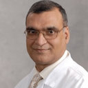 Ashokkumar Jain合作论文数Department of Surgery, Penn State Hershey Medical Center;Department of Surgery, Penn State College of Medicine85
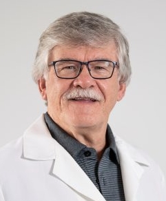 Angus W. Thomson合作论文数Department of Surgery, University of Pittsburgh84
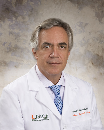 Camillo Ricordi合作论文数Division of Cellular Transplantation, DeWitt Daughtry Family Department of Surgery, Miller School of Medicine, University of Miami78Joeph l. Mccauley合作论文数69BW SHAW合作论文数64KA PORTER合作论文数63William Irish合作论文数Pittsburgh Transplantation Institute, University of Pittsburgh Health Science Center of Pittsburgh61Massimo Trucco合作论文数60George Mazariegos合作论文数59Thomas L. Marchioro合作论文数57
 Parmjeet S. Randhawa合作论文数Division of Transplantation Pathology, Department of Pathology, University of Pittsburgh;Thomas E. Starzl Transplantation Institute, University of Pittsburgh57
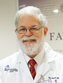 Ron Shapiro合作论文数Mount Sinai’s Recanati/Miller Transplantation Institute55Gilbert J. Burckart合作论文数53
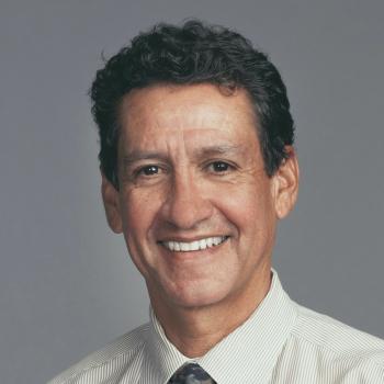 Carlos O. Esquivel合作论文数Division of Gastroenterology, Hepatology, and Nutrition, Department of Podiatric, School of Medicine, Stanford University;Department of Surgery, School of Medicine, Stanford University53Cw Putnam合作论文数52L Polimeno合作论文数52
 Velma P. Scantlebury合作论文数52TR HAKALA合作论文数50Van Thiel D H合作论文数50P Fontes合作论文数49C Vivas合作论文数48
 Michael A. Nalesnik合作论文数Division of Transplantation Pathology, Department of Pathology, University of Pittsburgh Medical Center48Cg Halgrimson合作论文数47J Wallis Marsh合作论文数47Yuichi Iwaki合作论文数47LA Valdivia合作论文数46ML JORDAN合作论文数45D VANTHIEL合作论文数44A Casavilla合作论文数43C.-G. Groth合作论文数43Robert R. Selby合作论文数41




