Conventional cardiac cine MRI methods rely on retrospective gating, which limits temporal resolution and the ability to capture continuous cardiac dynamics, particularly in patients with arrhythmias and beat-to-beat variations. To address these challenges, we propose a reconstruction framework based on subspace implicit neural representations for real-time cardiac cine MRI of continuously sampled radial data. This approach employs two multilayer perceptrons to learn spatial and temporal subspace bases, leveraging the low-rank properties of cardiac cine MRI. Initialized with low-resolution reconstructions, the networks are fine-tuned using spoke-specific loss functions to recover spatial details and temporal fidelity. Our method directly utilizes the continuously sampled radial k-space spokes during training, thereby eliminating the need for binning and non-uniform FFT. This approach achieves superior spatial and temporal image quality compared to conventional binned methods at the acceleration rate of 10 and 20, demonstrating potential for high-resolution imaging of dynamic cardiac events and enhancing diagnostic capability (Code available: https://github.com/wenqihuang/SubspaceINR-CMR ).
Objective Multiparametric MRI is a promising technique for noninvasive structural and functional imaging of the kidneys that is gaining increasing importance in clinical research. Still, there are no standardized recommendations for analyzing the acquired images and there is a need to further evaluate the accuracy and repeatability of currently recommended MRI parameters. The aim of the study was to evaluate the test-retest repeatability of functional renal MRI parameters using different image analysis strategies. Methods Ten healthy volunteers were examined twice with a multiparametric renal MRI protocol including arterial spin labeling (ASL), diffusion-weighted imaging (DWI) with intravoxel incoherent motion (IVIM), blood-oxygen-dependent (BOLD) imaging, T1 and T2 mapping, and volumetry with an interval of one week. The quantitative results of both kidneys were determined by manual organ segmentation, ROI analysis, and automatic segmentation based on the nnUNet framework. Test-retest repeatability of each parameter was computed using the within-subject coefficient of variance (wCV) and the intraclass coefficient (ICC). Segmentation accuracy and inter-reader agreement were evaluated using the dice score. Results Structural tissue parameters (T1, T2) showed wCV (%) between 4 and 11 and an ICC between 0.2 and 0.8. Functional parameters (ASL, BOLD and DWI) showed wCV (%) between 3 and 38 and an ICC between 0.0 and 0.7. The highest variances between test-retest scans were observed in perfusion measurements with ASL and IVIM (wCV: 17-37%). Quantitative analysis of the cortex and medulla showed a better repeatability when acquired using manual segmentation compared to ROI-based image analysis. Comparable repeatability was achieved with manual and automatic segmentation of the total kidney. Conclusion Reasonable repeatability was achieved for all MR parameters. Structural MR parameters showed better repeatability compared to functional parameters. ROI-based image analysis showed overall lower repeatability compared to manual segmentation. Comparable repeatability to manual segmentation as well as acceptable segmentation accuracy could be achieved with automatic segmentation.
Deep learning models can excel on medical tasks, yet often experience spurious correlations, known as shortcut learning, leading to poor generalization in new environments. Particularly in medical imaging, where multiple spurious correlations can coexist, misclassifications can have severe consequences. We propose MIMM-X, a framework that disentangles causal features from multiple spurious correlations by minimizing their mutual information. It enables predictions based on true underlying causal relationships rather than dataset-specific shortcuts. We evaluate MIMM-X on three datasets (UK Biobank, NAKO, CheXpert) across two imaging modalities (MRI and X-ray). Results demonstrate that MIMM-X effectively mitigates shortcut learning of multiple spurious correlations. The code is publicly available https://github.com/lab-midas/MIMM-X.
Background Since the introduction of TotalSegmentator CT, there has been demand for a similar robust automated MRI segmentation tool that can be applied across all MRI sequences and anatomic structures. Purpose To develop and evaluate an automated MRI segmentation model for robust segmentation of major anatomic structures independent of MRI sequence. Materials and Methods In this retrospective study, an nnU-Net model (TotalSegmentator MRI) was trained on MRI and CT scans to segment 80 anatomic structures relevant for use cases such as organ volumetry, disease characterization, surgical planning, and opportunistic screening. Images were randomly sampled from routine clinical studies to represent real-world examples. Dice scores were calculated between the predicted segmentations and expert radiologist segmentations to evaluate model performance on an internal test set and two external test sets and against two publicly available models and TotalSegmentator CT. The Wilcoxon signed rank test was used to compare model performance. The proposed model was applied to a separate internal dataset containing abdominal MRI scans to investigate age-dependent volume changes. Results A total of 1143 scans (616 MRI, 527 CT; median patient age, 61 years [IQR, 50-72 years]) were split into a training set (n = 1088; CT and MRI) and an internal test set (n = 55; MRI only). The two external test sets (AMOS and CHAOS) contained 20 MRI scans each, and the aging-study dataset contained 8672 abdominal MRI scans (median patient age, 59 years [IQR, 45-70 years]). The proposed model had a Dice score of 0.839 for the 80 anatomic structures in the internal test set and outperformed two other models (Dice score of 0.862 vs 0.759 for 40 anatomic structures and 0.838 vs 0.560 for 13 anatomic structures; P < .001 for both). On the TotalSegmentator CT test set (89 CT scans), the performance of the proposed model almost matched that of TotalSegmentator CT (Dice score, 0.966 vs 0.970; P < .001). The aging study demonstrated a strong correlation between age and organ volume (eg, age and liver volume: ρ = -0.096; P < .0001). Conclusion The proposed open-source, easy-to-use model allows for automatic, robust segmentation of 80 structures, extending the capabilities of TotalSegmentator to MRI scans from any MRI sequence. The ready-to-use online tool is available at https://totalsegmentator.com; the model, at https://github.com/wasserth/TotalSegmentator; and the dataset, at http://zenodo.org/records/14710732. © RSNA, 2025 Supplemental material is available for this article. See also the editorial by Kitamura in this issue.
In recent years, accelerated MRI reconstruction based on deep learning has led to significant improvements in image quality with impressive results for high acceleration factors. However, from a clinical perspective image quality is only secondary; much more important is that all clinically relevant information is preserved in the reconstruction from heavily undersampled data. In this paper, we show that existing techniques, even when considering resampling for diffusion-based reconstruction, can fail to reconstruct small and rare pathologies, thus leading to potentially wrong diagnosis decisions (false negatives). To uncover the potentially missing clinical information we propose "Semantically Diverse Reconstructions" (SDR), a method which, given an original reconstruction, generates novel reconstructions with enhanced semantic variability while all of them are fully consistent with the measured data. To evaluate SDR automatically we train an object detector on the fastMRI+ dataset. We show that SDR significantly reduces the chance of false-negative diagnoses (higher recall) and improves mean average precision compared to the original reconstructions. The code is available on https://github. com/NikolasMorshuis/SDR
PURPOSE:The emergence of new medications for fatty liver conditions has increased the need for reliable and widely available assessment of MRI proton density fat fraction (MRI-PDFF). Whereas low-field MRI presents a promising solution, its utilization is challenging due to the low SNR. This work aims to enhance SNR and enable precise PDFF quantification at low-field MRI using a novel locally low-rank deep learning-based (LLR-DL) reconstruction. METHODS:LLR-DL alternates between regularized SENSE and a neural network (U-Net) throughout several iterations, operating on complex-valued data. The network processes the spectral projection onto singular value bases, which are computed on local patches across the echoes dimension. The output of the network is recast into the basis of the original echoes and used as a prior for the following iteration. The final echoes are processed by a multi-echo Dixon algorithm. Two different protocols were proposed for imaging at 0.55 T. An iron-and-fat phantom and 10 volunteers were scanned on both 0.55 and 1.5 T systems. Linear regression, t-statistics, and Bland-Altman analyses were conducted. RESULTS:LLR-DL achieved significantly improved image quality compared to the conventional reconstruction technique, with a 32.7% increase in peak SNR and a 25% improvement in structural similarity index. PDFF repeatability was 2.33% in phantoms (0% to 100%) and 0.79% in vivo (3% to 18%), with narrow cross-field strength limits of agreement below 1.67% in phantoms and 1.75% in vivo. CONCLUSION:An LLR-DL reconstruction was developed and investigated to enable precise PDFF quantification at 0.55 T and improve consistency with 1.5 T results.
Cardiac Cine MRI is limited by prolonged acquisition times and motion-related artifacts. Existing deep learning-based reconstruction methods typically depend on fully-sampled ground truth, which is often difficult to acquire in practice. In this work, we propose SSL-MoCo, a fully self-supervised motion-compensated reconstruction framework for cardiac Cine MRI that eliminates the need for fully-sampled references. SSL-MoCo adopts a two-stage design: a transformer-based registration network estimates non-rigid inter-frame motion in a self-supervised manner, followed by a physics-based unrolled reconstruction network that integrates the estimated motion fields in the data consistency steps. Evaluations on an in-house dataset of 120 subjects (including 82 patients) demonstrate that SSL-MoCo significantly outperforms other self-supervised methods, particularly during challenging systolic phases. The integrated motion compensation enhances temporal coherence, resulting in more accurate myocardial morphology, which is crucial for clinical functional assessment. Our results suggest that SSL-MoCo provides an effective solution for dynamic MRI reconstruction in data-constrained settings.
AbstractRecent research in patients with functionally univentricular hearts (UVH) is focusing on pathologies of the lymphatic vessels. Morphology of the abdominal lymphatic vessels was analyzed by MRI in patients with UVH following total cavopulmonary connection (TCPC) and it was examined, if clinical and laboratory parameters correlate with changes after TCPC. We prospectively examined 33 patients at the age of 19.8 (14.6;30.2) years [median (Q1;Q3)] after TCPC (follow-up 14.3 years (9.7;24.9) with a heavily T2-weighted MRI sequence on a 3.0 T scanner. Examinations in coronal orientation were performed with respiratory gating, slice thickness 0.6 mm, TR 2400 ms, TE 692 ms, FoV 460 mm (covering thoracic and abdominal regions), scan time 14:41 min (13:18;16:30) after a solid meal and a cup of pineapple juice. The findings were classified according to delineation of abdominal lymphatic vessels. Type 1: <3 abdominal vessels (av) definable; type 2: 4–6 av definable; type 3: >6 av and/or oedematous changes or ascites. The results were correlated with parameters obtained at the annual routine check-up. Statistical analysis was performed using U-test and Chi-square test. Fifteen patients (group 1) showed type 3 lymphatic morphologies, two of which had ascites. Eighteen patients (group 2) showed lower grade morphologies (type 1–2). Image quality was rated considering the delineation of the common hepatic duct and did not differ between groups (p = 0.134). “Lymphatic burden” was automatically examined and was indexed to the number of delineated abdominal vessels and showed quantification according to the chosen categories type 1–3. Patients in group 1 were younger at MRI examination (17.4;14.3/18.9 vs. 26.2;18.2/32.3 years, p = 0.03). Superior cavopulmonary connection (SCPC) had been performed earlier in group 1 (9.9;7.9/25.5 vs. 29.2;13.7/66.6 months, p = 0.018). Laboratory examinations in group 1 showed lower levels for Immunoglobulin G (IgG), Lipase, α-Antitrypsin, Cystatin C and TSH. There were no significant differences for total protein, NTproBNP, lymphocytes or platelets. A history of chylothorax was present in 7/15 versus 2/18 p = 0.022. Protein-losing enteropathy (PLE) occurred in 4/15 versus 1/18 (p = 0.092). T2 weighted MRI is feasible for noninvasive delineation of abdominal lymphatic vessel in patients following TCPC. In the long-term follow-up, patients with more pronounced changes of the abdominal lymphatic vessels were younger at SCPC and were more likely to show a history of chylothorax and lower IgG values.
Purpose: To estimate pixel-wise predictive uncertainty for deep learning-based MR image reconstruction and to examine the impact of domain shifts and architecture robustness. Methods: Uncertainty prediction could provide a measure for robustness of deep learning (DL)-based MR image reconstruction from undersampled data. DL methods bear the risk of inducing reconstruction errors like in-painting of unrealistic structures or missing pathologies. These errors may be obscured by visual realism of DL reconstruction and thus remain undiscovered. Furthermore, most methods are task-agnostic and not well calibrated to domain shifts. We propose a strategy that estimates aleatoric (data) and epistemic (model) uncertainty, which entails training a deep ensemble (epistemic) with nonnegative log-likelihood (aleatoric) loss in addition to the conventional applied losses terms. The proposed procedure can be paired with any DL reconstruction, enabling investigations of their predictive uncertainties on a pixel level. Five different architectures were investigated on the fastMRI database. The impact on the examined uncertainty of in-distributional and out-of-distributional data with changes to undersampling pattern, imaging contrast, imaging orientation, anatomy, and pathology were explored. Results: Predictive uncertainty could be captured and showed good correlation to normalized mean squared error. Uncertainty was primarily focused along the aliased anatomies and on hyperintense and hypointense regions. The proposed uncertainty measure was able to detect disease prevalence shifts. Distinct predictive uncertainty patterns were observed for changing network architectures. Conclusion: The proposed approach enables aleatoric and epistemic uncertainty prediction for DL-based MR reconstruction with an interpretable examination on a pixel level.
Deep Learning methods can detect patterns in data such as MR images but are incapable of determining causal relationships. However, causal understanding is crucial in medical applications, since the presence of confounders (e.g. scan conditions) obscure the causal relationship and create spurious-correlations. State-of-the-art models purely rely on correlated patterns which can result in wrong conclusions or diagnoses when spurious-correlations change (e.g. new scanner). We propose a deep learning framework that is robust in the presence of spurious-correlations by decreasing mutual information between learned features of MR images and leads to improved performance under distribution shifts.
Cardiac Cine Magnetic Resonance Imaging (MRI) provides an accurate assessment of heart morphology and function in clinical practice. However, MRI requires long acquisition times, with recent deep learning-based methods showing great promise to accelerate imaging and enhance reconstruction quality. Existing networks exhibit some common limitations that constrain further acceleration possibilities, including single-domain learning, reliance on a single regularization term, and equal feature contribution. To address these limitations, we propose to embed information from multiple domains, including low-rank, image, and k-space, in a novel deep learning network for MRI reconstruction, which we denote as A-LIKNet. A-LIKNet adopts a parallel-branch structure, enabling independent learning in the k-space and image domain. Coupled information sharing layers realize the information exchange between domains. Furthermore, we introduce attention mechanisms into the network to assign greater weights to more critical coils or important temporal frames. Training and testing were conducted on an in-house dataset, including 91 cardiovascular patients and 38 healthy subjects scanned with 2D cardiac Cine using retrospective undersampling. Additionally, we evaluated A-LIKNet on the real-time 8x prospectively undersampled data from the OCMR dataset. The results demonstrate that our proposed A-LIKNet outperforms existing methods and provides high-quality reconstructions. The network can effectively reconstruct highly retrospectively undersampled dynamic MR images up to 24x accelerations, indicating its potential for single breath-hold imaging.
PURPOSE:Joint bright- and black-blood MRI techniques provide improved scar localization and contrast. Black-blood contrast is obtained after the visual selection of an optimal inversion time (TI) which often results in uncertainties, inter- and intra-observer variability and increased workload. In this work, we propose an artificial intelligence-based algorithm to enable fully automated TI selection and simplify myocardial scar imaging. METHODS:The proposed algorithm first localizes the left ventricle using a U-Net architecture. The localized left cavity centroid is extracted and a squared region of interest ("focus box") is created around the resulting pixel. The focus box is then propagated on each image and the sum of the pixel intensity inside is computed. The smallest sum corresponds to the image with the lowest intensity signal within the blood pool and healthy myocardium, which will provide an ideal scar-to-blood contrast. The image's corresponding TI is considered optimal. The U-Net was trained to segment the epicardium in 177 patients with binary cross-entropy loss. The algorithm was validated retrospectively in 152 patients, and the agreement between the algorithm and two magnetic resonance (MR) operators' prediction of TI values was calculated using the Fleiss' kappa coefficient. Thirty focus box sizes, ranging from 2.3mm2 to 20.3cm2, were tested. Processing times were measured. RESULTS:The U-Net's Dice score was 93.0 ± 0.1%. The proposed algorithm extracted TI values in 2.7 ± 0.1 s per patient (vs. 16.0 ± 8.5 s for the operator). An agreement between the algorithm's prediction and the MR operators' prediction was found in 137/152 patients (κ= 0.89), for an optimal focus box of size 2.3cm2. CONCLUSION:The proposed fully-automated algorithm has potential of reducing uncertainties, variability, and workload inherent to manual approaches with promise for future clinical implementation for joint bright- and black-blood MRI.
Medical imaging cohorts are often confounded by factors such as acquisition devices, hospital sites, patient backgrounds, and many more. As a result, deep learning models tend to learn spurious correlations instead of causally related features, limiting their generalizability to new and unseen data. This problem can be addressed by minimizing dependence measures between intermediate representations of task-related and non-task-related variables. These measures include mutual information, distance correlation, and the performance of adversarial classifiers. Here, we benchmark such dependence measures for the task of preventing shortcut learning. We study a simplified setting using Morpho-MNIST and a medical imaging task with CheXpert chest radiographs. Our results provide insights into how to mitigate confounding factors in medical imaging. The project’s code is publicly available ( https://github.com/berenslab/dependence-measures-medical-imaging ).
Background: Pathologies of the thoracic aorta are associated with chronic cardiovascular disease and can be of life-threatening nature. Understanding determinants of thoracic aortic morphology is crucial for precise diagnostics and preventive and therapeutic approaches. This study aimed to automatically characterize ascending aortic morphology based on 3D non-contrast-enhanced magnetic resonance angiography (NE-MRA) data from the large epidemiological cross-sectional German National Cohort (NAKO) and to investigate possible determinants of mid-ascending aortic diameter (mid-AAoD). Methods: Deep learning was used to automatically segment the thoracic aorta and extract ascending aortic length, volume, and diameter from 25,073 NE-MRAs. Descriptive statistics, correlation analyses, and multivariable regression were used to investigate statistical relationships between mid-AAoD and demographic factors, hypertension, diabetes, alcohol, and tobacco consumption. Additionally, automated causal discovery analysis using the Peter-Clark algorithm was performed to identify possible causal interactions. Results: Males exhibited significantly larger mid-AAoD than females (M: 35.5±4.8 mm, F: 33.3±4.5 mm). Age and body surface area (BSA) were positively correlated with mid-AAoD. Hypertensive and diabetic subjects showed higher mid-AAoD. Hypertension was linked to higher mid-AAoD regardless of age and BSA, while diabetes and mid-AAoD were uncorrelated across age-stratified subgroups. Daily alcohol consumption and smoking history exceeding 16.5 pack-years exhibited highest mid-AAoD. Causal analysis revealed that age, BSA, hypertension, and alcohol consumption are possibly causally related to mid-AAoD, while diabetes and smoking are likely spuriously correlated. Conclusions: Mid-AAoD varies significantly within the unique large-scale NAKO population depending on demographic factors, individual health, and lifestyle. This work provides a proof-of-concept for automated causal analysis which can help disentangle observed correlations and identify potential causal determinants of ascending aortic morphology. ### Competing Interest Statement The authors have declared no competing interest. ### Funding Statement This project was conducted with data from the German National Cohort (NAKO) (www.nako.de). The NAKO is funded by the Federal Ministry of Education and Research (BMBF) [project funding reference numbers: 01ER1301A/B/C, 01ER1511D and 01ER1801A/B/C/D], federal states of Germany and the Helmholtz Association, the participating universities, and the institutes of the Leibniz Association. We thank all participants who took part in the German National Cohort and the staff in this research program. This project was partially funded by the Deutsche Forschungsgemeinschaft (DFG, German Research Foundation) project number 428219130 and supported under Germany?s Excellence Strategy ? EXC 2064/1 ? project number 390727645. ### Author Declarations I confirm all relevant ethical guidelines have been followed, and any necessary IRB and/or ethics committee approvals have been obtained. Yes The details of the IRB/oversight body that provided approval or exemption for the research described are given below: The responsible ethics committees of individual studies approved all study-related analyses: GUIDE: 202/22 approved by the Ethics Committee of the Medical Faculty of the Rheinische Friedrich-Wilhelm-University Bonn ELISA: University of Luebeck (Az. 20-150) NAKO: The study is continuously approved by the responsible local ethics committees of the German Federal States where all study centers are located in (original ethics approvals of the leading ethics committee of the Bayerische Landesaerztekammer (protocol code 13023, Approval Date: 27 March 2013 and 14 February 2014 (rectification of documents, study protocol, consent form)). An external ethics advisory board has been established that accompanies NAKO over the full study period. A 'Code of Ethics' of NAKO (Ethikkodex) has been developed and the study is under steady surveillance by the ethics committees of the regional study centers (8). STAAB: Ethics committee of the Medical Faculty of the University Wuerzburg (STAAB: #98/13) MuSPAD: Ethics committee of Hannover Medical School (9086\_BO\_S\_2020 for MuSPAD), Dresden paedSAXCOVID: Ethics Committee of the Technische University (TU) Dresden (BO-EK-156042020). Bochum CorKID: Ethics Committee of the Ruhr University Bochum (Nr. 20-6927\_7) Wuerzburg Wue-KITa-CoV: Wuerzburg, Kennzeichen 105/21 IMMUNEBRIDGE_ED: Ethics Committee of University Medical Center Goettingen (21/6/22) I confirm that all necessary patient/participant consent has been obtained and the appropriate institutional forms have been archived, and that any patient/participant/sample identifiers included were not known to anyone (e.g., hospital staff, patients or participants themselves) outside the research group so cannot be used to identify individuals. Yes I understand that all clinical trials and any other prospective interventional studies must be registered with an ICMJE-approved registry, such as ClinicalTrials.gov. I confirm that any such study reported in the manuscript has been registered and the trial registration ID is provided (note: if posting a prospective study registered retrospectively, please provide a statement in the trial ID field explaining why the study was not registered in advance). Yes I have followed all appropriate research reporting guidelines, such as any relevant EQUATOR Network research reporting checklist(s) and other pertinent material, if applicable. Yes The data that support the findings of this study are available from the German National Cohort but restrictions apply to the availability of these data, which were used under license for the current study, and so are not publicly available.
Age plays an important role in shaping medical decisions, but the biological changes associated with aging do not solely depend on the chronological age. Genetics, lifestyle, and environment cause variations in age-related characteristics, even within the same chronological age group. Biological age (BA) was introduced to better capture an individual’s actual aging process, although its imprecise definition remains a challenge. Organ systems can age at different rates, necessitating organ-specific BA evaluation. To our knowledge, there have been no studies assessing age regionally across multiple organ systems. These age estimations, reflecting various body parts, enable a comprehensive patient-based analysis. We conducted brain, heart, kidney, liver, spleen, pancreas, and retinal fundus age estimations using MRI and OCT scans in 40,000 subjects of the UK Biobank with an uncertainty-aware ResNet-based network. Our results demonstrate the feasibility of organ-specific age estimation with cross-organ correlations of age-related changes. We achieve a mean age difference between predicted and chronological age of 2.72 years across all organs and an averaged Pearson correlation coefficient of 0.87.
In cardiac CINE, motion-compensated MR reconstruction (MCMR) is an effective approach to address highly undersampled acquisitions by incorporating motion information between frames. In this work, we propose a novel perspective for addressing the MCMR problem and a more integrated and efficient solution to the MCMR field. Contrary to state-of-the-art (SOTA) MCMR methods which break the original problem into two sub-optimization problems, i.e. motion estimation and reconstruction, we formulate this problem as a single entity with one single optimization. Our approach is unique in that the motion estimation is directly driven by the ultimate goal, reconstruction, but not by the canonical motion-warping loss (similarity measurement between motion-warped images and target images). We align the objectives of motion estimation and reconstruction, eliminating the drawbacks of artifacts-affected motion estimation and therefore error-propagated reconstruction. Further, we can deliver high-quality reconstruction and realistic motion without applying any regularization/smoothness loss terms, circumventing the non-trivial weighting factor tuning. We evaluate our method on two datasets: 1) an in-house acquired 2D CINE dataset for the retrospective study and 2) the public OCMR cardiac dataset for the prospective study. The conducted experiments indicate that the proposed MCMR framework can deliver artifact-free motion estimation and high-quality MR images even for imaging accelerations up to 20x, outperforming SOTA non-MCMR and MCMR methods in both qualitative and quantitative evaluation across all experiments.
 Sergios Gatidis合作论文数Department of Radiology, Stanford University62
 Konstantin Nikolaou合作论文数39
 Bin Yang (杨斌)合作论文数Institute of Signal Processing and System Theory, University of Stuttgart34
 Kerstin Hammernik合作论文数Graz University of Technology30
 Petros Martirosian合作论文数29
 Daniel Rueckert合作论文数Department of Computing, Imperial College London;Centre for Cardiac Engineering, Imperial College London;School of Computation, Information and Technology, Technical University of Munich;School of Medicine and Health, Technical University of Munich28Fritz Schick合作论文数23Claudia Prieto合作论文数20Tobias Hepp合作论文数Univ Tubingen, Dept Radiol, Tubingen, Germany18
 Ferdinand Seith合作论文数18
 René Botnar合作论文数Department of Biomedical Engineering, School of Biomedical Engineering and Imaging Sciences, Faculty of Life Sciences and Medicine, King’s College London;Wellcome EPSRC Centre for Medical Engineering, King’s College London;British Heart Foundation Centre, School of Cardiovascular and Metabolic Medicine and Sciences, Faculty of Life Sciences and Medicine, King’s College London;Teaching Department of Anatomy, Faculty of Life Sciences and Medicine, King’s College London16
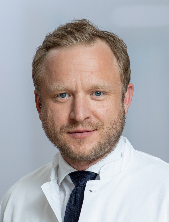 Fabian Bamberg合作论文数Department of Diagnostic and Interventional Radiology, University of Freiburg15Haikun Qi (齐海坤)合作论文数14
 Holger Schmidt合作论文数14
 Jiazhen Pan合作论文数Institutes of Radiology and Artificial Intelligence and Informatics in Medicine, Technical University of Munich13
 Gastao Lima da Cruz合作论文数University of Michigan12Marc Fischer合作论文数11
 Patrick Krumm合作论文数11Marcel Früh合作论文数10
 Nina Schwenzer合作论文数10
 Karim Armanious合作论文数Univ Stuttgart, Inst Signal Proc & Syst Theory, Stuttgart, Germany10
 Ahmed Othman合作论文数9
 Aurelien Bustin合作论文数Kings Coll London, Sch Biomed Engn & Imaging Sci, London, England9Martin A. Schwartz合作论文数Department of Chemistry, Florida State University9
 Christian la Fougère合作论文数Department für Radiologie, Faculty of Medicine, University of Tuebingen8
 Radhouene Neji合作论文数Laboratoire des Mathématiques Appliquées aux Systèmes, Ecole Sup8Mike Notohamiprodjo合作论文数8
 R. Pohmann合作论文数7Hajhosseiny Reza合作论文数7
 Jakob Weiss合作论文数Eberhard Karls University, Tuebingen University Hospital7Christina Schraml合作论文数7Jana Taron合作论文数6
 Annika Liebgott合作论文数Institute of Signal Processing and System Theory, University of Stuttgart6
 Klaus Scheffler合作论文数Department High-field Magnetic Resonance, Max-Planck-Institute for Biological Cybernetics;Department of Biomedical Magnetic Resonance, University of Tubingen6
 Jens Kubler合作论文数6
 Tobias Pischon合作论文数The Molecular Epidemiology Research Group, Max Delbrück Center for Molecular Medicine;Charité Universitätsmedizin Berlin;NAKO Gesundheitsstudie - German National Cohort, NAKO e.V.6Thoralf Niendorf合作论文数6Christina Pfannenberg合作论文数6Andreas Lingg合作论文数5
 Sherif Abdulatif合作论文数Institute of Signal Processing and System Theory, University of Stuttgart5
 Stefan Winzeck合作论文数Biomedical Image Analysis Group, Imperial College London5
 Wenqi Huang合作论文数Research Center for Medical AI, Shenzhen Institutes of Advanced Technology, Chinese Academy of Sciences, Shenzhen, China5Sebastian Gassenmaier合作论文数5
 Matthias Schulze合作论文数Department of Molecular Epidemiology, German Institute of Human Nutrition Potsdam-Rehbruecke;University of Potsdam5
 Fuin, N.合作论文数Inst. of Nucl. Med., Univ. Coll. London Hosp.;c;Inst. of Nucl. Med., Univ. Coll. London Hosp.5
 Christopher Gilliam合作论文数School of Engineering, RMIT University5Thomas Kröncke合作论文数5
 Ben Glocker合作论文数Department of Computing, Imperial College London5
 Hans-Ulrich Kauczor合作论文数Translational Lung Research Center Heidelberg, Universitätsklinikum Heidelberg;University of Heidelberg;German Center for Lung Research5




