Angstrom-scale polygonal rings in monolayer amorphous carbon (MAC) not only tailor its electronic and mechanical properties, but also endow the atomically thin membrane material with diverse angstrom pores for precise regulation of the subatomic species separation and transport, crucial for technological advances across various fields including catalysis, energy devices, and medical applications. However, the lack of industrial-scale synthesis route of intrinsic MAC has severely hindered the progress to harness their versatile angstrom polygons and properties for technological applications that are not offered by the state-of-the-art graphene or bulk amorphous materials. Herein, we report an industry-compatible disorder-to-disorder (DTD) synthetic approach to achieve wafer-scale intrinsic ultra-clean MAC (UC-MAC) within seconds-timescale, featuring optimized angstrom polygons without detectable metal contamination and nano-sized pores. Contrary to metal-contaminated MAC, angstroporous UC-MAC enables not only atomic-scale characterization of its intrinsic electronic properties, but also serve as an angstroporous membrane material, enabling the splitting of high-flux H2+ ions into a high-precision proton beam with minimal detrimental fragment proton scattering events to date, about twice and 40 times less than those from graphene and commercial carbon thin films, respectively. The membrane with minimum possible material thickness that can yield a highly sharpened proton beam with accurately modulated beam current is desired for proton therapy, especially for non-invasive tumor treatment.
Ångström-scale polygonal rings in monolayer amorphous carbon (MAC) enhance its electronic and mechanical properties while providing unique ångström pores for precise subatomic species separation, essential for advancements in catalysis, energy and medicine. However, the absence of an industrial-scale synthesis method for intrinsic MAC has limited its technological applications compared with graphene and bulk amorphous materials. Herein, we report an industry-compatible disorder-to-disorder synthesis approach to achieve wafer-scale ultraclean MAC (UC-MAC) within a timescale of seconds, featuring optimized ångström polygons without detectable metal contamination, and nanosized pores. In contrast to metal-contaminated MAC, UC-MAC allows atomic-scale characterization of intrinsic electronic properties and functions as an ångström-scale membrane, facilitating the splitting of high-flux H2+ ions into a high-precision proton beam with minimal detrimental fragment-proton scattering events, about half and 40 times less than those from single-crystal graphene and commercial carbon thin films, respectively. The minimum possible membrane material thickness that can yield a highly sharpened proton beam with accurately modulated beam current is desired for proton therapy.
Mechanically polished Gun-metal (G-metal) alloy specimens were irradiated using a 2 MeV Au+ beam of Pelletron Linear Accelerator. The range of Au+ fluence was 5 × 10¹¹ to 1 × 10¹⁴ Au+/cm2. XRD patterns revealed the crystallographic peaks of Cu, Zn, and Sn. Harris analysis demonstrated that the Au+ fluence strongly influenced the preference of crystal plane alignment. The structural attributes like crystallite size, strain, etc. change as the Au+ fluence varies. Due to the low depth of ion-induced defects region, the estimation of ion irradiated material surface hardening is difficult. So, the technique of nanoindentation used in this work proves helpful for estimating the surface hardness of ion irradiated material. The surface hardness of specimens varied with the Au+ fluence and indentation depth. A significant increase in surface roughness, nanoindentation surface hardness, and elastic modulus values was observed at 5 × 10¹³ Au+/cm2 irradiation. Moreover, the surface hardness improves with decreasing crystallite size, reflecting the Hall-Petch relation. Energy dispersive X-ray (EDX) and Proton-induced X-ray emission (PIXE) were used for elemental analysis of specimens before and after Au+ irradiation.
MeV ion microprobe measurements of the lesser and trace life element concentrations in biological tissues are challenging because of complex spatial inhomogeneities in these types of samples. Measurements on ex vivo tissue sections require determination of the matrix element composition and the tissue section thickness. For these reasons, in this work, we adapted the Dynamic Analysis approach known from literature, to interpret the MeV ion microscopy data to determine concentrations of H, C, O, and N as well as the thickness of different tissue regions in Non Human Primate (NHP) mesenteric lymph node section. The results showed no strong variations of the matrix element contents regardless of section thickness variations in the tissue. The matrix information was used to quantify total -Ca molarities and a significant similar to 30 mM Ca concentration hotspot was observed at the edge of sinus structure in the mesenteric lymph node as compared to the 3-4 mM total -Ca levels in the surrounding tissues. Thus, MeV ion microprobe imaging combined with dynamic analysis comprise a novel chemometric approach paving a way for quantitative analysis of similarly complicated animal and plant biological tissue sections.
Fourier ptychography (FP) is a high resolution wide-field imaging method based on the extended aperture in the Fourier space, which is synthesized from raw images with varying illumination angles. If FP is extended to coherent nonlinear optical imaging, the resolution could be further improved due to the increase of the cutoff frequency of the synthesized coherent optical transfer function (C-OTF) with respect to the order of nonlinear optical processes. However, there is a fundamental conflict between wide-field FP and nonlinear optical imaging, whereby the nonlinear optical imaging typically requires a focused excitation laser beam with high power density. To tackle the problem, in this work, a unique point-scanning FP (PS-FP) method is presented for super-resolution nonlinear optical imaging, in which the nonlinear optical signal is obtained by using focused laser beam, while the conventional FP algorithm can still be used to retrieve the super-resolution image. PS-FP coherent anti-Stokes Raman scattering (PS-FP-CARS) imaging on a variety of samples, where a 1.8-fold expansion of the OTF is achieved experimentally for enhancing vibrational imaging. Further theoretical calculation shows that the C-OTF of PS-FP higher-order CARS (PS-FP-HO-CARS) can be expanded up to ≈4.9-fold, thereby improving the spatial resolution by ≈3-fold in comparison with conventional point-scanning CARS with under tightly focused beams. The generality of PS-FP method developed in this work can be adapted to other coherent nonlinear optical imaging modalities for super-resolution imaging in tissue and cells.
Physiologically relevant concentrations in biological tissue, in the in vivo, state are of the order of μmol L −1 and mmol L −1 . Up to the present, mapping the major elements in the matrix and its thickness has been neglected, despite their importance for quantification of lesser and trace element concentrations. Ryan and Jamieson's dynamic analysis, statistical spectral decomposition approach, is developed to quantitatively measure ex vivo tissue sections cut using a cryomicrotome. This mitigates the problem that physical analysis methods require a vacuum environment. This approach is used to quantitatively image the major matrix elements H, C, N, and O as well as trace maps of Ca, Fe, and Zn in a tissue section of porcine intestine. This sample is selected as it exhibits a complex morphology with multiple tissue compartments (such as muscle and mucosa, as well as void areas from blood vessels, lymph ducts, sinuses crypts, and villi. In the results, it is demonstrated that different tissue types can have a different matrix composition and thickness. Using this information, quantitative maps and elemental molarities for the lesser and trace elements Ca, Fe, and Zn are obtained.
To maintain the scaling trends in the complementary metal oxide semiconductor (CMOS) technology, the thickness of barrier/liner systems used in back-end-of-line (BEOL) fabrication of metal interconnects needs to be sub-2 nm. However, reducing the thickness of the traditional barrier and liner systems necessary for the dimensional scaling of future interconnects is extremely challenging. Hence, ultrathin two-dimensional (2D) transition-metal dichalcogenide (TMD) films can be an alternative to current barrier/liner systems. However, the processes used to grow these films are generally not BEOL-compatible. Here, using the plasma-free metal-organic chemical vapor deposition (MOCVD) process, we grow BEOL-compatible tungsten disulfide (WS2) film, which has a clear advantage over current diffusion barrier/liner systems used in Cu-interconnects. Our results show that these WS2 films not only block Cu diffusion but also reduce the effective resistance of the Cu film by suppressing the grain boundary and interface scattering of electrons.
Changsha kiln ware represents an important status in the history of Chinese ceramics, well-known for its massive production capacity evidenced from extensive archaeological finds both domestically and abroad. It is long known or assumed that Changsha kiln is the pioneer for underglaze painted porcelains, however recent analytical studies suggest otherwise: overglaze, atypical underglaze, or cannot be ascertained microscopically. Changsha kiln glazing technique remains a convoluted issue debatable amongst scholars that is yet to be concluded. To investigate the glazing technique of Changsha kiln coloured porcelains, we track the colour distribution down the glaze using an improved methodological approach: thin cross-section light microscopy and elemental analysis with scanning electron microscopy-energy dispersive X-ray spectroscopy (SEM-EDS) and proton induced X-ray emission (PIXE). The samples selected for this study, Changsha bowls with Fe-brown and Cu-green decorations excavated from the Belitung shipwreck, and Changsha Cu-red pieces of a jar and box lid unearthed from the Changsha kiln site of Hunan province, have not been analysed before for the purpose of determining the glazing technique, thereby offering new data to contribute to this scholarly subject. Microscopy results: Thin cross-sections clearly enhance the visibility of colour distribution in the transparent glaze, especially for highly tinting browns and reds. The green glaze, however, is extremely homogeneous and requires elemental analysis to affirm its glazing technique. Elemental results: SEM-EDS line scan and mapping results show that Fe-brown decreases down the glaze (i.e. overglaze), whereas for Cu-green and Cu-red colourations, the levels of Cu are too low to detect an appreciable change down the glaze. With the more sensitive PIXE technique, the distribution of trace levels of Cu in the green and red glaze are mapped out for the first time, providing concrete evidence for Cu-green as overglaze and Cu-red as mid-glaze.
The spatial resolution plays a crucial role in determining the performance of a nuclear microprobe. However, the formation of spatial resolutions below 10 nm remains a challenge in nuclear microprobes. Here, we propose novel technologies (near-axis scanning transmission ion microscopy and double-fragment scattering) utilizing molecular ions to address this challenge and demonstrate a H2+ molecular beam with 6.0 x 10 nm2 lateral resolution and monolayer thickness resolution respectively. Using the improved nuclear microprobe, we directly demonstrate that the ionization of a H2+ can be efficiently achieved using one single layer graphene, and also that single and few layers of freestanding graphene can be clearly differentiated and identified. The precise control of fast molecular ions at sub-10 nm scales has the potential to unlock new avenues of applications.
Herein, the secondary electron emission (SEE) from 1D nanomaterials in the form of nanorods is investigated. The small beam of a 1.5 MeV +H2 hydrogen with a sub 70 nm in diameter allows studying the SEE with a very high resolution. A wide range of nanomaterials from various laboratories are studied, including thin ZnO and ZnO/GaN nanostructures grown on 1 μm thick Si3N4 membranes and thick InP, GaN and GaN/AlN nanorod structures grown on bulk Si substrates. By virtue of the small size of the exciting nanobeams, high‐resolution maps could be created presenting an SEE yield from various parts of the structures. This allows us to show that the top parts of nanorods in ZnO, ZnO/GaN, GaN, InP, and GaN/AlN nanostructures emit secondary electrons much more efficiently than the valley areas between nanorods. These results indicate that by a proper design and growth of 1D nanostructures, SEE properties could be improved over those of the traditionally used Au and CsI thin films. This work has been undertaken to find materials with the highest achievable SEE emission, which is a figure of merit for the detection efficiency relevant for the development and application of novel radiation detectors.
The onset and progression of many degenerative diseases including atherosclerosis, have been shown to directly link to the presence/absence of certain metal ions. Consequently, the detection of these ions in tissues may improve the understanding of the driving pathophysiology. The Cu content during atherosclerosis development has not been studied due to its low concentration involved. In this work, the Cu level in atherosclerotic rabbit tissue is determined using PIXE with a 3.5 MeV proton beam. The arteries of three animal groups fed with different diets were studied: group 1, rabbits on normal standard diet, group 2, on High Fat Diet (HFD) and group 3, on HFD + Zinc diet. Zinc supplement has been proven to inhibit the beginning of atherosclerotic lesion. The result of this study shows that the Cu levels in all the atherosclerotic lesions were lower than that in the arterial walls of the samples in HFD group.
Abstract Radiotherapy (RT) is a primary treatment modality that is used in 50% of cancer patients. Clinically, tumor radioresistance poses a challenge to achieve complete anti-tumor response following conventional photon-RT. Diverse mechanisms have been proposed to underpin radioresistance. However, targeting these aberrant pathways to overcome tumor radioresistance remains a clinical challenge. Here, we investigate if proton beam therapy (PBT) invokes differential cellular responses compared to photon-RT in a broad panel of radioresistant (RR) cancer cell lines. We generated isogenic RR human cancer cell lines: 22Rv1 prostate cancer, FaDu hypopharyngeal cancer and C666-1 nasopharyngeal cancer, by exposure of respective wildtype (wt) to 90 Gy photon-RT (2 Gy x 45 fr). RR was confirmed by clonogenic survival, with surviving fraction ratio (SFRR/SFwt) at 1-4 Gy (range: 1.2-1.8). Whole exome sequencing (100x; Illumina NovaSeq) was performed to profile RR-associated mutational drivers. We performed transcriptomic and proteome profiling of cellular response such as non-homologous end-joining (NHEJ) and homologous recombination (HR) repair at 1-24 h after 4 Gy PBT (2.5 MeV, 2 Gy/min) and photon-RT (0.66 MeV, 0.716 Gy/min). At baseline, we found common mutations and differential activation of several pro-survival (Akt, mTOR), epithelial-mesenchymal transition (EMT) (PDGF, TGFb) and DNA repair pathways (ATM, BRCA1, NHEJ- and HR-associated) genes in RR- relative to wt-22Rv1. Post-photon-RT, we observed residual persistent DNA repair in wt-cell lines up to 48 h. In contrast, repair in RR-cell lines was more proficient, as shown by recovery of γH2AX, ATM-Chk2 within 6 h, with significant NHEJ activation (DNA-PKcs). Next, we observed that RR-cell lines were more sensitive to PBT than photon-RT (mean SF4Gy PBT/SF4Gy photon-RT = 0.61), with delayed DNA repair (γH2AX) observed up to 48 h, which may be attributed to the difficulty in repairing PBT-induced DNA damage. This phenomenon, seems to be primarily orchestrated by decreased NHEJ activation combined with diminished cell cycle checkpoint arrest (p21) and anti-apoptotic (bcl-2) signaling. Radiosensitization by PBT was however not observed in wt (mean SF4Gy proton/SF4Gy photon = 1.14), corroborated by gene and protein expression of DDR-related pathways. We observed dysregulation of multiple pathways relating to EMT and DNA repair in our panel of RR-cancer cell lines, compared to the wt-counterparts. Additionally, we show the potential for PBT to overcome radioresistance in these models by targeting the DNA repair machinery. Due to the high cost of PBT, precise patient stratification is needed to ensure that PBT is applied only to patients who benefit significantly from this procedure over photon-RT. Our preliminary data represents a proof-of-concept for feasibility of patient stratification to PBT. Citation Format: Pek Lim Chu, Eugenia L.L. Yeo, Dennis J.J. Poon, Ce-belle Chen, Minqin Ren, Saumitra Vajandar, Dewi Susanti, Hong Qi Tan, Sung Yong Park, Thomas Osipowicz, Kwok Wai Lo, Stanley K. Liu, Khee Chee Soo, Melvin Lk Chua. Proton irradiation sensitizes cancer cell lines with acquired radioresistance by exploiting differentially dysregulated DNA damage response (DDR) pathways [abstract]. In: Proceedings of the American Association for Cancer Research Annual Meeting 2021; 2021 Apr 10-15 and May 17-21. Philadelphia (PA): AACR; Cancer Res 2021;81(13_Suppl):Abstract nr 3062.
Correlative imaging and quantification of intracellular nanoparticles with the underlying ultrastructure is crucial for understanding cell-nanoparticle interactions in biological research. However, correlative nanoscale imaging of whole cells still remains a daunting challenge. Here, we report a straightforward nanoscopic approach for whole-cell correlative imaging, by simultaneous ionoluminescence and ultrastructure mapping implemented with a highly focused beam of alpha particles. We demonstrate that fluorescent nanodiamonds exhibit fast, ultrabright and stable emission upon excitation by alpha particles. Thus, by using fluorescent nanodiamonds as imaging probes, our approach enables quantification and correlative localization of single nanodiamonds within a whole cell at sub-30 nm resolution. As an application example, we show that our approach, together with Monte Carlo simulations and radiobiological experiments, can be employed to provide unique insights into the mechanisms of nanodiamond radiosensitization at the single whole-cell level. These findings may benefit clinical studies of radio-enhancement effects by nanoparticles in charged-particle cancer therapy.
There is an increasing number of radiobiological experiments being conducted with low energy protons (less than 5 MeV) for radiobiological studies due to availability of sub-millimetre focused beam. However, low energy proton has broad microdosimetric spectra which can introduce dosimetric uncertainty. In this work, we quantify the impact of this dosimetric uncertainties on the cell survival curve and how it affects the estimation of the alpha and beta parameters in the LQ formalism. Monte Carlo simulation is used to generate the microdosimetric spectra in a micrometer-sized water sphere under proton irradiation. This is modelled using radiobiological experiment set-up at the Centre of Ion Beam Application (CIBA) in National University of Singapore. Our results show that the microdosimetric spectra can introduce both systematic and random shifts in dose and cell survival; this effect is most pronounced with low energy protons. The alpha and beta uncertainties can be up to 10% and above 30%, respectively for low energy protons passing through thin cell target (about 10 microns). These uncertainties are non-negligible and show that care must be taken in using the cell survival curve and its derived parameters for radiobiological models.
The radiation response, long-term performance, and reliability of HfO2-based gate dielectric materials play a critical role in metal oxide semiconductor (MOS) technology for space device applications. Al/HfO2/Si atomic layer-deposited devices were irradiated by gamma and swift heavy ions. An increase in the leakage current and charge trapping has been observed as the gamma irradiation dose varied from 25 to 100 krad. The density of oxide traps is found to increase with an increase in the gamma dose while the interface trap density is found to decrease. Another set of samples were irradiated by 120 MeV Au ions to study the SHI-induced defect annealing/creation of defects and intermixing effects in HfO2/Si-based devices. The formation of an interfacial layer of HfSiO at a fluence of at 5 × 1013 cm−2 is revealed by X-ray reflectivity analysis. The densities of interface- and oxide-trapped charges are found to decrease up to a critical fluence of 1 × 1012 cm−2 and then increase with further increase in fluence to 5 × 1013 cm−2. The presence of the interlayer, due to the swift heavy ion-induced intermixing, has been confirmed by X-ray photoelectron spectroscopy measurements. Various current conduction mechanisms in both substrate and gate injection cases were used to understand the basic mechanisms of direct, Fowler–Nordheim, and Poole–Frenkel tunneling, as well as Schottky emission in these devices. These studies elucidated the radiation tolerance and charge-trapping behavior of Al/HfO2/Si nMOS capacitors.
BiFeO3-based composite materials are important due to their versatile multiferroic properties which can be further tuned upon doping. 0.5BiNd(x)Fe(1-x)O(3)-0.5PbZrO(3) (x = 0.05, 0.1, and 0.2) polycrystalline ceramic samples were prepared by solid-state reaction technique at high temperature. X-ray diffraction (XRD) and Rietveld refinement process confirm the presence of mainly rhombohedral (R3c) phase. Dielectric studies reveal the presence of Maxwell-Wagner type polarization. Electric impedance and modulus values of the samples were studied over a wide range of temperature and frequency. Complex impedance and modulus studies shows the presence of non-Debye type of relaxation in the materials. ac conductivity results can be fitted with Jonscher's power law and indicates the dominance of correlated barrier hopping mechanism in charge transport. Furthermore, density of states calculated from ac conductivity shows variation as a function of dopant content which is further corroborated from density functional theory-based results.
A new beam line facility dedicated to investigations into the radiobiology of biological single cells has been constructed at the Centre for Ion Beam Applications, National University of Singapore. This facility, which has a horizontal layout, has some novel features including post lens magnetic ion deflection and scanning and provision for a diamond exit window and confocal fluorescence microscope for high performance online investigations. The radiobiology facility uses a system of Oxford Microbeam compact magnetic quadrupole lenses enabling high demagnifications (up to 123 x 51) at the cell plane, and a relatively short lens to cell distances (22.5 cm). Beam optics and SRIM calculations have indicated a theoretical design performance of up to 450 nm for the proton spot size at the cell surface. To measure the spatial resolution of the proton beam at the cell position, we positioned a gold calibration grid inside the external cell chamber at a position normally occupied by cells. Preliminary results were obtained by scanning a 2 MeV focused beam over a grid and using a surface barrier detector to construct a STIM image of the grid we have measured an in-air spot size of sub-500 nm at the cell position. In this paper, the design of the new radiobiology beam line, preliminary results of resolution tests, as well as presenting first investigations into the damage caused by focused proton beam irradiation of human liver cancer cells by detecting DNA damage markers gamma-H2AX and 53BP1.
Hydrogen is a crucial element for crystalline silicon solar cells due to its ability to passivate bulk defects in silicon. The introduction and distribution of hydrogen has gained a lot of interest due to its proposed involvement in the phenomenon termed "light and elevated temperature induced degradation" (LeTID) in multicrystalline silicon (mc-Si) solar cells. LeTID, which can cause an efficiency loss of about 6-14% (relative) for mc-Si PERC (passivated emitter and rear cell) devices upon exposure to elevated temperature and illumination, is a serious cause of concern for the silicon photovoltaic industry. Interaction of hydrogen with mc-Si is complex as mc-Si contains grain boundaries, dislocations, large concentrations of impurities and traps which may affect the diffusivity of hydrogen in silicon. Understanding the diffusion of hydrogen in mc-Si, and how it affects LeTID, is therefore of great interest. In this contribution, the concentration of hydrogen diffused into p-type mc-Si lifetime samples from hydrogen-rich passivation layers (SiNx:H and AlOx:H) fired at different peak firing temperatures is measured by elastic recoil detection analysis (ERDA) along with Rutherford backscattering (RBS). Also, experiments are done to study the impact of annealing in the presence of hydrogen on the extent of LeTID. A correlation is established between the hydrogen concentration diffused into silicon bulk and the extent of LeTID in lifetime samples fired at different peak firing profiles.
Relative Biological Effectiveness (RBE) is an important but complex quantity which is used in Proton Therapy to compare the efficacy of cell-killing of protons relative to x-ray. It is known to depend on many factors (such as proton’s energy, dose, and cell type) and varies along the track of a particle. The exact RBE value is also known to be subjected to a large amount of uncertainties. In light of these complexities and uncertainties, this paper presents the calculation of the biologically and clinically relevant parameters - RBE (for double strand breaks) and double strand breaks yield, using a mechanistic and radiobiology-inspired approach. This paper is divided into two main parts. The first features the debut of our in-house radiobiology simulation software known as Integrated Radiobiology Simulation with Geant4 and System Biology (IRSGS). The software and the methodology employed will be covered in details including chromosome geometry construction and processing of direct and indirect effect. The second part focuses on the derivation of the spatial distribution of strand breaks (especially double strand breaks) and RBE for protons.
 F. Watt合作论文数Centre for Ion Beam Applications, Department of Physics. National University of Singapore79
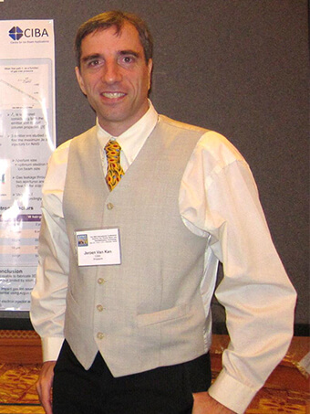 Jeroen Anton Van Kan合作论文数Department of Physics, Faculty of Science, National University of Singapore36
 Taw Kuei Chan合作论文数Department of Physics, Faculty of Science, National University of Singapore25
 Andrew A. Bettiol合作论文数Department of Physics, Faculty of Science, National University of Singapore;Centre for Ion Beam Applications, National University of Singapore25
 Saumitra K. Vajandar合作论文数Department of Mechanical Engineering, Vanderbilt University21
 H.L. Seng合作论文数Department of Physics, National University of Singapore20
 Piraviperumal Malar合作论文数Centre for Ion Beam Applications, National University of Singapore18Minqin Ren合作论文数18Jean-Louis Sanchez合作论文数16
 Ee Jin Teo合作论文数Department of Physics, National University of Singapore16
 Chua Soo Jin合作论文数A*STAR/Department of Electrical and Computer Engineering, College of Design and Engineering, National University of Singapore14
 Kin Leong Pey合作论文数14
 YEO, Yee Chia合作论文数College of Design and Engineering, National University of Singapore;Agency for Science, Technology and Research13
 Pooi See Lee合作论文数School of Materials Science and Engineering, Nanyang Technological University13Y. K. Lee合作论文数13
 Iva Orlic合作论文数Department of Physics, National University of Singapore13
 Dongzhi Chi合作论文数Institute of Materials Research and Engineering, A*STAR Research12
 Tok Eng Soon合作论文数Department of Physics, Faculty of Science, National University of Singapore12KP Lieb合作论文数11Harry J. Whitlow合作论文数10
 Mark B. H. Breese合作论文数Department of Physics, Faculty of Science, National University of Singapore10S.M. Tang合作论文数10
 Chong Kim Ong合作论文数Department of Physics, Faculty of Science, National University of Singapore10Sandhya Kasiviswanathan合作论文数9JCH PHANG合作论文数8
 J Ahn合作论文数8
 S.F. Yoon合作论文数8S. Kolachina合作论文数8
 Andreas Markwitz合作论文数7D. S. H. Chan合作论文数7
 Chaw Sing Ho合作论文数7K Maung Latt合作论文数7
 Khin Maung Latt合作论文数7
 Mukhtar Ahmed Rana合作论文数7Alex See合作论文数7
 Dominique Mangelinck合作论文数Faculté de Saint Jérôme, Aix Marseille Univ7
 Grace Huiqi Wang合作论文数7Xincai Wang合作论文数ASTAR, Singapore Inst Mfg Technol7WK Choi合作论文数6
 Mingqiang Ren (任明强)合作论文数Department of Physics, College of Science, Southern University of Science and Technology6Zmeck, M.合作论文数6
 Chorng Haur Sow合作论文数Nanomaterials Research Lab, Department of Physics, Faculty of Science, National University of Singapore6
 Ganesh Samudra合作论文数Department of Electrical & Computer Engineering, College of Design and Engineering, National University of Singapore6
 Anand P Pathak合作论文数School of Physics, University of Hyderabad6Ludwig Josef Balk合作论文数6Franz-Josef Niedernostheide合作论文数6
 Chin Yaw Tan合作论文数national university of singapore6
 Soma Venugopal Rao合作论文数Advanced Centre for Research on High Energy Materials, University of Hyderabad;School of Physics, University of Hyderabad6holger schulze合作论文数5




