Positron emission tomography combined with magnetic resonance imaging (PET/MR) has not yet achieved the level of adoption of PET/CT. This study aimed to harmonise PET imaging protocols across a national PET/MR network and to quantitatively assess whether PET/MR can achieve reliability comparable to PET/CT. While previous PET test-retest studies have demonstrated good repeatability, they have typically been limited to small cohorts or restricted site configurations. We conducted a multi-site harmonisation and rigorous test-retest study across the network of eight PET/MR scanners. Thirty-seven healthy older participants (65-90 years) underwent harmonised one-hour amyloid PET/MR scans using either [ ^18 F]flutemetamol or [ ^18 F]florbetaben on two occasions. Retest scans were performed under conditions of same-site repeatability or multi-site reproducibility. Harmonised acquisition and reconstruction protocols were applied, and amyloid burden was quantified on the Centiloid (CL) scale. CL values across 74 scans showed excellent test-retest agreement (ICC = 0.968), improving to 0.987 after exclusion of one attenuation correction related outlier. Mean test-retest variability was 2.58
Abstract Neuroinflammation is a hallmark of numerous neurodegenerative, psychiatric, and chronic pain disorders and can be assessed in vivo with 18 kDa translocator protein (TSPO) positron emission tomography (PET). However, conventional quantification methods of TSPO PET are limited and often overlook the spatial relationships between regional signals. The application of network-based approaches to TSPO PET imaging may provide a novel framework to capture disease-specific neuroinflammatory patterns. To address this question, here we developed a data-driven, network-based approach to generate individual brain-wide TSPO PET matrices, employing Euclidean distance to quantify inter-regional pharmacokinetics similarity. We applied this approach to a large multicenter dataset of 528 PET scans utilizing three different TSPO tracers ([11C]-PBR28, [18F]-DPA714, [11C]-PK11195), including healthy controls and patients with different diseases such as multiple sclerosis, traumatic brain injury, schizophrenia, depression, and chronic low back pain. Statistical modelling and machine learning classifiers were applied to evaluate the impact of experimental and biological factors on TSPO similarity patterns and to investigate their potential for capturing disease-specific signatures. TSPO similarity patterns demonstrated high biological specificity and reproducibility, with strong test–retest correlations (mean Spearman’s ρ = 0.84). Average precision of disease classification exceeded chance performance by 23–89% across conditions and was driven by condition-specific regional hubs whose topological distributions closely mirrored disease pathophysiology. This specificity was further supported by minimal overlap in feature importance values across conditions. Altogether, our findings show that network-based analysis of human TSPO PET data can detect disease-specific neuroinflammatory signatures. Such methodologies underscore the biological significance of TSPO PET and enhance its translational value, supporting precision medicine strategies for neuroinflammatory disorders.
Abstract Brain inflammation is a key feature of frontotemporal dementia (FTD). TSPO PET is widely used as an in vivo proxy for neuroinflammation, but whether the elevated signal reflects microglial, astrocytic, or vascular pathology is controversial. We paired ante mortem [¹¹C]PK11195 TSPO PET with post mortem neuropathology in 10 individuals with FTD (5 FTLD-tau, 5 FTLD-TDP) and 5 controls, combining CD68 immunohistochemistry across 17 regions, multiplex immunofluorescence pairing TSPO with microglial/macrophagic (IBA1, CD68), astrocytic (GFAP) and endothelial (CD31) markers, and three-dimensional single-cell reconstruction. CD68 burden was elevated in FTD, and correlated with regional TSPO PET binding across pathologies (β = 8.40, P < 0.001). The CD68-TSPO co-localised fraction tracked the PET signal, with CD68+ IBA1+ cells having increased TSPO expression over CD68-cells. The elevated TSPO PET signal in FTD likely reflects an increased burden of CD68+ microglia, supporting TSPO PET as a microglial-burden biomarker in both FTLD-tau and FTLD-TDP.
Neuroinflammation is a hallmark of various brain disorders, including neuropsychiatric conditions like major depressive disorder and schizophrenia. Increasing evidence suggests that both peripheral and central inflammation play a critical role in central nervous system dysfunction associated with such disorders. There is evidence, particularly in preclinical models, of a mediating role of the blood-brain barrier in the relationship between peripheral and central immunity. However, this relationship is poorly studied in human cohorts and, importantly, little is known about its effect on the quantification of radioligands used to monitor neuroinflammation in vivo. To address this gap, a recently developed non-invasive method for estimating the blood-to-brain influx rate constant (K1) (Maccioni et al., 2024) was applied to TSPO PET imaging, a putative marker of neuroinflammation. In total, 358 dynamic TSPO PET scans from three different radiotracers ([11C]-PK11195, [18F]-DPA714, and [11C]-PBR28) were reanalyzed using data from healthy controls, as well as patients with depression and schizophrenia. The relationship between brain-wide K1 estimates, peripheral inflammatory marker C-reactive protein (CRP), sex, anthropometric measures, and disease states was systematically evaluated. A brain-wide negative correlation between peripheral inflammation and K1 was observed (range: [-0.33, -0.25]). Notably, this association was not influenced by diagnostic labels. Additionally, significant effects of body weight (or body mass index) and sex on K1 were identified. The findings were consistent at both regional and voxel levels, as well as across the three radiotracers. Imaging transcriptomics analyses revealed that the effect of CRP on K1 was associated with the expression of genes involved in transport and homeostasis. The study confirms that increased peripheral inflammation is associated with reduced blood-to-brain transport of TSPO tracers, suggesting a role for blood-brain barrier permeability in the observed effect. This finding supports the emerging model of peripheral-to-central immune interactions via brain barriers by Turkheimer and colleagues (2023). By considering variables such as body mass, sex, and peripheral inflammatory status, this research provides crucial insights into the use of TSPO PET in psychiatry and the interpretability of its findings.
Dementia with Lewy bodies (DLB) is the second most common cause of neurodegenerative dementia, pathologically defined by the presence of Lewy bodies. Peripheral and central inflammation are increasingly recognized in DLB in clinical, post-mortem and animal studies. Finding clinically relevant biomarkers of inflammation in DLB will support the identification of novel pathways for disease-modifying therapies or use in clinical trials of immunomodulatory agents. Whilst there are cross-sectional studies of inflammation markers in DLB, there is limited evidence on the association between these markers and cognitive decline over time. Twenty participants with DLB underwent blood sampling for serum inflammatory markers, paired with PET imaging of the translocator protein (TSPO) and up to 4 years of longitudinal cognitive testing. Thirty participants with Alzheimer's disease-comprising both Alzheimer's dementia and/or mild cognitive impairment with biomarker evidence of amyloid pathology (AD/MCI+)-and 28 controls were also recruited for group comparisons. Data from 42 baseline cytokine immunoassays and TSPO PET were used as predictors of longitudinal cognitive scores in linear mixed-effects models. Partial least squares regression was used to test the association between peripheral and central inflammation. Using peripheral inflammatory markers as single predictors, we identified 14 associated with either a slower or faster rate of cognitive decline in DLB, whilst no single marker was predictive of decline in AD/MCI+. As many inflammatory markers were highly correlated, we used principal component analysis to identify a cytokine component associated with reduced cognitive decline in both DLB and AD/MCI+, that overlapped with the single markers identified in the previous analysis. A separate component was associated with cognitive decline in AD/MCI+ or DLB with Alzheimer's dementia co-pathology (ascertained by amyloid PET). Widespread TSPO binding was associated with reduced cognitive decline in DLB, whilst a fronto-temporal pattern was associated with more rapid cognitive decline in both DLB and AD/MCI+. There were associations between peripheral cytokines and TSPO PET in AD/MCI+, but these were not significant in DLB. Overall, peripheral and central inflammation predicted cognitive decline in DLB. Specific patterns associated with both faster and slower rates of decline were identified. These profiles had both overlapping and contrasting associations when compared to AD/MCI+. Collectively, these data add to a body of evidence suggesting clinically relevant levels of inflammation in DLB. Future studies in larger, multi-site cohorts with multiple biomarker sampling points are required to understand the impact and dynamics of inflammation across all stages of the disease.
INTRODUCTION:Lewy body dementia (LBD) shares genetic risk factors with Alzheimer's disease (AD), including apolipoprotein E (APOE), but is distinguishable at the genome-wide level. Polygenic risk scores (PRS) may therefore improve diagnostic classification. METHODS:We assessed diagnostic classification using AD-PRS excluding APOE (AD-PRSno APOE), APOE risk score (APOE-RS), and plasma phosphorylated tau 181 (p-tau181), in 83 participants with LBD, 27 with positron emission tomography amyloid beta (Aβ)positive mild cognitive impairment or AD (MCI+/AD), and 57 controls. RESULTS:Together AD-PRSno APOE and APOE-RS performed similarly to p-tau181 in discriminating MCI+/AD from controls (area under the curve 76% vs. 79%) and LBD (71% vs. 72%). In LBD, Aβ positivity was significantly associated with APOE-RS, but not with AD-PRSno APOE, or p-tau181. Combining AD-PRSno APOE, APOE-RS, and p-tau181 improved the discrimination of MCI+/AD from controls (81%) and LBD (75%), and the detection of Aβ in LBD (82%). DISCUSSION:Aβ deposition in LBD was associated with APOE, while MCI+/AD was also associated with AD-PRS beyond APOE. AD-PRS explains phenotypic variance not captured by APOE or p-tau181. HIGHLIGHTS:We investigated Alzheimer's disease (AD) polygenic risk score (PRS), apolipoprotein E (APOE), and plasma phosphorylated tau 181 (p-tau181) to classify AD and Lewy body dementia (LBD). AD-PRS with APOE achieved similar classification accuracy to p-tau181. AD-PRS without APOE significantly contributed to discriminating AD from LBD. Amyloid beta positivity in LBD was associated with APOE but not AD-PRS without APOE or p-tau181. Combining AD-PRS, APOE, and p-tau181 improved diagnostic classification accuracy.
The cerebellum is frequently used as the reference region for amyloid PET analysis. However, this reference region has been shown to demonstrate longitudinal variability, particularly with [ 18 F]florbetapir (FBP) PET (Landau, JNM 2015). For investigations in individuals with Down syndrome (DS), cerebellar atrophy and rapid disease progression may increase these longitudinal variabilities. Although white matter possesses different non-displaceable uptake properties, the relative lack of specific binding makes white matter a suitable reference region for longitudinal studies. This work compares the observed longitudinal change when using whole cerebellum and white matter reference regions in [ 18 F]FBP and [ 11 C]PiB scans of adults with DS. Participants with DS, recruited through the ABC-DS study, underwent longitudinal PiB or FBP PET imaging and T1w MRIs (Table 1 lists cohort differences). PET images were smoothed to 8mm resolution, summed 50-70 min, co-registered with the MRI, and normalized to a common DS MRI template (LeMerise, 2022). GAINN whole cerebellum (WC) VOI was applied to create SUVR WC . Whole brain white matter was segmented in native space using SPM, smoothed to PET resolution, and eroded to 90% tissue probability. The resulting eroded white matter (EWM) mask was used as reference to create SUVR EWM . Average SUVR was calculated for GAINN global cortex (CTX). Longitudinal scans were assessed for correlations between reference region strategies and average rate of SUVR change: % Change/year = (SUVR 2 -SUVR 1 )/(SUVR 1 *Δt). Figure 1 displays the averaged EWM reference template. Figure 2 displays longitudinal PET data and regressions between SUVRs. Across participants, SUVR WC shows 78/90 (PiB) and 50/83 (FBP) between-scan increases. SUVR EWM shows 66/90 (PiB) and 71/83 (FBP) between-scan increases. For A+ individuals (18CL cutoff), the average difference (% Change EWM - % Change WC )/year is -0.5%/year [-1.2,0.3] (PiB) and 1.9%/yr [0.5,3.3]** (FBP). FBP group SD in % Change/year decreases from 5.6% (WC) to 2.9% (EWM). As observed in LOAD, SUVR EWM demonstrates lower group variability and greater longitudinal change in FBP. SUVR EWM shows strong agreement with SUVR WC in PiB. These data suggest that an EWM reference region can reduce variability in longitudinal FBP studies in DS.
The link between regional tau load and clinical manifestation of Alzheimer's disease (AD) highlights the importance of characterizing spatial tau distribution across disease variants. In typical (memory-predominant) AD, the spatial progression of tau pathology mirrors the functional connections from temporal lobe epicentres. However, given the limited spatial heterogeneity of tau in typical AD, atypical (non-amnestic-predominant) AD variants with distinct tau patterns provide a key opportunity to investigate the universality of connectivity as a scaffold for tau progression. In this large-scale, multicentre study across 14 international sites, we included cross-sectional tau-PET data from 320 individuals with atypical AD (n = 139 posterior cortical atrophy/PCA-AD; n = 103 logopenic variant primary progressive aphasia/lvPPA-AD; n = 35 behavioural variant AD/bvAD; n = 43 corticobasal syndrome/CBS-AD), with a subset of individuals (n = 78) having longitudinal tau-PET data. Additionally, as an independent sample, we included regional post-mortem tau stainings from 93 atypical AD patients from two sites (n = 19 PCA-AD, n = 32 lvPPA-AD, n = 23 bvAD, n = 19 CBS-AD). Gaussian mixture modelling was used to harmonize different tau-PET tracers by transforming tau-PET standardized uptake value ratios to tau positivity probabilities (a uniform scale ranging from 0% to 100%). Using linear regression, we assessed whether brain regions with stronger resting-state functional MRI-based functional connectivity, derived from healthy elderly controls in the Alzheimer's Disease Neuroimaging Initiative (ADNI), showed greater covariance in cross-sectional and longitudinal tau-PET and post-mortem tau pathology. Furthermore, we examined whether functional connectivity of tau-PET epicentres (i.e. the top 5% of regions with the highest baseline tau load) and tau-PET accumulation epicentres (i.e. the top 5% of regions with the highest tau accumulation rates) was associated with cross-sectional and longitudinal tau patterns. Our findings show that tau-PET epicentres aligned with clinical variants, e.g. a visual network predominant pattern in PCA-AD ('visual AD') and left-hemispheric temporal predominance, particularly within the language network, in lvPPA-AD ('language AD'). Moreover, more strongly functionally connected regions showed correlated concurrent tau-PET levels (confirmed with post-mortem data) and tau-PET accumulation rates. The functional connectivity profile of tau-PET epicentres and accumulation epicentres corresponded to tau-PET progression patterns, with higher tau-PET levels and accumulation rates in functionally close regions, and lower tau-PET levels and accumulation rates in functionally distant regions. Our data are consistent with the hypothesis that tau propagation occurs along functional connections originating from local epicentres, across all AD clinical variants. Since tau proteinopathy is a major driver of neurodegeneration and cognitive decline, this finding may advance personalized medicine and participant-specific end points in clinical trials.
There is a strong link between tau and progression of Alzheimer’s disease (AD), necessitating an understanding of tau spreading mechanisms. Prior research, predominantly in typical AD, suggested that tau propagates from epicenters (regions with earliest tau) to functionally connected regions. However, given the constrained spatial heterogeneity of tau in typical AD, validating this connectivity-based tau spreading model in AD variants with distinct tau deposition patterns is crucial. We included 269 amyloid-β-positive (PET/CSF) individuals with clinically diagnosed atypical AD (113 posterior cortical atrophy, PCA-AD; 83 logopenic variant primary progressive aphasia, lvPPA-AD; 33 behavioural variant AD, bvAD; 40 corticobasal syndrome, CBS-AD) and 68 with typical AD from 12 international cohorts, who underwent tau-PET (54% [ 18 F]AV1451/[ 18 F]flortaucipir/Tauvid, 27% [ 18 F]MK6240, 19% [ 18 F]PI2620). Using Gaussian mixture modeling including amyloid-β-negative controls, cross-sectional tau-PET standardized uptake value ratios within Schaefer-200 atlas regions were transformed to tau positivity probabilities. Tau epicenters were defined as the 5% regions with highest tau positivity probabilities. For each variant, the association between functional connectivity-based distance (using the 30% strongest positive region-to-region connections of a group-average connectivity matrix from ADNI elderly controls) and tau-PET covariance (group-average correlation per region pair) was assessed through linear regression, adjusting for age, sex, site, and Euclidean distance. Regions were categorized based on functional proximity to the epicenter (quartiles 1-4) and tau positivity probabilities were assessed accordingly. Tau positivity probabilities matched clinical variants, with a posterior pattern in PCA-AD, left-hemispheric dominant pattern in lvPPA-AD, widespread pattern in bvAD, sensorimotor cortex involvement in CBS-AD, and temporo-parietal predominance in typical AD (Figure 1). In line with this, tau epicenters were highly heterogeneous across variants (Figure 1). In all variants, greater tau-PET covariance was associated with shorter functional connectivity-based distance (Figure 2). We observed that regions in closer functional proximity to the epicenter exhibited higher tau positivity probabilities than regions functionally further away (p<0.05, Figure 3). This multi-center study shows that the brain’s functional architecture serves as a universal predictor of tau spreading in AD. Since tau is a key driver of neurodegeneration and cognitive decline in AD, this finding holds potential for personalized medicine and defining participant-specific endpoints in clinical trials.
BACKGROUND:Increasingly, the immune system is implicated in the aetiology and progression of Parkinson's disease (PD). Immune activation is seen both peripherally in the blood, with a tendency towards a pro-inflammatory profile, and centrally in the cerebrospinal fluid and brain parenchyma, with microglial activation and increased numbers of immune cells in the central nervous system. However, the relationship between this peripheral and central immune profile, as well as the association with clinical measures of disease severity is not clear. METHODS:61 people with PD, within three years of diagnosis and no immune comorbidities, and 51 matched controls underwent detailed blood immunophenotyping using a flow cytometry panel with markers to characterise adaptive and innate immune populations. In the PD cohort, 35 also had cerebrospinal fluid (CSF) immune cell analysis and 31 underwent positron emission tomography (PET) brain imaging with the radioligand [11C]-PK11195 to assess microglial activation. PD participants were assessed with the Movement Disorder Society-Unified Parkinson's disease rating Scale (MDS-UPDRS) and the Addenbrooke's Cognitive Examination (ACE-III). The immune profiles of PD and control participants were compared. In the PD group, relationships between peripheral and CSF immune cell populations, [11C]-PK11195 binding, and clinical measures were investigated in exploratory analyses using multiple linear regression. RESULTS:Compared to controls, PD participants had a pro-inflammatory profile in the blood with an elevated Systemic Inflammatory Index (SII) (p = 0.049), a higher percentage of classical monocytes (p = 0.046), and decreased expression of functional markers of T regulatory cells (FoxP3 (p = 0.030) and Helios (p = 0.015)) and B regulatory cells (CD1d (p = 0.031)). Immune cell subset numbers in blood and CSF were correlated for CD8+ cells (rho = 0.42, p = 0.011), CD16+ NK cells (rho = 0.49, p = 0.004) and classical monocytes (rho = -0.38, p = 0.028). CSF immune populations were also correlated with [11C]-PK11195 binding in disease-relevant regions of interest. Several blood and CSF immune cell subsets and regional [11C]-PK11195 binding showed relationships with motor and cognitive scores, with a consistent trend of pro-inflammatory markers being related to a more severe disease phenotype. Increased Toll-like receptor 2 expression on classical monocytes in the CSF and [11C]-PK11195 binding in the substantia nigra independently predicted motor score (MDS-UPDRS-III). CONCLUSION:This exploratory study suggests that peripheral and central immune changes are closely linked in PD, and relevant to clinical disease severity. These findings warrant further validation and exploration to identify immune biomarkers linked to disease state, as well as candidate therapeutic targets.
Positron Emission Tomography (PET) of 18 kDa translocator protein (TSPO) has been investigated as putative marker of neuroinflammation but faces substantial methodological challenges. These include issues with arterial blood sampling for kinetic modeling, the absence of suitable reference regions, genetic polymorphisms affecting tracer affinity, altered blood-to-brain tracer delivery in inflammatory conditions, and high signal variability. This study presents a novel blood-free reference-free method for TSPO PET quantification, leveraging a logistic regression model to estimate the probability of TSPO overexpression across brain regions. Validation was performed on 323 human brain scans from five datasets and three radiotracers. The quantified TSPO topology in healthy controls showed strong concordance with constitutive TSPO gene expression for all tracers. When using [ 11 C]PBR28 PET data, the method replicated previous findings in schizophrenia, Alzheimer’s disease, chronic pain, and XBD173 blocking. However, model extension to [ 18 F]DPA-714 and [ 11 C]-(R)-PK11195 revealed small effect sizes and high variability, suggesting the need for tracer-specific model optimization. Finally, validation in a rat model of lipopolysaccharide-induced neuroinflammation confirmed previous evidence of increased brain TSPO uptake after systemic challenge. This novel non-invasive method provides individualized TSPO PET quantification, demonstrating broad applicability across TSPO PET tracers and imaging sites, assuming sufficient training data for model development.
INTRODUCTION:Adults with Down syndrome demonstrate striatum-first amyloid accumulation with [11C]Pittsburgh Compound-B (PiB) positron emission tomography (PET) imaging, which has not been replicated with [18F]florbetapir (FBP). Early striatal accumulation has not been temporally quantified with respect to global cortical measures. METHODS:Longitudinal PiB (n = 175 participants) and FBP (n = 92 participants) data from the Alzheimer Biomarkers Consortium-Down Syndrome (ABC-DS) were used to measure cortical and striatal binding. Generalized temporal models for cortical and striatal amyloid accumulation were created using the sampled iterative local approximation (SILA) method. RESULTS:PiB demonstrated greater striatal-to-cortical ratios than FBP. SILA analysis revealed striatal amyloid burden occurs 3.40 (2.39) years earlier than the cortex in PiB. There was no difference between the cortex and striatum in FBP. DISCUSSION:Among adults with Down syndrome, the striatum consistently accumulates amyloid earlier than the cortex when measured with PiB. This suggests the striatum is more sensitive to the onset of PiB PET-detectable amyloid in Down syndrome. HIGHLIGHTS:Striatal amyloid is detectable 3.4 years before the cortex using PiB PET in DS. Florbetapir PET does not detect early striatal amyloid accumulation in DS. White matter can be used as reference region in longitudinal florbetapir PET. SILA trajectory models can be used to compare regional estimates for age of onset.
AbstractNeuroinflammation is a feature of many neurodegenerative diseases, and can be quantifiedin vivoby PET imaging with radioligands for the translocator protein (TSPO, e.g. [11C]-PK11195). TSPO radioligand binding correlates with clinical severity and predicts clinical progression. However, the cellular substrate of altered TSPO binding is controversial and requires neuropathological validation.We used progressive supranuclear palsy (PSP) as a demonstrator condition, to test the hypothesis that [11C]-PK11195 PET reflects microglial changes. We included people with PSP-Richardson’s syndrome who had undergone [11C]-PK11195 PET in life. Inpost-mortembrain tissue from the same participants we characterised cell-type specific TSPO expression with double-immunofluorescence labelling and quantified microgliosis in eight cortical and eleven subcortical regions with CD68 immunohistochemistry.Double-immunofluorescence labelling for TSPO and cell markers showed TSPO expression in microglia, astrocytes, and endothelial cells. Microglial TSPO expression was higher in donors with PSP compared to controls, which was not the case for astrocytic TSPO expression. There was a significant positive correlation between regional [11C]-PK11195 binding potentialante-mortemand the density ofpost-mortemCD68+ phagocytic microglia, as well as microglial TSPO expression.We conclude that [11C]-PK11195 bindingin vivois driven by microglia and can be interpreted as a biomarker of microglia-mediated neuroinflammation in tauopathies.
Neuroinflammation is a feature of many neurodegenerative diseases and is quantified in vivo by PET imaging with radioligands for the translocator protein (TSPO, e.g. 11C-PK11195). TSPO radioligand binding correlates with clinical severity and predicts clinical progression. However, the cellular substrate of altered TSPO binding is controversial and requires neuropathological validation. We used progressive supranuclear palsy (PSP) as a demonstrator condition, to test the hypothesis that 11C-PK11195 PET reflects microglial changes. We included people with PSP-Richardson's syndrome who had undergone 11C-PK11195 PET in life (n = 8). In post-mortem brain tissue from the same participants, we characterized cell-type specific TSPO expression and quantified microgliosis in eight cortical and 11 subcortical regions. Double-immunofluorescence labelling for TSPO and cell markers showed TSPO expression in microglia, astrocytes and endothelial cells. Microglial (and not astrocytic) TSPO levels were higher in donors with PSP compared to control subjects (n = 3), and correlated with changes in microglial burden. There was a significant positive correlation between regional 11C-PK11195 binding potential ante-mortem and the burden of post-mortem CD68+ phagocytic microglia, as well as microglial TSPO levels. We conclude that in vivo disease-related changes in 11C-PK11195 binding is largely driven by microglia and can be interpreted as a biomarker of microglia-mediated neuroinflammation in tauopathies.
Plasma biomarkers have emerged as powerful tools to identify Alzheimer’s disease (AD) pathology and are increasingly used for diagnosis and monitoring. However, their wider differential diagnostic and prognostic value in neurodegenerative diseases other than AD remains unclear. This study tested and compared the diagnostic and prognostic performance of neurofilament light chain (NfL), p-tau217, Aβ42/40 and GFAP, in a large cohort of participants recruited from memory clinics and parkinsonism services, with survival data and neuropathology confirmation. Participants with AD and non-AD neurodegenerative diseases were recruited from memory and parkinsonism secondary healthcare services (n=646, 94 with mild cognitive impairment, 130 with Alzheimer’s dementia, 58 with Lewy-body dementia, 58 with behavioural variant frontotemporal dementia, 56 with primary progressive aphasia, 110 with progressive supranuclear palsy, 58 with corticobasal syndrome and 82 with motor neuron disease), and age-/sex-matched healthy volunteers (n=133). Out of 779 participants, 102 patients had amyloid-positivity also assessed by CSF and/or PET, and 48 patients donated their brains for neuropathological assessment. Group differences and differential performance of plasma biomarkers were analysed using non-parametric tests, ROC analyses, and Cox regression for survival prognosis. Plasma p-tau217 showed high accuracy in discriminating patients with AD versus controls (AUC=0.82), and patients with positive vs negative amyloid markers (AUC=0.80). However, plasma p-tau217 and p-tau231 were also elevated in patients with Lewy body dementia ( versus controls AUC=0.73) and motor neuron disease ( versus controls AUC=0.72). The plasma NfL/p-tau217 ratio showed better performance in differentiating patients with AD versus frontotemporal lobar degeneration (FTLD) pathologies at post-mortem (AUC=0.94, while p-tau217 AUC=0.82). Plasma NfL was the strongest predictor of survival (HR=1.18 [1.01-1.37], p=0.036), over and above other plasma markers and diagnoses. Our findings demonstrate the utility of plasma p-tau217 as diagnostic marker for AD pathology (noting elevation in the clinically distinct MND), and plasma NfL for prognosis across multiple neurodegenerative diseases. The NfL/p-tau217 ratio was the most sensitive and specific marker in differentiating AD from non-AD groups. We propose that the combination of plasma p-tau217 and NfL can enhance diagnostic and prognostic precision by leveraging their complementary strengths.
Progressive supranuclear palsy (PSP) is a primary tauopathy characterized by atrophy and neuroinflammation of the brainstem, the basal ganglia and, to a lesser degree, the cortex. This study investigates the association of regional atrophy (structural MRI), neuroinflammation ([11C]-PK11195 PET), peripheral markers of neurodegeneration [plasma neurofilament light chain (NfL)] and clinical severity [PSP rating scale (PSPRS)] with survival in people with PSP. Fifty-nine people with PSP underwent longitudinal structural MRI, surviving on average 3.2 years from the first scan (MRI cohort). Sixteen participants (PET cohort) within this cohort underwent cross-sectional [11C]-PK11195 PET and blood sampling for plasma NfL. We applied modality-specific principal component analyses on imaging data and ran partial correlations, multivariate regressions and Bayesian models to evaluate the association between survival and imaging patterns, clinical severity and plasma NfL. In the PET cohort, higher levels of localized inflammation in subcortical regions [rho = -0.49, P = 0.02, Bayes factor (BF) = 8.07] and plasma NfL (rho = -0.57, P = 0.01, BF = 4.63) were associated with shorter survival, while PSPRS scores were not significant predictors of survival. Subcortical atrophy was associated with shorter survival in the larger cohort (r = -0.38, P = 0.001; β = -0.66, P = 0.001). Spearman's correlations, multivariate regressions and Bayesian models converged to the same results. Regional subcortical atrophy is a robust biomarker associated with survival in people with PSP that can be utilized in large-scale clinical trials. Translocator protein (TSPO) PET and plasma NfL offer promising complementary markers for smaller-scale trials, where they may prove more sensitive than clinical scores or structural MRI alone. By linking neuroinflammation to survival, our results also highlight immunotherapy as a promising avenue for disease-modifying treatment in PSP.
 Franklin Aigbirhio合作论文数Yusuf Hamied Department of Chemistry, University of Cambridge;Molecular Imaging Chemistry Laboratory, University of Cambridge194
 Young T. Hong合作论文数Addenbrooke's Hospital, University of Cambridge143
 John T. O’Brien合作论文数Department of Psychiatry, School of Clinical Medicine, University of Cambridge;Cambridge Centre for Parkinson-Plus, University of Cambridge;Faculty of Medical Sciences, Newcastle University81
 David Menon合作论文数Division of Anaesthesia, Department of Medicine, Queens' College, University of Cambridge74
 James B. Rowe合作论文数Cambridge Centre for Frontotemporal Dementia and Related Disorders, Department of Clinical Neurosciences, University of Cambridge;Dementias Platform UK;Alzheimer's Research UK;Medical Research Council Cognition and Brain Sciences Unit, University of Cambridge66
 Jonathan Coles合作论文数Department of Medicine, University of Cambridge65
 Jean-Claude Baron合作论文数University of Cambridge;INSERM65
 John Pickard合作论文数Department of Clinical Neurosciences, St Catharine's College, University of Cambridge56
 Peter Jones合作论文数Wolfson College, University of Cambridge;Department of Psychiatry, School of Clinical Medicine, University of Cambridge54
 Mak Elijah合作论文数Department of Psychiatry, University of Cambridge50
 Luca Passamonti合作论文数Lantheus49W Richard Bevan-Jones合作论文数Dept Psychiat, Univ Cambridge43
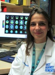 Maura Malpetti合作论文数Cambridge University Hospitals National Health Service Foundation Trust43
 James Rudd合作论文数Department of Medicine, University of Cambridge;Addenbrooke’s Hospital37
 Elizabeth Warburton合作论文数Neurology Unit, University of Cambridge34Guy B. Williams合作论文数Addenbrooke's Hospital, Cambridge University34
 Roido Manavaki合作论文数Department of Radiology, University of Cambridge33John C. Clark合作论文数32Timothy Rittman合作论文数29
 Anthony Davenport合作论文数Department of Medicine, University of Cambridge29A. T. C. Carpenter合作论文数29
 Su Li合作论文数Department of Psychiatry, University of Cambridge28John C Clark合作论文数27
 Ajenthan Surendranathan合作论文数27
 Patricia Vazquez Rodriguez合作论文数Department of Clinical Neurosciences, University of Cambridge27
 George Savulich合作论文数25Peter J. Nestor合作论文数25
 Jonathan Gillard合作论文数Department of Radiology, University of Cambridge24
 Peter Smielewski合作论文数Division of Neurosurgery, University of Cambridge22
 Peter J A Hutchinson合作论文数Department of Clinical Neurosciences, Robinson College, University of Cambridge22Elizabeth A. Warburton合作论文数21
 Negin Holland合作论文数Univ Cambridge, Cambridge Univ Hosp NHS Trust20
 Martin J Graves合作论文数Department of Radiology, University of Cambridge19Shahid H. Zaman合作论文数MRC Molecular Neurobiology Unit, MRC Centre, Cambridge, UK18Peter Johnström合作论文数18David Williamson合作论文数17
 Stephen Sawiak合作论文数Wolfson Brain Imaging Centre, Fitzwilliam College;Department of Psychology, University of Cambridge17
 Thomas E Cope合作论文数Centre for Frontotemporal Dementia and Related Disorders, Department of Clinical Neurosciences, University of Cambridge16
 Anthony Holland合作论文数Department of Psychiatry, School of Clinical Medicine, University of Cambridge16
 Jason Tarkin合作论文数University of Cambridge16o barret合作论文数14
 Trevor W. Robbins合作论文数Department of Psychology, University of Cambridge;Behavioural and Clinical Neuroscience Institute, University of Cambridge;Institute of Science and Technology for Brain-inspired Intelligence, Fudan University14Hugh Richards合作论文数14Deepa Gopalan合作论文数14
 Young Hong合作论文数Cooper University Hospital, Rowan University12
 Kamen A. Tsvetanov合作论文数Department of Clinical Neurosciences, Cambridge Centre for Frontotemporal Dementia and Related Disorders, University of Cambridge;Department of Psychology, University of Cambridge12Peter Weissberg合作论文数12
 David Izquierdo-Garcia合作论文数Mass General Research Institute, Massachusetts General Hospital;Universidad Carlos III de Madrid;Harvard Medical School12Da Chatfield合作论文数12




