BACKGROUND:The cholecystokinin-2/gastrin receptor (Cck2r) is expressed in corpus isthmus progenitor, enterochromaffin-like and parietal cells, regulating acid secretion and cell turnover. However, the role of gastrin on Cck2r progenitors during mucosal regeneration remains unexplored. OBJECTIVE:To study the role of gastrin-Cck2r axis and corpus progenitors during gastric injury and regeneration. DESIGN:We generated Cck2r-CreERT2; Gastrin-DTR-p2A-TdTomato; Rosa26-ZsGreen mice to trace corpus Cck2r+ progenitors during homeostasis and injury, under conditions of hypogastrinaemia and hypergastrinaemia. Injury models included acute ulceration, chronic H. pylori gastritis and N-Nitroso-N-Methylurea (MNU) exposure. RESULTS:Hypergastrinaemia significantly expanded Cck2r+ isthmus progenitors, whereas hypogastrinaemia reduced them. Gastric ulceration induced a twofold elevation in plasma gastrin by day 14, antral G-cell expansion and complete ulcer healing by day 28. Gastrin infusion or proton pump inhibitor (PPI) treatment further elevated gastrin and promoted complete ulcer healing by day 14, whereas G-cell ablation minimised gastrin, impaired healing and abrogated the benefits of PPI (p<0.05). The vagus nerve, through the muscarinic receptor 3, mediated both gastrin elevations and Cck2r+ progenitor expansion during ulcer healing. G-cell ablation in H. pylori-infected mice increased colonisation and exacerbated inflammation, atrophy, metaplasia and dysplasia (p<0.05), while hypergastrinaemia was protective. Similarly, in the MNU model, G-cell ablation worsened gastric pathology while hypergastrinaemia mitigated it. CONCLUSIONS:We report a novel role for G-cell-derived gastrin in ulcer healing. Hypogastrinaemia is a risk factor for poor ulcer healing, corpus atrophy and potentially cancer, while physiological gastrin responses are protective. PPI-induced hypergastrinaemia plays a key role in ulcer healing, and gastrin signalling may prevent gastric preneoplasia.
Co-culture with stellate ganglia (SG) promotes the growth of mouse SCLC
Sympathetic and parasympathetic nerve fibers in mouse SCLC
Abstract Background: Mammalian tissues can “remember” prior inflammatory or injurious events and respond more rapidly to later stimuli. Whether vagus-derived sensory neurons encode such experiences and convert them into tumor-promoting signals remains unknown. We show that inflammatory memory within vagal sensory neurons drives gastric regeneration and tumorigenesis through ILC2-mediated epigenetic reinforcement and CGRP/RAMP1 signaling. Methods: Gastric injury was induced by high-dose tamoxifen (HDT) or Helicobacter pylori infection/eradication. Orthotopic (ACKP) and spontaneous (Iqgap3-CreERT2; KRASG12D) tumor models assessed memory-driven initiation. Trpv1-Cre; hM3Dq and Trpv1-Cre; DTA mice enabled chemogenetic activation or ablation of nodose ganglion (NG) neurons. An AAV-C-Fos-tTA/TetO-Cre-DTA TRAP system labeled and ablated neurons activated during the first injury. Ramp1 was deleted in gastric stem cells (Iqgap3-CreERT2; Ramp1 flox), and IL25R+ ILC2s were depleted genetically or pharmacologically. Chromatin of sorted NG neurons was analyzed by ChIP-qPCR for H3K4me3 enrichment at memory loci. Results: Prior injury produced persistent “neuronal memory” marked by expanded CGRP+ fibers that accelerated mucosal recovery but enhanced metaplasia and tumorigenesis. Activation of Trpv1+ NG neurons was required; activation mimicked memory, while ablation or vagotomy abolished it. TRAP tracing showed reactivated neurons during reinjury overlapped with and exceeded those from the first; ablating the initial cohort erased the phenotype. MRI and c-Fos in the brainstem revealed stronger vagal signals after reinjury, indicating peripheral memory transmission to the central axis. During memory formation, subsets of sensory terminals directly innervated gastric isthmus stem cells; anterograde and retrograde tracing verified bidirectional connectivity, providing a structural basis for rapid signal recall. Memory neurons released CGRP acting on Ramp1+ isthmus stem cells; deleting or antagonizing Ramp1 suppressed dysplasia and tumorigenesis. IL25R+ ILC2s infiltrated the isthmus and maintained NG memory via IL-5-triggered CGRP release, Wnt5a-guided terminal attraction, and IL-13-induced SMYD4 with enhanced H3K4me3 in NG neurons. These cues reinforced the feedback sustaining the memory state. Conclusions: We identify a vagal sensory neuronal inflammatory memory circuit linking prior injury to gastric tumor initiation. ILC2-mediated IL-13/SMYD4/H3K4me3 signaling stabilizes memory within NG neurons, while CGRP/RAMP1 and neuro-epithelial contacts transmit it to the mucosa, forming a neuro-epigenetic feedback loop driving inflammation-associated gastric cancer. Citation Format: Yi Zeng, Puran Zhang, Ruhong Tu, Feijing Wu, Xiaofei Zhi, Jin Qian, Biyun Zheng, Hualong Zheng, Shuang Li, Hiroki Kobayashi, Yosuke Ochiai, Masahiro Hata, Juli Lin, Junya Arai, Leah B. Zamechek, Timothy C. Wang. Vagal sensory neuronal inflammatory memory promotes gastric tumorigenesis through ILC2-mediated epigenetic signaling and the CGRP/Ramp1 axis [abstract]. In: Proceedings of the American Association for Cancer Research Annual Meeting 2026; Part 1 (Regular Abstracts); 2026 Apr 17-22; San Diego, CA. Philadelphia (PA): AACR; Cancer Res 2026;86(7 Suppl):Abstract nr 4078.
Abstract Background: Aging is associated with a chronic low-grade inflammatory state that profoundly influences tumor development and progression, known as inflammaging. While this is due to increased myelopoiesis, the exact mechanisms leading to increased myeloid production and the effects on the gastric tumor microenvironment remain poorly understood. Methods: We conducted comprehensive analyses of young (3 months) and aged (18 months) Hdc-GFP mice, evaluating expression of the peptide trefoil factor family 2 (TFF2) and abundance of GFP+ MDSCs in the stomach, circulation and other sites. The ACKP orthotopic model and Mist1CreERT; RhoAY42CCDH1ff; Hdc-GFP mouse (GEM) model were used to test whether aging promotes gastric cancer progression and to explore the underlying mechanisms. TFF2-MSA was given by i.p. injection twice weekly for 2 weeks before ACKP tumor inoculation, to test whether pretreatment could reverse the aging-related pro-tumor effects. Results: Aging led to a significant reduction in TFF2 levels in both the stomach and circulation, as confirmed by IHC, ELISA, and qPCR. The decline in circulating TFF2 was accompanied by elevated IL-1β expression in bone marrow, peripheral blood, and gastric neutrophils, together with increased circulating IL-1β levels. Enhanced IL-1β signaling contributed to a myeloid-biased hematopoietic phenotype, leading to the increased GFP+ MDSCs in the stomach, spleen, and peripheral blood. Elevated IL-1β in the aged stomach induced fibroblast activation with a senescence-associated secretory phenotype (SASP)-like profile, including increased IL-6, CXCL1 and CXCL2 expression. In the ACKP orthotopic and model and GEM model, with tumorigenesis initiated at 3mo or 18mo, aging significantly accelerated tumor growth and shortened survival. Immunofluorescence analyses of aged ACKP tumor identified an increased subset of IL-1R1+ cancer-associated fibroblasts (CAFs) and CGRP+ sensory nerves. Functionally, pretreatment with TFF2-MSA prior to ACKP tumor implantation decreased IL-1R1+ CAF and sensory nerves, reduced MDSC accumulation and IL-1β expression, and suppressed gastric tumor growth in aged mice. Conclusion: Our findings uncover a systemic aging axis linking reduced gastric TFF2 production to myelopoiesis and thus to gastric cancer susceptibility. Aging-associated IL-1β signaling from expanded myeloid cells reprograms gastric fibroblasts toward a pro-inflammatory SASP phenotype, fostering immune suppression and tumor growth. Restoring youthful TFF2 levels may provide effective strategies to mitigate the aging-related tumor risk and other associated inflammaging syndromes. Citation Format: Shuang Li, Hualong Zheng, Jin Qian, Puran Zhang, Feijing Wu, Yi Zeng, Biyun Zheng, Juli Lin, Hiroki Kobayashi, Yosuke Ochiai, Mashahiro Hata, Arai Junya, Leah B. Zamechek, Bruce Daugherty, Seth Lederman, Timothy C. Wang. TFF2 deficiency amplifies IL-1β-driven inflammation and promotes aging-associated gastric tumor progression [abstract]. In: Proceedings of the American Association for Cancer Research Annual Meeting 2026; Part 1 (Regular Abstracts); 2026 Apr 17-22; San Diego, CA. Philadelphia (PA): AACR; Cancer Res 2026;86(7 Suppl):Abstract nr 6822.
Figure S8 shows that innervation is increased in PDAC liver metastasis and that NTN1 overexpression in Panc02 cells promotes the growth of PDAC liver metastasis in a splenic injection model.
Nerves and cancer-associated fibroblasts (CAFs) have each been shown to regulate cancer progression directly. However, whether these cells interact to control tumor progression remains largely unknown. We show that in colorectal cancer (CRC), cholinergic stimulation induces CHRM3/Gq-dependent NTN1 secretion from CAFs, which in turn enhances intratumor cholinergic innervation. Within this feedforward loop, cholinergic stimulation promotes CRC growth directly through tumoral CHRM3/Gq-mediated YAP activation, while CAF-derived NTN1 promotes CRC growth and epithelial-to-mesenchymal transition-like programs via UNC5B-PI3K/AKT signaling. Chemogenetic activation of cholinergic neurons or fibroblast activation of the M3 receptor/Gq promotes tumoral YAP and AKT signaling and CRC progression. Conversely, blocking CHRM3 or NTN1 suppresses these pathways and improves mouse survival. In human CRC, high NTN1 expression is associated with a mesenchymal-like subtype and poor patient outcomes. These findings suggest that the neuro-mesenchymal interaction is central to CRC progression and could be therapeutically targeted with a CHRM3 antagonist or NTN1-blocking antibody.
High levels of ADRB2 promote the growth of mouse SCLC
Abstract Background: Nociceptive sensory nerves are emerging as active regulators of tumor progression and immune suppression within the tumor microenvironment (TME). We previously showed that gastric cancer cells establish a bi-directional electrical circuit with CGRP+ sensory neurons, in which cancer depolarization triggers neuronal firing and CGRP release. Given that sensory afferents serve as the afferent limb of the inflammatory reflex transmitting peripheral signals to the brainstem, we hypothesized that cancer may similarly exploit neural circuits and neuropeptide signaling to regulate systemic immunity. Methods: A chemogenetic receptor (PSAM4-5HT3) was introduced into a syngeneic murine (ACKP) gastric cancer cell line to evoke cancer depolarization in vivo. Cancer-activated neuronal projections were mapped in TRAP2 (Fos-CreERT2) mice using Cre-dependent AAV reporters. Immune profiling was performed to characterize antigen-experienced T cells in the tumor-draining lymph node (TDLN) and various CD8+ T-cell subsets within the TME. In vivo imaging was used to monitor T-cell motility dynamics, and GCaMP6s was expressed in immune cells to visualize neuron-triggered calcium flux. Results: Cancer depolarization activated nociceptive sensory neurons, which established synapse-like contacts with CD8+ TILs preferentially at the perivascular niche and tertiary lymphoid structures. The cancer-coupled sensory nerves upregulated Cxcl10, attracting T cells toward their terminals to form these connections. In a 3-D co-culture system comprising cancer spheroids, dorsal root ganglia, and CD8+ TILs, optogenetic stimulation of cancer cells induced a calcium flux in sensory nerve-connected T cells, confirming functional transmission. Local activation of this cancer-nerve-T-cell circuit enhanced T-cell migration toward nerve terminals but diminished effector capacity, evidenced by reduced TNFα and IFNγ expression and limited infiltration into the tumor core. Sensory afferents relayed signals to higher brainstem areas, including the nucleus tractus solitarius (NTS) and rostral ventrolateral medulla (RVLM), eliciting sympathetic output to the TDLN. This afferent-brain-efferent loop retained and immobilized antigen-experienced CD44+PD-1+ T cells within the TDLN, thereby restraining systemic immunity in a norepinephrine-Adrb2-dependent manner. Anti-PD-1 therapy paradoxically strengthens the cancer-sensory nerve coupling. Ablatio of this cancer-evoked neural circuit restored T-cell effector function systemically and synergized with anti-PD-1 therapy. Conclusions: These findings reveal that cancer depolarization activates CGRP+ sensory neurons to form synaptic contacts with CD8+ T cells, linking local peptidergic signaling to systemic immune suppression via the NTS-RVLM-TDLN axis. Disrupting this circuit restores T-cell function and enhances anti-PD-1 response. Citation Format: Puran Zhang, Feijing Wu, Yi Zeng, Shuang Li, Jin Qian, Hualong Zheng, Biyun Zheng, Juli Lin, Hiroki Kobayashi, Junya Arai, Yosuke Ochiai, Masahiro Hata, Leah B. Zamechek, Timothy C. Wang. Electrical activity powered neural circuit sustains systemic immune evasion in gastric cancer [abstract]. In: Proceedings of the American Association for Cancer Research Annual Meeting 2026; Part 1 (Regular Abstracts); 2026 Apr 17-22; San Diego, CA. Philadelphia (PA): AACR; Cancer Res 2026;86(7 Suppl):Abstract nr 7384.
Figure S2 shows that KrasG12D mutation and β-adrenergic stimulation upregulate Ntn1 and Neo1 through MAPK pathway-mediated interaction of ELK1 (for Ntn1) or c-JUN (for Neo1) with their promoter regions.
Abstract Background Esophagogastric junction (EGJ) adenocarcinoma is rising in developed countries. While diet and reflux contribute, microbial-neuronal mechanisms remain poorly defined. Methods Metabolomic and 16S rRNA sequencing were performed on 40 paired human EGJ tumors and adjacent tissues, and single-cell RNA sequencing on five pairs. Mechanistic studies used the pL2-IL1B mouse model of Barrett’s esophagus, Cck2r-CreERT2 mice to label EGJ progenitors, and crosses to p53R172H; pL2-IL1B to provide a tumor-prone background. Dclk1-CreERT2 targeted Tuft cells, while Trpv1-Cre, hM3Dq, and Ramp1 flox examined sensory signaling. Additional lines (Cck2r-CreERT2; Ramp1flox/flox, Dclk1-CreERT2; Cox1flox/flox; Cox2flox/flox, Pou2f3-CreERT2; DTA) dissected the Tuft-neuron-epithelial circuit. P. melaninogenica colonization and deoxycholic acid (DCA) supplementation modeled microbial-metabolic effects. Calcium imaging, phospho-ERK staining, and Tuft-neuron co-cultures assessed functional signaling. Results EGJ tumors were enriched in Firmicutes/Bacteroidota and Prevotella, while adjacent tissues contained Proteobacteria and Helicobacter. Prevotella abundance correlated positively with arachidonic-acid and negatively with tryptophan metabolites. Single-cell data showed epithelial, immune, and metabolic clusters, with bile- and fatty-acid pathways enriched in proliferative cells. P. melaninogenica, linked to bile-acid-rich, cancer-prone environments, was selected for mechanistic study. DCA alone enhanced EGJ dysplasia in pL2-IL1B mice. Oral P. melaninogenica colonized the EGJ, further increased by DCA, forming a bile-acid niche. Colonization amplified CGRP+ neuronal signaling, expanded Tuft cells, and aggravated dysplasia. Tumor-prone Cck2r-CreERT2; p53R172H; pL2-IL1B mice showed more Tuft-CGRP+ contacts, and pseudotime analysis indicated enhanced Cck2r+-to-Tuft differentiation. In ex vivo organoids, activation of Dclk1+; hM3Dq+ Tuft cells elevated PGE2/NGF secretion, strengthened Tuft-nerve contacts, and induced calcium influx in co-cultured Trpv1+; GCaMP6s+ DRG neurons. Trpv1+ neurons co-expressed CGRP and EP4. Combined DCA + CNO treatment in Trpv1-Cre; hM3Dq; pL2-IL1B mice further increased dysplasia. Tuft ablation (Pou2f3-CreERT2; DTA), Tuft-specific Cox1/2 loss, or Ramp1 deletion in Cck2r+ cells suppressed P. melaninogenica-induced dysplasia, demonstrating that blocking the Tuft-neuron-progenitor axis halts tumor progression. Conclusions We define a microbiota-bile-acid-Tuft-neuron feedback loop driving EGJ tumorigenesis. P. melaninogenica and DCA activate Tuft cells to release PGE2/NGF, stimulating EP4-CGRP/Ramp1 signaling and promoting epithelial dysplasia. Disrupting this circuit suppresses tumor growth, highlighting a metabolic-neuronal target for EGJ cancer prevention. Citation Format: Yi Zeng, Feijing Wu, Puran Zhang, Ruhong Tu, Xiaofei Zhi, Biyun Zheng, Jin Qian, Hualong Zheng, Shuang Li, Hiroki Kobayashi, Yosuke Ochiai, Masahiro Hata, Juli Lin, Junya Arai, Leah B. Zamechek, Zaisheng Ye, Timothy C. Wang. Microbiotas activate a tuft cell-sensory neuron feedback loop to promote esophagogastric junction (EGJ) tumorigenesis [abstract]. In: Proceedings of the American Association for Cancer Research Annual Meeting 2026; Part 1 (Regular Abstracts); 2026 Apr 17-22; San Diego, CA. Philadelphia (PA): AACR; Cancer Res 2026;86(7 Suppl):Abstract nr 6222.
Figure S7 shows that, in an mT4 orthotopic transplantation model, tumoral Ntn1 overexpression increases tumor burden, FAK phosphorylation, and the expression of ZEB1, SOX9, and Ki-67, as well as PGP9.5+ and TH+ nerve density.
ICI treatment reduces the growth of SCLC
PKA inhibition (PKAi) reduces the growth of primary human SCLC cells
ADRB2 regulates the growth of human SCLC cells
 James G. Fox合作论文数Department of Biological Engineering, Massachusetts Institute of Technology137
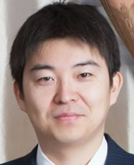 Yoku Hayakawa (早河翼)合作论文数Faculty of Medicine, The University of Tokyo112
 Michael Quante合作论文数Universität Münster85
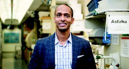 Samuel Asfaha合作论文数Department of Pharmacology & Therapeutics;University of Calgary;Department of Pharmacology & Therapeutics, University of Calgary78
 Daniel Worthley合作论文数Colonoscopy Clinic67
 Anil K Rustgi合作论文数Herbert Irving Comprehensive Cancer Center, Columbia University Irving Medical Center64
 Benedikt Westphalen合作论文数61
 Hiroki Kobayashi合作论文数Adelaide Med Sch, Univ Adelaide61Shigeo Takaishi合作论文数54
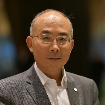 Duan Chen合作论文数Department of Clinical and Molecular Medicine, Faculty of Medicine and Health Sciences, Norwegian University of Science and Technology53
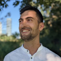 Ermanno Malagola合作论文数Univ Utrecht, Fac Vet Med, Dept Clin Sci Compan Anim, NL-3584 CM Utrecht, Netherlands50
 Sureshkumar Muthupalani合作论文数StageBio50Bi-Yun Zheng合作论文数49
 Shuiping Tu (涂水平)合作论文数Renji Hospital, Shanghai Jiao Tong University School of Medicine45Julian A. Abrams合作论文数44
 Tailor Yagnesh合作论文数Department of Medicine and Irving Cancer Research Center, Columbia University43
 Kenneth P. Olive合作论文数Division of Digestive and Liver Diseases, Department of Medicine, Columbia University Irving Medical Center;Herbert Irving Comprehensive Cancer Center, Columbia University Irving Medical Center43
 Bernhard W. Renz合作论文数Department of General, Visceral and Transplant Surgery, Ludwig-Maximilians-University of Munich;Klinik für Allgemein-, Viszeral- und Transplantationschirurgie, Ludwig-Maximilians-University of Munich Klinikum42
 Chun-Mei Zhao合作论文数Department of Clinical and Molecular Medicine, Norwegian University of Science and Technology (NTNU),41Ruth A. White合作论文数39
 Zhi Xiaofei合作论文数The First Affiliated Hospital, Nanjing Medical University35
 Graham Dockray合作论文数Institute of Systems, Molecular & Integrative Biology, University of Liverpool;Faculty of Health and Life Sciences, University of Liverpool33Tj Koh合作论文数31
 Arlin B Rogers合作论文数31Zhongming Ge合作论文数30Richard A Friedman合作论文数29Whary Mark T合作论文数29Andrea Varro合作论文数29
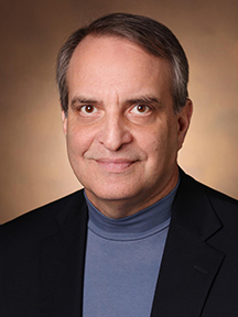 James R. Goldenring合作论文数Department of Surgery, School of Medicine, Vanderbilt University;Section of Surgical Sciences, Vanderbilt University Medical Center;Epithelial Biology Center, School of Medicine, Vanderbilt University28Que Jianwen合作论文数28Hua-Long Zheng合作论文数26Wataru Shibata合作论文数Department of Gastroenterology, University of Tokyo26Takayuki Tanaka合作论文数25Yosuke Ochiai合作论文数24Nagar Karan合作论文数Department of Medicine and Irving Cancer Research Center, Columbia University24
 David Sabatini合作论文数Institute of Organic Chemistry and Biochemistry, Czech Academy of Sciences24Xiangdong Yang合作论文数24
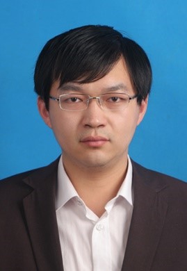 Zhengyu Jiang (姜正羽)合作论文数China Pharmaceutical University24Kelly S. Betz合作论文数24
 Junya Arai合作论文数23
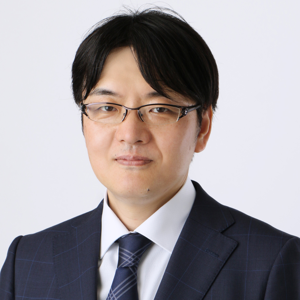 Atsushi Enomoto (榎本篤)合作论文数Center for Neurological Diseases, Graduate School of Medicine, Nagoya University23
 András Varró合作论文数Department of Pharmacology and Pharmacotherapy, University of Szeged23Feijing Wu合作论文数22Ruhong Tu合作论文数Department of General Surgery, Fujian Medical University22Masaki Sunagawa合作论文数21Leah B Zamechek合作论文数21Woosook Kim合作论文数Columbia Univ, Coll Phys & Surg, Dept Med, Div Digest & Liver Dis, New York, NY 10032 USA21
 Roland M. Schmid合作论文数Klinik und Poliklinik für Innere Medizin II, TUM Universitätsklinikum;School of Medicine and Health, Technical University of Munich21Guangchun Jin合作论文数21




