Using a selective plating strategy for staphylococci, we surveyed the local community wastewater and purified 16 independent isolates representing the following seven species of Staphylococcus: S. cohnii, S. equorum, S. lentus, S. nepalensis, S. sciuri, S. shinii, and S. xylosus. Staphylococcus aureus was not detected. The wastewater also served as a source to identify a bacteriophage (phage), referred to here as JS1, that could infect all these species of Staphylococcus, as well as a range of clinical S. aureus strains, including methicillin-resistant isolates. The class Caudoviricetes are tailed phages, and classification systems recognize the following three major morphotypes: the Myo-like (medium-to-long, straight, contractile tails), Sipho-like (long, flexible, non-contractile tails), and Podo-like (very short, rigid tails). Electron microscopy showed that JS1 virions have 252 nm long, curved, contractile tails. Curvature analysis showed that this represented a range with a 1/R value of 7.6 ± 1.3 μm-1, where R is the radius of curvature. Phage JS1 also encodes hydrolases that are assembled onto the phage virions. One of these hydrolases, JS1_0224, was biochemically characterized and found to etch regions from the Staphylococcal cell wall. The possibility that these on-board hydrolases and the curvature of the long contractile tails are advantageous to the phage for navigating through the cell wall of these various species of Staphylococcus is discussed.IMPORTANCEPast work has seen over-representation of Staphylococcus aureus clinical isolates in genome and biology studies on staphylococci. Here, we show by a selective plating analysis of municipal wastewater that independent isolates representing seven other species of Staphylococcus were recovered (S. cohnii, S. equorum, S. lentus, S. nepalensis, S. sciuri, S. shinii, and S. xylosus), as readily identified in the samples. Genome sequence analysis revealed some species-specific antibiotic resistance profiles across the strains, and a bacteriophage was isolated that had a cross-species host range. Using this broad biological approach to analyze staphylococci has identified a phage with a broad killing range, and this phage is morphologically distinct from the three known types of tailed phages.
The outer membrane is the interface through which Gram-negative bacteria - a broad classification of organisms including Escherichia coli and a number of deadly pathogens - interact with the environment. Two decades of work on the process of outer membrane biogenesis have led to the discovery of the components that mediate this process, and the characterisation of structure and function of these component parts of the bacterial cell machinery. However, neither current experimental methods, nor conventional molecular dynamics (MD) simulation approaches are capable of investigating this membrane machinery on the time scale of the cell division cycle. This leaves crucial questions unanswered, such as how this lipid-poor, largely static environment is organised to permit ongoing membrane growth. Here, we introduce a semi-quantitative agent-based model to explore the molecular-scale dynamics of Gram-negative outer membrane as it grows. Model simulations across a broad region of parameter space suggest that protein incorporation into the membrane by the β-barrel assembly machinery (BAM complex) is a process which is prone to stalling, and may take place only in short bursts. We also find suggestions that BAM complexes work collaboratively with each other, and with the lipopolysaccharide-inserting Lpt complex when in close proximity. The agent-based framework we introduce provides a means to assess and generate hypotheses on outer membrane biogenesis on previously inaccessible time scales.
Natural environments are a key reservoir for many opportunistic pathogens, yet mechanisms enabling persistence within complex microbial communities remain poorly defined. Current methods do not adequately link molecular and functional understanding of bacterial persistence to the activities of bacteria within their natural microbial community. Here we combine in situ culture-independent techniques, including metagenomics and metatranscriptomics, with isolate-level functional genomics and mutant studies to understand how opportunistic pathogens from the genera Escherichia , Klebsiella , and Enterococcus survive in urban-adjacent freshwater ecosystems. . Our results show that potential pathogens from each of these genera are both present and active within freshwater microbial communities. We further investigate a mouse-virulent isolate of the E. coli UPEC lineage ST73 isolated directly from this ecosystem, showing that this isolate persists in freshwater microcosms for at least one month. Paired transcriptomics and genome-scale fitness screening in creek water containing autochthonous microbiota identified E. coli genes required for persistence, including those involved in amino acid metabolism, nucleotide biosynthesis and biogenesis of curli. Isolate-resolved metatranscriptomics analysis supported these findings by revealing that many of these genes were highly expressed in situ . Deletion of curli structural genes resulted in reduced biofilm formation and a fitness defect specifically in the presence of freshwater microbiota, indicating that these structures promote environmental survival by improving E. coli competitiveness. Our study deepens our understanding of E. coli survival in waterways, while providing a broadly applicable framework for interrogating mechanisms of pathogen persistence in complex environments and communities.
Biofilm formation by Klebsiella pneumoniae is mediated by the type 3 fimbriae Mrk, and regulated by MrkH and 3’,5’-cyclic diguanylic acid (c-di-GMP). We sought to identify specific chemical inhibitors of K. pneumoniae biofilm formation that reduced the activity of MrkH. A compound N-(3-cyano-5,6,7,8-tetrahydro-4H-cyclohepta[b]thien-2-yl)-2-methoxybenzamide, JT71, reduced K. pneumoniae mrkA promoter activity and biofilm formation by 50% without affecting cell viability. Western blot analysis, hemagglutination assays, electron microscopy and qPCR showed that JT71 reduced type 3 fimbriae production, and transcription of mrkA and mrkH. JT71 demonstrated activity against other clinical and multi-drug resistant K. pneumoniae isolates, and a type 3 fimbriate-positive Citrobacter koseri strain. In silico molecule docking was used to illustrate that JT71 could bind directly to the MrkH protein and block its activity. JT71 possesses promising drug-likeness properties and is non-toxic to mammalian cells. Chemical inhibition of transcriptional regulators that control fimbriae expression can inhibit bacterial biofilm formation.
Antimicrobial resistance (AMR) has been declared one of the top 10 global public health challenges of our age by the World Health Organization, and the World Bank describes AMR as a crisis affecting the finance, health, and agriculture sectors and a major threat to the attainment of Sustainable Development Goals. But what is AMR? It is a phenotype that evolves in microbes exposed to antimicrobial molecules and causes dangerous infections. This suggests that scientists and healthcare workers should be on the frontline in the search for sustainable solutions to AMR. Yet AMR is also a societal problem to be understood by everyone. This review aims to explore the need to address the problem of AMR through a coherent, international strategy with buy-in from all sectors of society. As reviewed here, the sustainable solutions to AMR will be driven by better understanding of AMR biology but will require more than this alone to succeed. Some advances on the horizon, such as the use of bacteriophage (phage) to treat AMR infections. However, many of the new technologies and new therapeutics to address AMR require access to biodiversity, where the custodians of that biodiversity-and the traditional knowledge required to access it-are needed as key partners in the scientific, clinical, biotechnological, and international ventures that would treat the problem of AMR and ultimately prevent its further evolution. Many of these advances will be built on microbial assessments to understand the extent of AMR in our environments and bioprospecting to identify microbes that may have beneficial uses. Genuine partnerships for access to this biodiversity and sharing of benefits accrued require a consideration of ethical practice and behavior. Behavior change is needed across all sectors of culturally diverse societies so that rapid deployment of solutions can be implemented for maximum effect against the impacts of AMR.
Bacteriophages (phages) are viruses that kill bacteria, with potential as antibacterial agents in industrial settings, agriculture, and human health. Here, we identified two phages, PIN1 and PIN2, that can kill clinical isolates of the human pathogen Klebsiella pneumoniae. The phages are highly stable; PIN2 in particular resisted multiple freeze-thaw cycles over 12 months without loss of activity. PIN1 and PIN2 are related to flagellotropic phages, an idiosyncratic group of viruses that bind to bacterial flagellae, but K. pneumoniae is an immotile pathogen that does not have flagellae. Genetic mosaicism is observed, wherein the long, flexible tail fiber of the flagellotropic phages has been substituted by a more compact tail fiber that binds the Klebsiella host through cell-surface capsular polysaccharide and lipopolysaccharide. PIN1 and PIN2 belong to the Yonseivirus group of phages, with initial analyses across the group suggesting further recent diversification in the tail-fiber cassette in the Yonseivirus genomes.
Bacteriophages (phages) are viruses that can kill bacteria, thereby editing and shaping microbial communities. The telomere phages are a curious form using telomere-like structures to replicate their genomes as linear extrachromosomal elements. Here, we find that telomere phages are widely distributed in bacteria, being highly prevalent in Klebsiella species. We establish a model system to investigate telomere phage biology by isolating the virions of telomere phages and infecting naïve strains to create isogenic lines with and without a phage. We find that only a small set of telomere phage proteins is expressed in phage-host cells, including a toxin-the telocin-that kills other Klebsiella strains. We identify and validate a set of telocins in the genomes of other prevalent Klebsiella telomere phages. Thus, telomere phages are widespread elements encoding diverse antibacterial weapons and we discuss the prospect of using telocins for precision editing of microbial populations.
Antimicrobial-resistant (AMR) bacteria and genes are continually exchanged among humans, animals, and environmental reservoirs. Disparate and siloed surveillance methods present a major challenge for tracing and disrupting AMR transmission, with clinical monitoring focusing on detecting specific pathogens or specific genes of interest, and environmental surveys often relying on inferences drawn from indicator organisms. Here, we demonstrate that, following sample-specific pre-processing, common surveillance approaches can be applied consistently to profile AMR abundance, distribution, and phenotypes across diverse reservoirs, including soil, sediment, water, wastewater, and faecal samples from both urban and agricultural settings. Across all sample types, three core methods provided complementary insights: (i) quantitative PCR (qPCR) arrays to measure multiple AMR genes, (ii) gene- and genome-centric metagenomics for comprehensive resistome profiling, and (iii) culture-based genomics with susceptibility testing to link genotypes to phenotypes. We applied this approach to profile 1,032 metagenome-assembled genomes, 66 bacterial isolate genomes, and 78 and 6,442 AMR genes/reference sequences via qPCR and metagenomics, respectively. This integrated framework revealed a moderate prevalence but high diversity of resistance mechanisms in both pathogens and non-pathogenic bacteria with potentially transmissible genes, with wastewater especially enriched in AMR genes. We detected mismatches between genotype and phenotype predictions and a prevalence of intermediate resistance phenotypes, highlighting how many mechanisms of environmental resistance remain poorly understood. Overall, this study demonstrates that unified field-leading surveillance methods can be extended beyond clinical contexts into diverse environmental and animal samples, while highlighting that multiple methods are needed to capture the diverse AMR genotypes and phenotypes in these settings to enable comprehensive monitoring and adaptive solutions to restrict transmission. ### Competing Interest Statement The authors have declared no competing interest.
Antimicrobial resistance (AMR) is the poster child for the need for a 'One Health' approach that develops solutions across the human, agricultural and environmental sectors. This article provides a viewpoint of where AMR research is heading in the future, from the perspective of three Australian initiatives specifically established to combat AMR: the Centre to Impact AMR, the ARC Training Centre for Environmental and Agricultural Solutions to Antimicrobial Resistance, and the Community for Open Antimicrobial Drug Discovery.
Ventilator-associated pneumonia is defined as pneumonia that develops in a patient who has been on mechanical ventilation for more than 48 hours through an endotracheal tube. It is caused by biofilm formation on the indwelling tube, which introduces pathogenic microbes such as Pseudomonas aeruginosa, Klebsiella pneumoniae and Candida albicans into the patient's lower airways. Currently, there is a lack of accurate in vitro models of ventilator-associated pneumonia development. This greatly limits our understanding of how the in-host environment alters pathogen physiology and the efficacy of ventilator-associated pneumonia prevention or treatment strategies. Here, we showcase a reproducible model that simulates the biofilm formation of these pathogens in a host-mimicking environment and demonstrate that the biofilm matrix produced differs from that observed in standard laboratory growth medium. In our model, pathogens are grown on endotracheal tube segments in the presence of a novel synthetic ventilated airway mucus medium that simulates the in-host environment. Matrix-degrading enzymes and cryo-scanning electron microscopy were employed to characterize the system in terms of biofilm matrix composition and structure, as compared to standard laboratory growth medium. As seen in patients, the biofilms of ventilator-associated pneumonia pathogens in our model either required very high concentrations of antimicrobials for eradication or could not be eradicated. However, combining matrix-degrading enzymes with antimicrobials greatly improved the biofilm eradication of all pathogens. Our in vitro endotracheal tube model informs on fundamental microbiology in the ventilator-associated pneumonia context and has broad applicability as a screening platform for antibiofilm measures including the use of matrix-degrading enzymes as antimicrobial adjuvants.
Hypervirulent Klebsiella pneumoniae (hvKp) can infect healthy individuals, in contrast to classical strains that commonly cause nosocomial infections. The recent convergence of hypervirulence with carbapenem-resistance in K. pneumoniae can potentially create ‘superbugs’ that are challenging to treat. Understanding virulence regulation of hvKp is thus critical. Accumulating evidence suggest that posttranscriptional regulation by small RNAs (sRNAs) plays a role in bacterial virulence, but it has hardly been studied in K. pneumoniae. We applied RIL-seq to a prototypical clinical isolate of hvKp to unravel the Hfq-dependent RNA-RNA interaction (RRI) network. The RRI network is dominated by sRNAs, including predicted novel sRNAs, three of which we validated experimentally. We constructed a stringent subnetwork composed of RRIs that involve at least one hvKp virulence-associated gene and identified the capsule gene loci as a hub target where multiple sRNAs interact. We found that the sRNA OmrB suppressed both capsule production and hypermucoviscosity when overexpressed. Furthermore, OmrB base-pairs within kvrA coding region and partially suppresses translation of the capsule regulator KvrA. This agrees with current understanding of capsule as a major virulence and fitness factor. It emphasizes the intricate regulatory control of bacterial phenotypes by sRNAs, particularly of genes critical to bacterial physiology and virulence.
The assembly of β-barrel proteins into the bacterial outer membrane is an essential process enabling the colonization of new environmental niches. The TAM was discovered as a module of the β-barrel protein assembly machinery; it is a heterodimeric complex composed of an outer membrane protein (TamA) bound to an inner membrane protein (TamB). The TAM spans the periplasm, providing a scaffold through the peptidoglycan layer and catalyzing the translocation and assembly of β-barrel proteins into the outer membrane. Recently, studies on another membrane protein (YhdP) have suggested that TamB might play a role in phospholipid transport to the outer membrane. Here we review and re-evaluate the literature covering the experimental studies on the TAM over the past decade, to reconcile what appear to be conflicting claims on the function of the TAM.
Polymyxins are often the only effective antibiotics against the "Critical"pathogen Acinetobacter baumannii . Worryingly, highly polymyxin-resistant A. baumannii displaying dependence on polymyxins has emerged in the clinic, leading to diagnosis and treatment failures. Here, we report that arginine metabolism is essential for polymyxin-dependent A. baumannii . Specifically, the arginine degradation pathway was significantly altered in polymyxin-dependent strains compared to wild -type strains, with critical metabolites (e.g., L-arginine and L -glutamate) severely depleted and expression of the astABCDE operon significantly increased. Supplementation of arginine increased bacterial metabolic activity and suppressed polymyxin dependence. Deletion of astA , the first gene in the arginine degradation pathway, decreased phosphatidylglycerol and increased phosphatidylethanolamine levels in the outer membrane, thereby reducing the interaction with polymyxins. This study elucidates the molecular mechanism by which arginine metabolism impacts polymyxin dependence in A. baumannii , underscoring its critical role in improving diagnosis and treatment of life -threatening infections caused by "undetectable"polymyxin-dependent A. baumannii .
Outer membrane proteins (OMPs) are essential components of the outer membrane of Gram-negative bacteria. In terms of protein targeting and assembly, the current dogma holds that a ‘β-signal’ imprinted in the final β-strand of the OMP engages the β-barrel assembly machinery (BAM) complex to initiate membrane insertion and assembly of the OMP into the outer membrane. Here, we revealed an additional rule that signals equivalent to the β-signal are repeated in other, internal β-strands within bacterial OMPs, by peptidomimetic and mutational analysis. The internal signal is needed to promote the efficiency of the assembly reaction of these OMPs. BamD, an essential subunit of the BAM complex, recognizes the internal signal and the β-signal, arranging several β-strands and partial folding for rapid OMP assembly. The internal signal-BamD ordering system is not essential for bacterial viability but is necessary to retain the integrity of the outer membrane against antibiotics and other environmental insults.
Bacteriophages (phages), viruses capable of infecting and lysing bacteria, are a promising alternative for treating infections from hypervirulent, antibiotic-resistant pathogens like Klebsiella pneumoniae, though narrow host range and phage resistance remain challenges. In this study, the hypervirulent K. pneumoniae NTUH-K2044 was used to purify phage Phi K2044, while two Phi K2044-resistant strains were used to purify two further phages: Phi KR1, and Phi KR8 from hospital sewage. A detailed characterization showed that Phi K2044 specifically killed KL1 capsule-type K. pneumoniae, while Phi KR1 and Phi KR8 targeted 13 different capsular serotypes. The phage cocktail (Phi K2044 + Phi KR1 + Phi KR8) effectively killed K. pneumoniae in biofilms, pre-treatment biofilm formation, and delayed phage-resistance. The phage cocktail improved 7-day survival in Galleria mellonella and mouse models and showed therapeutic potential in a catheter biofilm model. In summary, this proof-of-principle phage cocktail has a broad host range, including hypervirulent and highly drug-resistant K. pneumoniae, and serves as a promising starting point for optimizing phage therapy.
In many bacteria, membrane proteins account for around one-third of the proteome and can represent much more than half of the mass of a membrane. Classic techniques in cell biology can be applied to characterise bacterial membranes and their membrane protein constituents. Here we describe a protocol for the purification of outer and inner membranes from Escherichia coli. The procedure can be applied with minor modifications to other bacterial species, including those carrying capsular polysaccharide attached to the outer membrane.
In a survey of a waterway on Wurundjeri land, two sub-populations of the bacteriophage Merri-merri-uth nyilam marra-natj (phage MMNM) were isolated on a permissive host, Klebsiella B5055 of capsule-type K2, but were distinguished by minor phenotypic differences. The variant phage MMNM(Ala(134)) showed an inhibited activity against Klebsiella AJ174-2, and this was used as a basis to select for further variation through experimental evolution. Over the course of an evolution experiment, 20 phages that evolved distinct phenotypes in terms of the morphologies of plaques formed when they infected host Klebsiella were subject to whole-genome sequencing. The evolved phages had mutations in a small set of proteins that contribute to the baseplate portion of the phage virion. Phages MMNM and MMNM(Ala134) are minimalist phages, with baseplates formed from only five predicted subunits, akin to other minimalist phages Pam3 and XM1. The homology between all three minimalist phages provided a structural framework to interpret the two classes of mutations derived through evolution in the presence of the semi-permissive host: those that affect the interfacial surfaces between baseplate subunits, and those in a base-plate associated tail-fiber. This study evidences that multiple small mutations can be fixed into a sub-population of phage to provide a basis for phenotypic variation that we suggest could ultimately provide for a shift of virus properties, as an alternative evolutionary scenario to the major genetic events that result in more well-studied evolutionary mechanism of phage mosaicism. IMPORTANCE Bacteriophages (phages) are viruses that prey on bacteria. This study sampled natural phage populations to test the hypothesis that untapped genetic variation within a population can be the basis for the selection of phages to diver sify their host-range. Sampling of a freshwater site revealed two populations of the phage Merri-merri-uth nyilam marra-natj (phage MMNM), differing by a variant residue (Val134Ala) in the baseplate protein MMNM_26. This sequence variation modulated bacterial killing in plaques, and further evolution of the phages on a semi-permissive bacterial host led to a new generation of phages with more diverse phenotypes in killing the bacterium Klebsiella pneumoniae.
To kill bacteria, bacteriophages (phages) must first bind to a receptor, triggering the release of the phage DNA into the bacterial cell. Many bacteria secrete polysaccharides that had been thought to shield bacterial cells from phage attack. We use a comprehensive genetic screen to distinguish that the capsule is not a shield but is instead a primary receptor enabling phage predation. Screening of a transposon library to select phage-resistant Klebsiella shows that the first receptor-binding event docks to saccharide epitopes in the capsule. We discover a second step of receptor binding, dictated by specific epitopes in an outer membrane protein. This additional and necessary event precedes phage DNA release to establish a productive infection. That such discrete epitopes dictate two essential binding events for phages has profound implications for under-standing the evolution of phage resistance and what dictates host range, two issues critically important to translating knowledge of phage biology into phage therapies.
Recent studies applying advanced imaging techniques are changing the way we understand bacterial cell surfaces, bringing new knowledge on everything from single-cell heterogeneity in bacterial populations to their drug sensitivity and mechanisms of antimicrobial resistance. In both Gram-positive and Gram-negative bacteria, the outermost surface of the bacterial cell is being imaged at nanoscale; as a result, topographical maps of bacterial cell surfaces can be constructed, revealing distinct zones and specific features that might uniquely identify each cell in a population. Functionally defined assembly precincts for protein insertion into the membrane have been mapped at nanoscale, and equivalent lipid-assembly precincts are suggested from discrete lipopolysaccharide patches. As we review here, particularly for Gram-negative bacteria, the applications of various modalities of nanoscale imaging are reawakening our curiosity about what is conceptually a 3D cell surface landscape: what it looks like, how it is made and how it provides resilience to respond to environmental impacts.
Antibiotic resistance is driven by selection, but the degree to which a bacterial strain's evolutionary history shapes the mechanism and strength of resistance remains an open question. Here, we reconstruct the genetic and evolutionary mechanisms of carbapenem resistance in a clinical isolate of Klebsiella quasipneumoniae. A combination of short- and long-read sequencing, machine learning, and genetic and enzymatic analyses established that this carbapenem-resistant strain carries no carbapenemase-encoding genes. Genetic reconstruction of the resistance phenotype confirmed that two distinct genetic loci are necessary in order for the strain to acquire carbapenem resistance. Experimental evolution of the carbapenem-resistant strains in growth conditions without the antibiotic revealed that both loci confer a significant cost and are readily lost by de novo mutations resulting in the rapid evolution of a carbapenem-sensitive phenotype. To explain how carbapenem resistance evolves via multiple, low-fitness single-locus intermediates, we hypothesised that one of these loci had previously conferred adaptation to another antibiotic. Fitness assays in a range of drug concentrations show how selection in the antibiotic ceftazidime can select for one gene (bla(DHA-1)) potentiating the evolution of carbapenem resistance by a single mutation in a second gene (ompK36). These results show how a patient's treatment history might shape the evolution of antibiotic resistance and could explain the genetic basis of carbapenem-resistance found in many enteric-pathogens.
 Richard (Dick) Strugnell合作论文数The Peter Doherty Institute for Infection and Immunity, The University of Melbourne;Department of Microbiology and Immunology, The University of Melbourne38
 Rhys A Dunstan合作论文数Department of Microbiology, Monash University31
 Stubenrauch Christopher合作论文数29
 Webb Chaille T合作论文数Department of Biochemistry &Molecular Biology, Monash University;Department of Biochemistry & Molecular Biology, Monash University28
 Jonathan Wilksch合作论文数Department of Microbiology &The Peter Doherty Institute for Infection and Immunity, University of Melbourne25
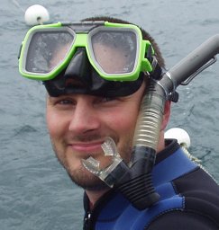 Andrew J. Perry合作论文数Bio21 Institute of Molecular Science and Biotechnology, University of Melbourne24
 Jiangning (John) Song合作论文数Bioinformatics and Biomedicine Laboratory, Monash Biomedicine Discovery Institute, Monash University22
 Vladimir A. Likić合作论文数Department of Biochemistry and Molecular Biology, University of Melbourne;Bio21 Molecular Science and Biotechnology Institute, University of Melbourne20
 Rebecca S. Bamert合作论文数20
 Matthew Belousoff合作论文数Monash Institute of Pharmaceutical Sciences, Monash University;CCeMMP20
 Eva Heinz合作论文数Faculty of Life Sciences, University of Vienna19
 Iain Hay合作论文数Bacterial Cell Biology Laboratory, School of Biological Sciences, Faculty of Science, University of Auckland18
 Takuya Shiota合作论文数Graduate School of Science, Nagoya University18
 Traude H. Beilharz合作论文数St Vincent's Clinical School and School of Biotechnology and Biomolecular Sciences, University of New South Wales18
 Rhys Grinter合作论文数Department of Microbiology, School of Biomedical Sciences, Faculty of Medicine, Nursing and Health Sciences, Monash University17
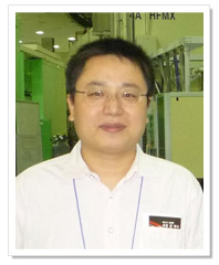 Jiawei Wang (王佳伟)合作论文数School of Life Sciences, Tsinghua University14
 Tieli Zhou (周铁丽)合作论文数The First Affiliated Hospital,Wenzhou Medical College14Jiahui Li合作论文数13
 Pavel Dolezal合作论文数Charles Univ Prague, Dept Parasitol, Fac Sci, Prague, Czech Republic13Gabriel Kipros合作论文数13
 Gordon Dougan合作论文数Department of Medicine, University of Cambridge;Cambridge Institute for Therapeutic Immunology & Infectious Disease, University of Cambridge13
 Anthony Purcell合作论文数Monash Biomedicine Discovery Institute, Monash University13
 Tatiana T Marquez-Lago合作论文数13André Leier合作论文数School of Medicine, University of Alabama at Birmingham13Kher Shing Tan合作论文数12
 Hsin-Hui Shen合作论文数Department of Biochemistry and Molecular Biology, School of Biomedical Sciences, Faculty of Medicine, Nursing and Health Sciences, Monash University12
 Yue Qu合作论文数Department of Infectious Diseases, Central Clinical School, Monash University12
 Ana Traven合作论文数Traven Laboratory, Monash Biomedicine Discovery Institute, Monash University12
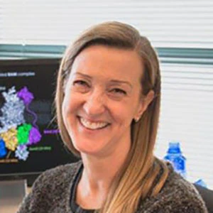 Susan Buchanan合作论文数Laboratory of Molecular Biology, National Institute of Diabetes and Digestive and Kidney Diseases11Von Vergel L Torres合作论文数10
 Jian Li (李健)合作论文数Biomedicine Discovery Institute, Monash University;Data Futures Institute, Monash University;Centre to Impact Antimicrobial Resistance, Monash University10Rocker Andrea合作论文数10
 Tatsuya Akutsu (阿久津達也)合作论文数Bioinformatics Center, Institute for Chemical Research, Kyoto University10Joanne M. Hulett合作论文数10G Schatz合作论文数9
 Schittenhelm Ralf B合作论文数Institute of Zoology, University of Zurich9
 Thomas Naderer合作论文数Department of Biochemistry and Molecular Biology, School of Biomedical Sciences, Faculty of Medicine, Nursing and Health Sciences, Monash University9
 Laura Perlaza-Jimenez合作论文数Laboratory of Mycology and Plant Pathology, Universidad de los Andes9
 Victoria L Hewitt合作论文数Department of Biochemistry and Molecular Biology and Victorian Bioinformatics Consortium, Monash University9
 Geoff Webb合作论文数School of Information Technology, Monash University;Department of Data Science and Artificial Intelligence, Monash University;G I Webb & Associates Pty. Ltd9
 Paul R. Gooley合作论文数Bio21 Institute of Biotechnology and Molecular Science, University of Melbourne9
 Travis Beddoe合作论文数School of Biomedical Sciences, Monash University9Dejan Bursać合作论文数8Yanju Zhang合作论文数Guilin Univ Elect Technol, Sch Comp Sci & Informat Secur, Guilin 541004, Peoples R China8
 Wang Nancy合作论文数8
 André Schneider合作论文数Department of Chemistry, Biochemistry and Pharmaceutical Sciences, University of Bern/Deparmtent of Chemistry and Biochemistry, University of Bern8
 Francesca L. Short合作论文数Department of Biochemistry, University of Cambridge8
 Peter N. Walsh合作论文数Temple University8Mark R. Davies合作论文数The Queensland Institute of Medical Research8




