Bystander killing by anti-PTPRZ1 CAR-T cells using additional tumor cell lines and scFv
Cytotoxic T cell killing is executed at the immunological synapse, whose nanoscale organization underlies function but remains difficult to resolve in native states. Here, we apply cryo-expansion microscopy (cryo-ExM) to visualize the near-native three-dimensional architecture of human T cell synapses and cytotoxic organelles. Cryo-ExM preserves actin, microtubules, membranes, and fine membrane protrusions with high fidelity, enabling volumetric quantification of synapse morphogenesis. We identify an adhesion-dependent, dome-like membrane architecture beneath activated T cells that collapses upon ICAM-1 engagement, linking synapse topology to adhesive cues. Cryo-ExM further resolves intact lytic granules in primary human CD4 and CD8 T cells, revealing single-core and multi-core ultrastructures, spatial organization, and perforin and granzyme loading. Using tissue-adapted expansion microscopy, we map cytotoxic granule content in tumor-infiltrating T cells in FFPE human brain tumors. Together, these data establish a near-native structural framework for human T cell cytotoxicity and an imaging workflow bridging cell models and clinical tissues.
CAR-T-cell-associated on-target off-tumor (OTOT) toxicity represents a major safety concern, as recognition of target antigens on healthy tissues can trigger severe and potentially life-threatening complications. Predicting OTOT toxicity remains a challenge because current preclinical models fail to capture the complexity of native human tissues. Here, we developed a human organotypic tissue platform that enables functional assessment of CAR-T-cell activity in intact human tissues across organ-specific and inflammatory contexts. Using a panel of clinically relevant CAR-T-cell products with known OTOT toxicities, we demonstrate that the platform faithfully recapitulates clinically observed tissue-specific toxicity profiles. CAR-T cells targeting EGFR, HER2, and mesothelin induced inflammatory and cytotoxic responses in healthy human lung tissue, whereas CD19 CAR-T cells remained inactive. We further show that OTOT toxicity cannot be reliably predicted from antigen abundance alone but instead results from the integration of multiple target-dependent determinants, including CAR affinity, inflammatory context, antigen accessibility, and effector-cell dose. The platform also enables quantitative assessment of inflammatory and cytotoxic responses and supports evaluation of pharmacological and CAR design-based strategies to mitigate toxicity. Together, this work establishes the first human organotypic platform for functional modeling of CAR-T-cell-associated OTOT toxicity, providing a clinically relevant framework for preclinical safety evaluation and the rational development of safer engineered cell therapies.
Glioblastoma (GBM) is characterized by a profoundly immunosuppressive tumor microenvironment (TME) that constrains the efficacy of chimeric antigen receptor (CAR)-T cell therapy. Here, we show that surgical resection in both male mice and human GBM ex vivo induces a rapid and sustained remodeling of the TME, marked by upregulation of TREM2 in myeloid cells followed by emergence of T cell exhaustion-like phenotypes. In male mice, targeting TREM2 reshapes the perioperative TME and potentiates tumor antigen-specific CAR-T cell responses, improving intratumoral persistence, proliferation, and effector differentiation, and resulting in enhanced survival. In parallel, we identify the timing of CAR-T cell administration as a critical determinant of therapeutic outcome, with neoadjuvant outperforming adjuvant treatment by preserving CAR-T cell effector function in mice. These findings establish perioperative myeloid cell remodeling and treatment timing as key determinants of CAR-T cell efficacy in GBM.
Anti-PTPRZ1 CAR-T cell effector function after incubation with the Ge1302_PTPRZ1-KI cells
Vertebrates rely on a network of blood vessels to meet organ demands for oxygen and nutrients. While endothelial cells are known to transport excess nutrients to white adipose tissue (WAT) for energy storage, how their metabolic state impacts this process remains unclear. Here, we identify MYCT1 as a conserved, pan-endothelial protein essential for WAT expansion. Endothelial-specific MYCT1 deletion limited WAT expansion independently of angiogenesis, adipogenesis, or systemic metabolic parameters. Mechanistically, MYCT1 interacted with the transmembrane endolysosomal proteins IFITM2/3 to restrict nutrient consumption by the vascular barrier. Loss of MYCT1 caused IFITM2/3 accumulation in early endosomes, promoting excessive endolysosomal degradation and mTORC1 hyperactivation, limiting the WAT energy storage capacity. Notably, endothelial-specific mTORC1 activation through TSC1 deletion phenocopied the fat storage defects of MYCT1 deficiency. Our findings establish the MYCT1-IFITM2/3 complex as endothelial metabolic checkpoint regulating systemic energy storage. Targeting MYCT1-IFITM2/3 may offer new therapeutic options for obesity and metabolic disorders.
Bystander killing by 471_28z CAR-T cells depends on soluble mediators and does not affect macrophages
Expression of effector/memory and activation/exhaustion markers by CAR-T cells in pre- vs. post-thawing samples
PTPRZ1 expression at the mRNA level according to different GBM clinicopathologic features
SUMMARY:Cruz Cobo et al. identify small molecules that reprogram dendritic cells toward an immuno-stimulating secretory phenotype, revealing endoplasmic reticulum calcium signaling as a druggable axis to boost vaccine-based and T cell-mediated cancer immunotherapies. The study links broad changes in calcium-induced secretion to improved immune activation and therapeutic potency.
Glioblastoma (GBM) harbors a profoundly immunosuppressive microenvironment that blunts chimeric antigen receptor (CAR) T-cell therapy. Here, we implemented strategies to reprogram the resection-induced tumor microenvironment (TME) and support CAR T-cell activity. In SB28 and CT2A murine GBM, resection rapidly remodeled the TME, evidenced by TREM2 upregulation across myeloid populations and T-cell exhaustion. Delivery of TREM2 CAR T cells into the resection cavity depleted TREM2⁺ myeloid cells and recruited activated neutrophils. Co-administration of TREM2 and GD2 CAR T cells enhanced survival and reduced exhaustion of both GD2 CAR and endogenous T cells. Additional checkpoint blockade altered CAR T-cell phenotype but did not improve survival. Finally, neoadjuvant GD2 CAR T-cell delivery outperformed adjuvant administration, preserving GD2 CAR T cell effector function and promoting a proinflammatory TME. Collectively, these findings establish TREM2⁺ myeloid cells as critical regulators of perioperative CAR T-cell efficacy and highlight the use of neoadjuvant strategies to overcome post-resection immunosuppression in GBM.
Locally advanced rectal cancer (LARC) is treated with neoadjuvant chemoradiotherapy (nCRT), but only a minority of patients achieve a pathological complete response (pCR). Predictive biomarkers of response could help guide treatment decisions, yet none have reached clinical practice. In this exploratory study, we integrated six publicly available transcriptomic datasets and applied machine learning to derive a 186-gene signature predictive of nCRT response. The signature showed good performance in cross-validation (AUC 0.80) and was associated with consensus molecular (CMS4) and immune (iCMS3) subtypes enriched in responders. Gene set enrichment analyses highlighted pathways involved in tumor growth, immune regulation, and resistance. Spatial transcriptomic profiling of pre-treatment biopsies further identified compartment-specific markers, with tumor-associated genes showing greater predictive value. These results provide biological insights into response mechanisms and generate hypotheses for future validation. Larger prospective studies are required to assess the clinical utility of this approach.
BACKGROUND:Glioblastoma (GBM) is an aggressive brain tumor associated with poor outcome and limited treatment options. Chimeric antigen receptor (CAR) T cells targeting cell surface antigens were shown to induce tumor regression in patients with GBM, although efficacy was transient. To broaden the range of tumor-restricted antigens, we developed CAR T cells targeting Tenascin-C (TNC), a secreted extracellular matrix protein that is overexpressed in GBM and plays a critical role in tumor progression. METHODS:Second-generation CAR T cells were engineered to target the alternatively spliced fibronectin type III (FNIII)-D domain of TNC using a single-chain variable fragment isolated from the R6N antibody and coupled to a CD28 costimulatory domain. TNC-CAR T cells were evaluated in vitro for antigen specificity, activation, and cell proliferation using TNC-expressing patient-derived GBM cell lines cultured as adherent cells or as neurospheres. Reactivity toward purified TNC protein, tumor supernatant, and ex vivo patient tumor samples was also assessed. Cytotoxic CAR T-cell activity was tested against TNC-positive and TNC-negative GBM cell lines, including bystander effects mediated by secreted TNC. In vivo efficacy and safety were determined in NOD scid gamma mice bearing patient-derived GBM tumors. RESULTS:TNC-CAR T cells demonstrated activation when exposed to TNC-positive GBM cells, cell-derived supernatants, or purified TNC protein. They exhibited potent cytotoxicity against TNC-expressing, GBM-derived adherent cells and neurospheres, and induced bystander killing of TNC-negative cells in the presence of either TNC-secreting cells or purified TNC. In vivo, TNC-CAR T cells efficiently infiltrated tumors, triggered cancer cell apoptosis, and significantly extended survival of mice bearing patient-derived GBM, with no evidence of off-tumor toxicity. Notably, TNC-CAR T cells were activated exclusively in the presence of tumor samples and showed no reactivity toward patient-derived non-tumor tissues. CONCLUSIONS:Targeting the alternatively spliced FNIII-D domain of TNC with CAR T cells offers a promising therapeutic approach for GBM. TNC-CAR T cells demonstrated specific tumor recognition, robust antitumor activity and the ability to induce bystander effects mediated by secreted TNC. Their efficacy in preclinical models, combined with a favorable safety profile, underscores their potential for clinical translation.
Immunotherapy is facing a revolution with the advent of immune cell engineering. Chimeric antigen receptor (CAR)-T cell therapy has shown unprecedented efficacy in B cell malignancies and is now being evaluated in other disease areas. Viral transduction is the most common method for immune cell genetic engineering, but presents important limitations, such as high reagent costs and regulatory concerns due to mutagenesis risk. One prevailing non-viral gene delivery strategy relies on the electroporation of non-integrating RNA. However, most modern electroporation technologies also require high reagent costs and rely on the use of proprietary software and transfection buffers. Nanoparticle-sensitized optoporation represents an alternative method for transient permeabilization of cells. Here, we introduce magnetic bead-sensitized optoporation, in which commercially available superparamagnetic beads coupled with anti-human CD3 and CD28 antibodies are used as photosensitizers for efficient genetic cargo delivery into human primary T cells and other immune cells. We show that magnetic bead-sensitized optoporation of human T cells generates functional mRNA-based CAR-T cells without affecting T cell product memory phenotype or activation potential. Importantly, optoporated T cells exhibited a greater proliferation capacity relative to electroporated T cells. In conclusion, our findings suggest that magnetic bead-sensitized optoporation holds promise as mRNA delivery strategy for immune cell therapy.
Immunotherapy offers a promising alternative approach for patients with glioma. In this study, we conducted a phase I/II clinical trial to evaluate the safety and tolerability of a multipeptide therapeutic vaccine (IMA950) comprising nine GBM-associated HLA-A2-restricted and two tumor-associated MHC class II-restricted peptides administered in combination with the adjuvant Poly-ICLC with or without pembrolizumab in patients with relapsing GBM or grade IV astrocytoma. Patients received up to nine subcutaneous injections of IMA950/Poly-ICLC without (Arm 1) or with (Aim 2) pembrolizumab (200 mg IV every 3 weeks). Primary endpoints were safety and tolerability; secondary endpoints included overall survival (OS), progression-free survival (PFS) as well as analysis of vaccine-induced T cell responses in the peripheral blood and tumor and treatment-induced changes in the tumor microenvironment (TME). This clinical trial showed that the IMA950/Poly-ICLC vaccine administered with or without pembrolizumab was safe and well tolerated. No differences in OS and PFS were observed between the two arms. Vaccine-induced CD8 and CD4 T cell responses were detected in peripheral blood samples collected before, during and after vaccination in some individuals but were of low magnitude. Tumor samples collected before treatment and after the 4th vaccine were submitted to single-nuclei (n=11) and bulk (n=19) RNA sequencing for transcriptomic analysis. Expression of the vaccine antigen in tumors was heterogeneous across patients, and no significant loss of antigen expression was observed following treatment in either arm. Single-nuclei RNA sequencing revealed that tumor cells predominantly exhibited astrocyte-like, oligodendrocyte progenitor-like and neural progenitor-like states. Exploratory analysis of the TME revealed the presence of SPP1⁺ and M2-like macrophages. Additionally, a distinct cluster of CD8⁺ IL7Rhi T cells was identified in both pre- and post-treatment samples across both treatment arms. Altogether, the IMA950/Poly-ICLC vaccine administered with or without pembrolizumab is safe but did not significantly impact patient survival.
Glioblastoma (GBM) is an aggressive primary brain tumor with dismal clinical prognosis and resistance to current therapies. GBM progression is facilitated by the tumor microenvironment (TME), with an immune infiltrate dominated by tumor-associated microglia/macrophages (TAMs) and regulatory T cells (Tregs). The TME is also characterized by hypoxia and the expression of hypoxia-inducible factors (HIFs), with HIF-2α emerging as a potential regulator of tumor progression. However, its role in GBM immunosuppression remains unknown. Here, we investigate HIF-2α and the use of the HIF-2α inhibitor PT2385 to modulate the TME in the immunocompetent GL261 mouse GBM model. PT2385 administration in vivo decreased tumor volume and prolonged survival of tumor-bearing mice, without affecting GL261 viability in vitro. Notably, HIF-2α inhibition alleviated the immunosuppressive TME and synergized with immune checkpoint blockade (ICB) using αPD-1 and αTIM-3 antibodies to promote long-term survival. Comprehensive analysis of the immune infiltrate through single-cell RNA sequencing and flow cytometry revealed that combining PT2385 with ICB reduced numbers of pro-tumoral macrophages and Tregs while increasing numbers of microglia, with a corresponding transcriptional modulation towards an anti-tumoral profile of these TAMs. In vitro, deletion of HIF-2α in microglia impeded their polarization towards a pro-tumoral M2-like profile, and its inhibition impaired Treg migration. Our results show that targeting HIF-2α can switch an immunosuppressive TME towards one that favors a robust and sustained response to ICB based immunotherapy. These findings establish that clinically relevant HIF-2α inhibitors should be explored not only in malignancies with defects in the HIF-2α axis, but also in those exhibiting an immunosuppressive TME that limits immunotherapy responsiveness.
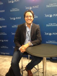 Migliorini Denis合作论文数Centre of Oncology, Geneva University Hospitals &University of Geneva;Geneva University Hospitals, University of Geneva49
 Pierre-Yves Dietrich合作论文数47
 Walker Paul合作论文数Translational Research Centre in Oncohaematology, Faculté De Médecine, University of Geneva;Division of Oncology, University of Geneva36
 Danila Valmori合作论文数31Eliana Marinari合作论文数26Jean-Charles Cerottini合作论文数Department of Immunology, Swiss Institute for Experimental Cancer Research25Philippe Guillaume合作论文数23Verena Rubio-Godoy合作论文数18Donata Rimoldi合作论文数Ludwig Center for Cancer Research of the UNIL, University of Lausanne17Danielle Lienard合作论文数Ludwig Institute for Cancer Research, Centre Hospitalier Universitaire Vaudois17P Romero合作论文数14Schaller Karl合作论文数Dept Neurosurg, Univ Hosp Geneva14
 Pedro Romero合作论文数Département d'Oncologie, Centre Hospitalier Universitaire Vaudois, Université de Lausanne13Darel Martinez Bedoya合作论文数12
 Daniel Speiser合作论文数Department of Fundamental Oncology, Faculty of Biology and Medicine, Université de Lausanne12Shahan Momjian合作论文数12Py Dietrich合作论文数10Philippe Hammel合作论文数9Lana E. Kandalaft合作论文数9
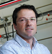 Nicolas Winssinger合作论文数Department of Organic Chemistry, University of Geneva9
 Philippe Bijlenga合作论文数University of Geneva Medical Center, University of Geneva9Sofia Barluenga合作论文数9
 Pierre Cosson合作论文数Centre Médical Universitaire, Département De Physiologie Cellulaire Et Métabolisme, Université De Genève9
 Mikael Pittet合作论文数Department of Pathology and Immunology, Faculty of Medicine, University of Geneva8Cosset Érika合作论文数8Thibaud Koessler合作论文数8Norbert Hilf合作论文数8
 Hans-Georg Rammensee合作论文数Interfaculty Institute for Cell Biology, University of Tübingen;Department of Immunology, University of Tübingen7Valérie Widmer合作论文数7Ayyoub Maha合作论文数6
 Hideho Okada合作论文数School of Medicine, University of California, San Francisco6
 Ugur Sahin合作论文数Institut für Immunologie, Universitätsmedizin der Johannes Gutenberg-Universität Mainz;University Medical Center Mainz;Biontech Se6
 Petros Tsantoulis合作论文数6Thomas A. Mckee合作论文数Geneva University Hospitals;Department of Pathology and Immunology, Faculté de Médecine, University of Geneva6
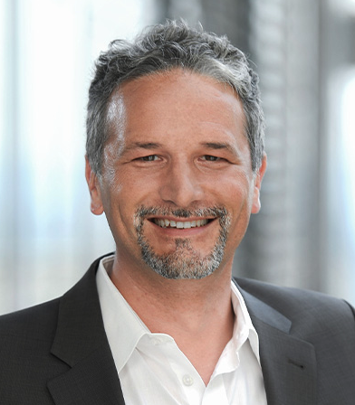 Toni Weinschenk合作论文数Immatics Biotechnologies GmbH6Jordi Rodon合作论文数5berta ponsati obiols合作论文数5Katrin Frenzel合作论文数5Colette Song合作论文数5
 Ulrik Niels Lassen合作论文数Department of Clinical Medicine, Faculty of Health and Medical Sciences, University of Copenhagen;Department of Oncology, Rigshospitalet5F Lejeune合作论文数5
 Tabatabai Ghazaleh合作论文数Universitätsklinikum und Medizinische Fakultät Tübingen;Department of Neurology & Interdisciplinary Neuro-Oncology, University Hospital Tübingen5
 Andreas Von Deimling合作论文数University of Heidelberg;German Cancer Research Center5
 Richard A. Houghten合作论文数Torrey Pines Institute for Molecular Studies5
 Sjoerd van der Burg合作论文数Department of Clinical Oncology, Leiden University Medical Center;Oncode Institute5
 Wolfgang Wick合作论文数Department Kopfklinik, Universitätsklinikum Heidelberg;Universität Heidelberg5C. Pinilla合作论文数5
 Doron Merkler合作论文数Institut für Neuropathologie, Universitätsklinik Göttingen5
 Per Thor Straten合作论文数Department of Immunology and Microbiology, Faculty of Health and Medical Sciences, University of Copenhagen5




