Chronic Insomnia Disorder (ID) is characterized by hyperarousal, a key pathophysiological feature. While Cognitive-Behavioral Therapy for Insomnia (CBT-I) is the first-line treatment, its physiological effects on sleep-related hyperarousal remain underexplored. This study assessed the impact of CBT-I on cortical hyperarousal using quantitative EEG (qEEG) during non-REM (NREM) sleep, with the delta/beta ratio as the primary outcome. Secondary aims included evaluating changes in sleep stability and exploring phenotypic differences in treatment response. Ninety-eight ID patients across five centers completed a 6-8-week CBT-I program. Pre-and post-treatment assessments included polysomnography (PSG), sleep diaries, and Insomnia Severity Index (ISI). Cortical hyperarousal was indexed by the NREM delta/beta ratio; sleep stability (Sstab) was derived from a transition probability matrix. Patients were categorized as insomnia with short (ISSD) or normal sleep duration (INSD) based on PSG-derived total sleep time (median TST = 347.3 min). CBT-I significantly improved ISI and sleep parameters (sleep onset latency, wake after sleep onset, time in bed, sleep efficiency) in both self-reported and PSG, with smaller effects in the latter. qEEG analyses revealed a significant increase in the delta/beta ratio post-CBT-I (baseline:13.4 ± 4.9, end-of-treatment:14.6 ± 5.9; p = 0.002), indicating reduced cortical hyperarousal, with no center effects. Sstab improved significantly (p = 0.005), though it was not correlated with delta/beta changes. ISSD showed greater delta/beta improvements than INSD (p = 0.014), suggesting phenotypic differences. CBT-I reduces cortical hyperarousal in ID, as reflected by increased delta/beta ratio. The dissociation from sleep stability suggests distinct mechanisms. These findings support qEEG biomarkers as valuable tools for understanding the neurophysiological mechanisms of insomnia treatment and guiding precision medicine approaches.
OBJECTIVE:This study investigated the electrophysiological features of the alpha band and Individual Alpha Frequency (IAF) before the Sleep Onset (SO) period in Insomnia Disorder patients (ID) and Healthy Controls (HCs). We hypothesized that IAF, as an individualized electrophysiological marker rather than broadband alpha power, could better capture individual sleep-initiation alterations characterizing ID and Sleep State Misperception (SSM). METHODS:Polysomnographic recordings were analyzed to assess changes in alpha activity and IAF Before-SO in occipital regions. Correlational analyses were conducted to investigate associations between the alpha activity, IAF, sleep parameters, and SSM. RESULTS:Compared to HC, ID patients showed higher occipital IAF power during the Before-SO, while no group differences emerged in the broadband alpha activity. Correlational analyses revealed that faster IAF was associated with longer sleep latency and delayed transition to deep sleep, whereas higher IAF power was related to greater insomnia severity and stronger SSM. CONCLUSIONS:These findings suggest that IAF during the period preceding the SO is a sensitive marker of cortical arousal, contributing to impaired sleep initiation and distorted sleep perception. SIGNIFICANCE:These findings underscore the potential role of the IAF as an electrophysiological marker of ID pathophysiology and a target for personalized therapeutic interventions.
Mood states may impact the response to Cognitive-Behavioral Therapy for Insomnia (CBT-I), the first-line treatment for chronic insomnia. Indeed, literature reported a bi-directional relationship between depressive and insomnia disorder (ID) symptoms. The aim of this study was to evaluate the psychometric properties of the Italian version of the Profile of Mood States (POMS) scale and to investigate the role of affective states in a large sample of patients with ID following CBT-I. 448 insomnia patients (59
Background/Objectives: Sleep state misperception (SSM) is a common phenomenon in insomnia disorder (ID), characterized by a discrepancy between subjective and objective sleep metrics. Recent studies have revealed microstructural EEG alterations specifically in misperceiving ID patients, yet clinically accessible SSM markers remain limited. This study aimed to characterize SSM within ID by integrating standard polysomnography (PSG) features and cognitive-affective traits, focusing on accessible clinical tools. Methods: Twenty patients with ID and twenty healthy controls (HC) underwent a night of PSG recording and completed both sleep diaries and a comprehensive psychological assessment. SSM was quantified using the Total Sleep Time misperception index (TSTm), analyzed both dimensionally and categorically Results: IDs reported significantly altered sleep parameters compared to HCs, both subjectively and objectively. Within the ID sample, although underestimators and normoestimators had similar objective TST, underestimators showed significantly more cortical arousal density (CAd), a higher percentage of sleep stage 1 and higher non-acceptance of emotions. Notably, none of the HC reached the threshold for being classified as underestimators. Regression analyses identified CAd, latency to sleep stage 3 and to REM, percentage of REM sleep and lack of emotional clarity, as key predictors of TSTm. Conclusions: SSM in insomnia reflects a dimensional vulnerability grounded in subtle sleep fragmentation and emotional dysregulation. Recognizing SSM as a clinically meaningful phenomenon may guide more targeted, emotion-focused, interventions for insomnia.
Major depressive disorder (MDD) and Insomnia disorder (ID) are characterized by sleep alterations. To define their polysomnographic profiles, we conducted a Network Meta-Analysis comparing MDD and ID patients versus healthy controls (HCs). The literature search, conducted from 2008 up to January 2023 and following PRISMA guidelines, covered PubMed, Web of Science, Scopus, and Embase databases. We addressed publication bias using funnel plot asymmetry inspection and Egger’s test, evaluated statistical heterogeneity with I2, and local and global inconsistencies with the separate indirect from direct evidence method and Q between designs, respectively. Pairwise meta-analyses employed a fixed-effects model, while network analysis utilized a random-effect approach. We evaluated 86 ID and 17 MDD studies, comparing sleep parameters for 636 MDDs versus 491 HCs, and 3661 IDs versus 2792 HCs. The network meta-analysis reported that patients with MDD have greater rapid eye movement (REM) sleep duration and REMs density, and lower REM sleep latency compared to IDs. ID patients instead exhibited lower total sleep time and time in bed, and greater wake after sleep onset and non-REM sleep stage 3 than MDD patients. This work emphasized sleep depth and continuity alterations in both MDD and ID, with major involvement of REM sleep in MDD.
Insomnia disorder (ID) is characterized by electroencephalographic indexes of hyperarousal, often associated with the underestimation of sleep duration (i.e. sleep state misperception). Albeit non-rapid eye movement sleep K-complexes (KCs) are involved in sleep protection and arousal, only a few studies investigated their alterations in ID with heterogenous findings, and results about their possible relationship with sleep state misperception are missing. The study aims to assess KCs in ID and their relationship with sleep state misperception, also considering their correlation with sleep architecture (i.e. the large-scale organization of sleep). Nineteen ID patients (12 F; age: 42.4 +/- 12.1 years) and 18 healthy controls (HC; 10 F; age: 41.6 +/- 11.9 years) underwent a night of home polysomnography and completed sleep diaries upon awakening. KC density, amplitude, and area under the curve were assessed in midline frontal, central, and parietal derivations. Sleep state misperception was investigated by considering polysomnographic and subjective total sleep time (TST). We found reduced anterior KC morphology (i.e. amplitude and area under the curve) in ID patients compared to HCs, which was associated with TST underestimation. KC morphology was negatively associated with N3 latency, sleep fragmentation and arousal indexes, and positively related with N3 percentage and sleep efficiency. Our findings suggest an impaired sleep protection mechanism expressed by altered KCs morphology in ID involved in sleep state misperception. The observed correlations support the view of KC as the forerunner of slow-wave sleep and protector of sleep continuity. A better understanding of sleep-protecting mechanisms alteration as a predisposing and/or maintaining factor of ID is needed.
IntroductionObstructive sleep apnea (OSA) is the most common breathing-related sleep disorder with a considerable economic burden, low diagnosis and treatment rates. Continuous positive airway pressure (CPAP/PAP) is the principal therapy for OSA treatment; nevertheless, effectiveness is often limited by suboptimal adherence.The present network meta-analysis aims to systematically summarize and quantify different interventions' effects on CPAP/PAP adherence (such as mean usage CPAP or PAP in hours per night) in OSA patients, comparing Behavioral, Educational, Supportive and Mixed interventions in Randomized Control Trials (RCT).MethodsWe conducted a computer-based search using the electronic databases of Pubmed, Psycinfo, Scopus, Embase, Chinal and Medline until August 2022, selecting 50 RCT.ResultsBy means of a random effect model network meta-analysis, results suggested that the most effective treatment in improving CPAP/PAP adherence was the Supportive approach followed by Behavioral Therapy focused on OSA treatment adherence.ConclusionThis network meta-analysis might encourage the most experienced clinicians and researchers in the field to collaborate and implement treatments for improving CPAP/PAP treatment adherence. Moreover, these results support the importance of multidisciplinary approaches for OSA treatment, which should be framed within a biopsychological model.
Investigating the mechanisms of action of cognitive-behavioural therapy for insomnia (CBT-I), the first-line treatment for chronic insomnia disorder (ID), can contribute to the overall understanding of insomnia and its treatment. To date, no study has examined the relationship between K-complexes (KC) and CBT-I, despite the known homeostatic and protective function of this relevant sleep brainwave. This retrospective multicentre study aims to explore the relationship between electroencephalographic (EEG) indices and CBT-I, with a particular focus on evaluating an index of sleep homeostasis identified by KC. This research is designed to assess the predictive value of this index for treatment outcomes and to examine its variations before and after intervention. Ninety eight patients with ID underwent a 6-8 week in-person CBT-I programme, with pre-and post-treatment evaluation conducted using polysomnography (PSG) and the Insomnia Severity Index (ISI). The main outcome was determined by calculating the slope of the linear equation indexing the KC density (number of KC/minutes of N2) in each non-artifacted NREM stage 2 epoch throughout the night (KCSlope). Furthermore, the sample was categorised into Responders (ISIdecrease ≥8) and non-Responders (ISIdecrease <8). The results indicate that the KC Slope is effective not only to predict treatment response (one-way ANOVA, F = 7.831 p = 0.007; Responders = -2.954*10-5 ± 3.346*10-5, non-Responders = -5.583*10-5 ± 5.305*10-5; adjusted for PSG wake after sleep onset at the baseline), but also to detect a statistically significant improvement in sleep pressure following CBT-I (Wilcoxon signed-rank test W = 3074.000 p = 0.022; KCSlope pre-treatment = -4.054*10-5 ± 4.446*10-5, KCSlope post-treatment = -4.797*10-5 ± 5.710*10-5). These findings suggest that CBT-I increases sleep pressure in patients with chronic insomnia, highlighting a novel and relevant biomarker in this context.
Purpose Sleep State Misperception (SSM) is described as the tendency of Insomnia Disorder (ID) patients to overestimate Sleep Latency (SL) and underestimate Total Sleep Time (TST). Literature exploring topographical components in ID with SSM is scarce and does not allow us to fully understand the potential mechanisms underlying this phenomenon. This study aims to evaluate the existence of sleep EEG topography alterations in ID patients associated with SSM compared to Healthy Controls (HC), focusing on two distinct periods: the Sleep Onset (SO) and the whole night. Methods Twenty ID patients (mean age: 43.5 ± 12.7; 7 M/13F) and 18 HCs (mean age: 41.6 ± 11.9; 8 M/10F) underwent a night of Polysomnography (PSG) and completed sleep diaries the following morning upon awakening. Two SSM indices, referring to the misperception of SL (SLm) and TST (TSTm), were calculated by comparing objective and subjective sleep indices extracted by PSG and sleep diary. According to these indices, the entire sample was split into 4 sub-groups: ID +SLm vs HC –SLm; ID +TSTm vs HC –TSTm. Results Considering the SO, the two-way mixed-design ANOVA showed a significant main effect of Groups pointing to a decreased delta/beta ratio in the whole scalp topography. Moreover, we found a significant interaction effect for the sigma and beta bands. Post Hoc tests showed higher sigma and beta power in anterior and temporo-parietal sites during the SO period in IDs +SLm compared to HC –SLm.Considering the whole night, the unpaired t-test revealed in IDs +TSTm significantly lower delta power during NREM, and lower delta/beta ratio index during NREM and REM sleep compared to HCs –TSTm.Finally, we found diffuse significant negative correlations between SSM indices and the delta/beta ratio during SO, NREM, and REM sleep. Conclusion The main finding of the present study suggests that higher SL overestimation and TST underestimation are both phenomena related to diffuse cortical hyperarousal interpreted as a sleep state-independent electrophysiological correlate of the SSM, both during the SO and the whole night.
Objective: In this Italian population-based study, we aimed to evaluate the neurological complications after the first and/or second dose of COVID-19 vaccines and factors potentially associated with these adverse effects. Methods: Our study included adults aged 18 years and older who received two vaccine doses in the vaccination hub of Novegro (Milan, Lombardy) between 7 and 16 July 2021. The NEURO-COVAX questionnaire was able to capture the neurological events, onset and duration. That data that were digitized centrally by the Lombardy region were used to match the demographic/clinical characteristics and identify a vulnerability profile. Associations between vaccine lines and the development of complications were assessed. Digital healthcare system matching was also performed to evaluate severe neurological complications (Guillain-Barrè syndrome, Bell’s palsy, transverse myelitis, encephalitis) and the incidence of hospital admissions and/or the mortality rate after two doses of the vaccines. Results: The NEURO-COVAX-cohort included 19.108 vaccinated people: 15.368 with BNT162b2, 2077 with mRNA-1273, 1651 with ChAdOx1nCov-19, and 12 with Ad26.COV2.S who were subsequently excluded. Approximately 31.2% of our sample developed post-vaccination neurological complications, particularly with ChAdOx1nCov-19. A vulnerable clinical profile emerged, where over 40% of the symptomatic people showed comorbidities in their clinical histories. Defining the neurological risk profile, we found an increased risk for ChAdOx1nCov-19 of tremors (vs. BNT162b2, OR: 5.12, 95% CI: 3.51–7.48); insomnia (vs. mRNA-1273, OR: 1.87, 95% CI: 1.02–3.39); muscle spasms (vs. BNT162b2, OR: 1.62, 95% CI: 1.08–2.46); and headaches (vs. BNT162b2, OR: 1.49, 95% CI: 0.96–1.57). For mRNA-1273, there were increased risks of parethesia (vs. ChAdOx1nCov-19, OR: 2.37, 95% CI: 1.48–3.79); vertigo (vs. ChAdOx1nCov-19, OR: 1.68, 95% CI: 1.20–2.35); diplopia (vs. ChAdOx1nCov-19, OR: 1.55, 95% CI: 0.67–3.57); and sleepiness (vs. ChAdOx1nCov-19, OR: 1.28, 95% CI: 0.98–1.67). In the period that ranged from March to August 2021, no one was hospitalized and/or died of severe complications related to COVID-19 vaccinations. Discussion: This study estimates the prevalence and risk for neurological complications potentially associated with COVID-19 vaccines, thus improving the vaccination guidelines and loading in future personalized preventive medicine.
 Luigi Ferini Strambi合作论文数Universita Vita-Salute San Raffaele70
 Marco Zucconi合作论文数68Ferini L. Strambi合作论文数43Sara Marelli合作论文数41Andrea Galbiati合作论文数33ME SOBEL合作论文数31Akeila Bellahcene合作论文数31Alessandro Oldani合作论文数31
 Marco Sforza合作论文数26M. Manconi合作论文数25
 Francesca Casoni合作论文数Neurology-Sleep Disorders Center, IRCCS San Raffaele Scientific Institute23L Ferini-Strambi合作论文数20
 Galbiati Andrea合作论文数Sleep Disorders Center, San Raffaele Scientific Institute;Faculty of Psychology, Vita-Salute San Raffaele University;San Raffaele Scientific Institute, Vita-Salute San Raffaele University19
 Cappa Stefano Francesco合作论文数IUSS Scuola Universitaria Superiore Pavia19
 David Waltregny合作论文数Département Des Sciences Cliniques, Faculté de Médecine, Université de Liège;Department of Urology, University Hospital of Liège;Clinique Sainte Thérèse16M Sforza合作论文数15
 Elisabetta Fasiello合作论文数Department of Psychology, Sapienza - University of Rome14Sylvie Ménard合作论文数14J.-M. Foidart合作论文数Estetra SRL, Mithra Pharmaceuticals14
 Mauro Manconi合作论文数13
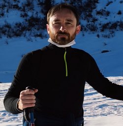 Nicola Canessa合作论文数San Raffaele Scientific Institute13Laura Giarolli合作论文数13
 Fu-Tong Liu (劉扶東)合作论文数Institute of Biomedical Sciences, Academia Sinica;Department of Dermatology, Keck School of Medicine, University of Southern California;Department of Dermatology, School of Medicine, University of California, Davis12Nathalie Clausse合作论文数11M. Fantini合作论文数10
 Caterina Leitner合作论文数“Vita-Salute, San Raffaele University10Marc Mareel合作论文数10
 Luigi De Gennaro合作论文数Department of Psychology, University of Rome “La Sapienza”,10Cedric Detry合作论文数10
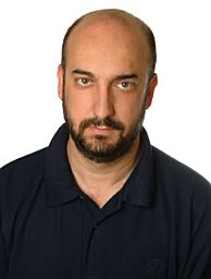 Pedro L. Fernandez合作论文数Department of Pathology Hospital Clinic C/Villarroel10Raffaele Ferri合作论文数9A Falini合作论文数8T. Kuo合作论文数8Dorina Belotti合作论文数8
 Gorgoni Maurizio合作论文数Department of Psychology, "Sapienza"University of Rome;Department of Psychology, "Sapienza" University of Rome8Chaplet M合作论文数8René Lambotte合作论文数8
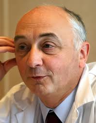 Jean-Michel Foidart合作论文数Département Des Sciences Cliniques, Faculté de Médecine, Université de Liège8
 Lance Liotta合作论文数Center for Applied Proteomics and Molecular Medicine, College of Science, George Mason University;School of Systems Biology, College of Science, George Mason University8Mark S. Aloia合作论文数8
 Elías Campo合作论文数Hospital Clínic de Barcelona, IDIBAPS, University of Barcelona;Facultad de Medicina, Universidad de Barcelona;Institut d'Investigacions Biomèdiques August Pi i Sunyer8Andrea Salibba合作论文数7M. Aloia合作论文数7Marc Bracke合作论文数7F van den Brûle合作论文数7
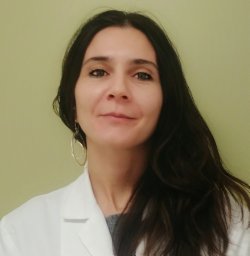 Anna Castelnovo合作论文数Department of Health Sciences, Università degli Studi di Milano Milan;Department of Psychiatry, University of Wisconsin-Madison Madison;Università degli Studi di Milano Milan, University of Wisconsin-Madison Madison7
 Alexandros N. Vgontzas合作论文数Sleep Research and Treatment Center, Department of Psychiatry, Penn State College of Medicine;University of Crete7Pascale Jackers合作论文数7
 Alain Chariot合作论文数Centre Hospitalier Universitaire de Liège, University of Liège7




