INTRODUCTION:Early diagnosis of premalignant gastric condition, such as chronic atrophic gastritis and intestinal metaplasia, allow us to assess and stratify the risk of gastric cancer. Gastric mucosal cleaning, mucosal exploration, photo-documentation, and advanced technologies like optical enhancement, electron chromoendoscopy and magnification endoscopy improve lesion detection rates. OBJECTIVE:This study compares the detection rate of premalignant gastric lesions between biopsies guided Electron chromoendoscopy combined with optical enhancement and magnification endoscopy versus random biopsies (Sydney Protocol) with optical enhancement. METHODS:This is a prospective, observational, blinded comparative study. Outpatients were divided into two groups: group 1 (162 patients), biopsies were performed either under Electron chromoendoscopy combined magnification endoscopy guidance; and group 2 (160 patients), biopsies were performed under blinded mapping (updated Sydney system) with optical enhancement. The following clinical outcomes were assessed in each group: age, gender, adequate gastric clearance rate, gastric atrophy rate, intestinal metaplasia rate. RESULTS:A total of 322 patients were randomized, where 76 were excluded from the study. Detection rate for Intestinal metaplasia was significantly higher with Electron chromoendoscopy combined with optical enhancement and magnification endoscopy (37.6% versus 23.7%; p < 0.0001). Extensive Intestinal metaplasia was significantly more prevalent with Electron chromoendoscopy combined magnification endoscopy compared to mapping with optical enhancement (p = 0.029; p = 0.048, respectively). CONCLUSION:Electron chromoendoscopy combined with optical enhancement and magnification endoscopy demonstrated a comparative superiority over Sydney System random biopsy protocol in the identification of patients with premalignant gastric conditions. By enabling more precise endoscopic targeting, directed biopsies help reduce sampling error; however, in high-risk populations, a complementary approach integrating both targeted and systematic random biopsies remains advisable to optimize diagnostic accuracy.
Antecedentes: la esofagitis inducida por fármacos es una entidad clínica subdiagnosticada debido al inadecuado abordaje al que suelen ser sometidos estos pacientes, y la elaboración de una historia clínica exhaustiva es un elemento fundamental en el algoritmo diagnóstico de esta patología. Es importante identificar a estos pacientes para un correcto manejo y una adecuada orientación con el fin de prevenir nuevos eventos similares que puedan afectar significativamente su calidad de vida. El apoyo de la endoscopia digestiva alta y el análisis histopatológico son necesarios para aclarar el diagnóstico y excluir otras entidades clínicas similares. El infiltrado eosinofílico severo evidenciado en algunos casos de esofagitis inducida por fármacos es un hallazgo histológico poco frecuente que a menudo se confunde con otras entidades, como la esofagitis eosinofílica. Caso clínico: presentamos el caso de un paciente joven que desarrolló disfagia severa después de la exposición a antiinflamatorios no esteroideos (AINES). La endoscopia digestiva alta mostró un marcado componente inflamatorio asociado a un infiltrado eosinofílico severo en la evaluación histopatológica. Estos síntomas remitieron luego de la suspensión de los AINES y el inicio de inhibidores de la bomba de protones sin recurrencias a mediano-largo plazo. Conclusiones: la esofagitis inducida por fármacos es una entidad clínica que debe ser considerada como una de las etiologías generadoras de un infiltrado eosinofílico severo, y que incluso podría producir un perfil endoscópico-histológico similar al tradicionalmente conocido en otras patologías, como la esofagitis eosinofílica.
BACKGROUND AND AIMS:Zenker's diverticulum (ZD) is the most common type of esophageal diverticulum. We conducted a systematic review and meta-analysis to compare the effectiveness and safety of endoscopic submucosal tunneling techniques (ESTTs) and flexible endoscopic septotomy (FES) for treating patients with ZD, including subgroup analyses by follow-up duration (<12 months and ≥12 months), diverticulum size (<2.5 cm and ≥2.5 cm), ESTT used (Zenker's peroral endoscopic myotomy and peroral endoscopic septotomy), and publication format (full text and abstract). METHODS:We searched PubMed, EMBASE, and Cochrane Library databases until June 20, 2024. Mean differences (MDs) and risk ratios (RRs) with 95% confidence intervals (CIs) were pooled for continuous and binary outcomes, respectively. Heterogeneity was assessed with the I2 statistics. RESULTS:We included 9 studies (759 patients). Compared with FES, ESTT had a higher probability of clinical success (RR, 1.15; 95% CI, 1.04-1.28). The results were similar in both groups for clinical recurrence (RR, .56; 95% CI, .29-1.07), technical success (RR, .99; 95% CI, .97-1.01), operative time (MD, 7.22 minutes; 95% CI, -.33 to 14.76), hospital stay (MD, .47 days; 95% CI, -1.25 to 2.19), and overall adverse events (RR, 1.19; 95% CI, .44-3.18). Subgroup analyses showed consistent results. CONCLUSIONS:ESTT demonstrated a higher probability of clinical success and showed a trend toward a lower recurrence rate compared with FES; however, both groups had similar technical success, operative time, length of hospital stay, and overall adverse events. These findings underscore ESTT as an effective and safe method for treating patients with ZD.
Esophageal squamous cell carcinoma (ESCC) remains a significant global health challenge, being the sixth leading cause of cancer mortality with pronounced geographic variability. The incidence rates range from 125 per 100,000 in northern China to 1-1.5 per 100,000 in the United States, driven by environmental and lifestyle factors such as tobacco and alcohol use, dietary habits, and pollution. Major modifiable risk factors include tobacco and alcohol consumption, with a synergistic risk increase when combined. Nonmodifiable risk factors include previous diagnoses of head and neck squamous cell carcinoma (H&N SCC), achalasia, and prior radiotherapy. Prevention strategies must be tailored to specific regional burdens to efficiently allocate medical and financial resources. Gastrointestinal endoscopy is crucial in reducing ESCC burden through early detection and characterization of neoplastic changes, such as high-grade dysplasia. Early diagnosis significantly improves survival rates, while endoscopic resection of noninvasive dysplasia can prevent ESCC onset, reducing treatment burden for advanced disease. Postresection surveillance can detect high-risk metachronous lesions. Despite these benefits, endoscopic prevention faces challenges, including the lack of high-level evidence supporting its efficacy, opportunity costs, the need for specialized training and techniques, and the requirement for advanced technology investments. This Position Statement from the World Endoscopy Organization (WEO) aims to address these challenges, supplying recommendations for the exploitation of endoscopic resources regarding the possible role of screening, quality, and training for the detection, characterization, resection, and surveillance of ESCC.
Aims To develop and validate a new scale for mucosal visualization of the upper gastrointestinal tract during esophagogastroduodenoscopy (EGD), the Gastroscopy RAte of Cleanliness Evaluation (GRACE), as a quality standard tool through the application of a standardized, reliable, and validated scoring system.
ABSTRACT Background: In the past decades, endoscopic ultrasound has developed from a diagnostic tool to a platform for many therapeutic interventions. Various technological advancements have emerged since the last Brazilian Consensus, demanding a review and update of the recommendations based on the best scientific evidence. Methods: A group of 32 renowned echoendoscopists selected eight relevant topics to be discussed to generate clinical questions. After that, a literature review was conducted to answer these questions based on the most updated evidence. Results: Thirty-three statements were formulated and voted on by the experts to reach a consensus. The Oxford System was used to grade the level of evidence. Conclusion: There is moderate evidence to support that the needle shape, gauge, or aspiration technique does not influence the yield of endoscopic ultrasound (EUS)-guided tissue sampling of pancreatic solid lesions. There is moderate evidence to support using EUS-TTNB of the cyst wall to differentiate between mucinous and non-mucinous cystic neoplasms. There is little evidence to support the EUS-guided treatment of gastric varices. There is a high level of evidence to support that EUS-guided biliary drainage and ERCP present similar outcomes in patients with distal malignant biliary obstruction. There is a high level of evidence for using EUS to diagnose neoplastic pancreatic cysts and detect necrosis before indicating drainage. There is moderate evidence to support EUS-GE over duodenal stent for malignant gastric outlet obstruction in patients with a life expectancy higher than 2 months. There is a high level of evidence to support the use of RFA in treating both functioning and non-functioning types of NET.
Background The use of antifoaming and mucolytic agents prior to upper gastrointestinal (GI) endoscopy and a thorough systematic review are essential to optimize lesion detection. This study evaluated the effect of simethicone and N-acetylcysteine on the adequate mucosal visibility (AMV) of the upper GI tract by an innovative systematic method. Methods This randomized, double -blind controlled trial included consecutive patients who underwent diagnostic upper GI endoscopy for screening for early neoplasms between August 2019 and December 2019. The upper GI tract was systematically assessed by systematic alphanumericcoded endoscopy. Patients were divided into 4 groups: 1) water; 2) only simethicone; 3) N-acetylcysteine + simethicone; and 4) only N-acetylcysteine. The following parameters were assessed in each group: age, sex, body mass index, level of adequate mucosal visibility, and sideeffects. Results A total of 4564 images from upper GI areas were obtained for evaluation. The mean AMV in the 4 groups was 93.98 +/- 7.36%. The N-acetylcysteine + simethicone group had a higher cleaning percentage compared with the other groups (P=0.001). There was no significant difference among the remaining groups, but several areas had better cleaning when a mucolytic or antifoam alone was used. No side-effects were found in any group. Conclusion The combination of N-acetylcysteine plus simethicone optimizes the visibility of the mucosa of the upper GI tract, which could potentially increase diagnostic yield.
Background/Aims Polyethylene glycol (PEG) is considered the gold standard regimen for bowel preparation; however, due to the necessity of a large volume, patient tolerance is impaired. Therefore, lactulose is a novel alternative for colonoscopy preparation. This study aimed to investigate the efficacy and safety of lactulose-based bowel preparations in comparison with PEG for colonoscopy. Methods This is a prospective, non-blinded, comparative study. Outpatients were randomly divided into two groups: group 1 (111 patients), PEG; and group 2 (111 patients), lactulose. The following clinical outcomes were assessed in each group: degree of bowel clearance using the Boston bowel preparation score, colorectal polyp detection rate, adenoma detection rate, tolerability, and side effects. Results The rate of inadequate bowel preparation was 8.1% and 1.8% for the PEG and lactulose groups, respectively (p=0.030). The Boston bowel preparation score for the entire colon was 7.34±1.17 and 8.36±1.09 for the PEG and lactulose groups, respectively (p<0.001). The satisfactory overall experience rates were 27.9% and 62.2% for the PEG and lactulose groups, respectively (p<0.001). Conclusions The novel bowel preparation with oral lactulose was superior to that with PEG in terms of colon cleansing, adenoma detection rate, tolerance, and patient experience.
Background:The standard of practice when a superficial lesion was identified during upper GI endoscopy is to take an endoscopic forceps biopsy (EFB) of the lesion. The histopathologic findings then will determine the management plan. Endoscopic submucosal dissection (ESD) enables en-bloc resection for early neoplasms of the gastrointestinal tract and provides an adequate specimen that permits a more reliable histopathologic assessment. The objective of this study was to determine the rate of histopathologic discrepancy between EFB and specimens resected by ESD, and to identify the predisposing risk factors for this discordance. Materials and Methods:This is a retrospective study, enrolling patients with superficial gastric neoplasms that underwent EFB followed by ESD. We divided cases to concordant or discordant group according to the histopathologic diagnosis of EFB and ESD specimens. We also analyzed the features that may have influenced the occurrence of histopathologic discordance and the association between discordant samples of adenocarcinoma and neoplastic invasion to deeper layers. Results:A total of 115 gastric ESD procedures were performed with 84 patients meeting the inclusion criteria. Histopathologic discordance between EFB and ESD specimens were observed in 35.8% of cases (30/84 lesions). The univariant-bivariant analysis and multivariate logistic regression analysis showed that histologic discordance was closely related to the size of the lesions (P=0.028). Conclusion:Histopathologic discrepancy between EFB and ESD specimens may occur in approximately one-third of cases, particularly for lesions over 20 mm, which may lead to crucial delays in gastric cancer precise diagnosis and treatment.
In light-emitting diode (LED) and LASER colonoscopy, linked color imaging (LCI) and blue light/laser imaging (BLI) are used for lesion detection and characterization worldwide. We analyzed the difference of LCI and BLI images of colorectal lesions between LED and LASER in a multinational study. We prospectively observed lesions with white light imaging (WLI), LCI, and BLI using both LED and LASER colonoscopies from January 2020 to August 2021. Images were graded by 27 endoscopists from nine countries using the polyp visibility score: 4 (excellent), 3 (good), 2 (fair), and 1 (poor) and the comparison score (LED better/similar/LASER better) for WLI/LCI/BLI images of each lesion. Finally, 32 lesions (polyp size: 20.0 ± 15.2 mm) including 9 serrated lesions, 13 adenomas, and 10 T1 cancers were evaluated. The polyp visibility scores of LCI/WLI for international and Japan-expert endoscopists were 3.17 ± 0.73/3.17 ± 0.79 (p = 0.92) and 3.34 ± 0.78/2.84 ± 1.22 (p < 0.01) for LED and 3.30 ± 0.71/3.12 ± 0.77 (p < 0.01) and 3.31 ± 0.82/2.78 ± 1.23 (p < 0.01) for LASER. Regarding the comparison of lesion visibility about between LED and LASER colonoscopy in international endoscopists, a significant difference was achieved not for WLI, but for LCI. The rates of LED better/similar/LASER better for brightness under WLI were 54.5
Cayetano Heredia University, Peru; Alfa Institute of Gastroenterology, Minas gerais, Brazil.
•This study aimed to assess the learning curve effect on patient's clinical outcome for EESD. Retrospective observational study, enrolling patients that underwent EESD from 2009 to 2021, divided in 2 groups. Mean procedure time was 111.8 min and 103.6 min for T1 and T2, respectively (P=0.004). The learning curve in esophageal ESD could be overcomed effectively and safely by an adequately trained Western endoscopist. Background - Esophageal endoscopic submucosal dissection (EESD) is a complex and time-consuming procedure at which training are mainly available in Japan. There is a paucity of data concerning the learning curve to master EESD by Western endoscopists. Objective - This study aimed to assess the learning curve effect on patient's clinical outcome for EESD. Methods - This is a retrospective observational study. Enrolling patients that underwent EESD from 2009 to 2021. The analysis was divided into two periods; T1: case 1 to 49 and T2: case 50 to 98. The following features were analyzed for each group: patients and tumors characteristics, en-bloc, complete and curative resection rates, procedure duration and adverse events rate. Results - Ninety-eight EESD procedures were performed. Mean procedure time was 111.8 min and 103.6 min for T1 and T2, respectively (P=0.004). En bloc resection rate was 93.8% and 97.9% for T1 and T2, respectively (P=0.307). Complete resection rate was 79.5% and 85.7% for T1 and T2, respectively (P=0.424). Curative resection rate was 65.3% and 71.4% for T1 and T2, respectively (P=0.258). Four patients had complications; three during T1 period and one during T2 period. Overall mortality rate: 0%. Conclusion - The esophageal endoscopic submucosal dissection could be performed effectively and safely by an adequately trained Western endoscopist.
Universidade Federal de Minas Gerais, Brazil; Universidad Peruana Cayetano Heredia, Peru.
 Josue Aliaga Ramos合作论文数21
 Walton Albuquerque合作论文数18Takashi Toyonaga合作论文数14
 Fauze Maluf-Filho合作论文数Endoscopic Unit of Hospital das Clínicas, São Paulo University Medical School12elias alfonso forero pineros合作论文数11
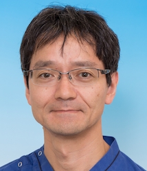 Noriya Uedo合作论文数Department of Gastrointestinal Oncology, Osaka Medical Center for Cancer and Cardiovascular Diseases8Fernanda Macedo合作论文数7Nicoly Dias合作论文数7
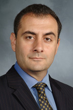 Michel Kahaleh合作论文数Weill Cornell Medical College7
 Everson Luiz de Almeida Artifon合作论文数Centro de Treinamento em Procedimentos Minimamente Invasivos, Faculdade de Medicina, Universidade de São Paulo;Departamento de Cirurgia, Faculdade de Medicina, Universidade de São Paulo;Departamento de Cirurgia e Anatomia, Faculdade de Medicina, Universidade de São Paulo7
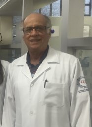 Luiz Gonzaga Vaz Coelho合作论文数Alfa Institute of Gastroenterology, Federal University of Minas Gerais7Luiz Caputo合作论文数6Fernando Carvalho合作论文数6Gabriel Garcia合作论文数6Tyberg Amy合作论文数6
 Fabian Emura合作论文数6Carlos Alberto Freitas Dias合作论文数6Moises Salgado Pedrosa合作论文数6
 Monica Gaidhane合作论文数Digestive Health, University of Virginia6Roberto Guimarães合作论文数5W Albuquerque合作论文数5
 Rafiz Abdul Rani合作论文数5
 Luiz Ronaldo Alberti合作论文数Universidade Federal de Minas Gerais5
 Paulo Sakai合作论文数University of Sao Paulo5Dalton M. Chaves合作论文数Stony Brook University, SUNY-Downstate University Medical Center5
 Adolfo Parra-Blanco合作论文数Fujigaoka Hospital, Showa University5
 Simone Guaraldi合作论文数Instituto Nacional do Câncer5Naohisa Yoshida合作论文数5
 Manoop S. Bhutani合作论文数Department of Gastroenterology Hepatology and Nutrition, Division of Internal Medicine, The University of Texas MD Anderson Cancer Center;Department of Radiation Oncology, Division of Radiation Oncology, The University of Texas MD Anderson Cancer Center;Department of Endoscopic Research and Development, The University of Texas MD Anderson Cancer Center5Amaury T. Xavier合作论文数4Raúl Araya合作论文数4Juan C. Carames合作论文数4
 Eduardo Moura合作论文数4Moises S Pedrosa合作论文数4
 Jose Celso Ardengh合作论文数Faculdade de Medicina de Ribeirão Preto, Universidade de São Paulo4Sergio Matuguma合作论文数4Gustavo Andrade De Paulo合作论文数Universidade Federal de São Paulo4
 Lucio Rossini合作论文数Santa Casa Medical School4
 Teresa Cristina A Ferrari合作论文数Departamento de Clínica Médica, Faculdade de Medicina, Universidade Federal de Minas Gerais4Arthur V. Alvares合作论文数3Sérgio Spinosa合作论文数3Marcus Clarêncio Silva合作论文数3Vera Helena Mello合作论文数3Carlos Marcelo Dotti合作论文数3Jairo S. Alves合作论文数3Sohaib Faruqi合作论文数3Fernando Casasrodriguez合作论文数3Paulo Bittencourt合作论文数3Bismarck Castillo合作论文数3




