The concept of assisted circulation began with the development of cardiopulmonary bypass (CPB). As the techniques for extracorporeal circulation utilizing CPB were perfected during the 1950s, the era of open heart surgery began. As open heart surgery became more commonplace patients with postoperative ventricular dysfunction were encountered creating a need for a device capable of supporting the patient's circulation. Blood contacting surfaces have to be nonthrombogenic in order to avoid thromboembolic complications during the period of mechanical circulatory support. In order to understand the role of mechanical circulatory support in the continuum of care provided to patients with heart failure it is helpful to briefly review therapeutic modalities available to heart failure patients. The medical management of heart failure has improved dramatically with the introduction of beta-blockers and angiotensin-converting enzyme inhibitors. The ultimate goal of blood pump development is to construct a completely implantable device designed for permanent use.
This chapter provides the reader with a proven method of device assembly and technique for implantation. The Abiomed ventricular assist device (VAD) comes prepackaged with inlet and outlet tubing and integral cannulae connectors. The atrial cannula is inserted in a manner similar to that used for centrifugal pump inflow cannulation. The inflow cannula is inserted through pursestring sutures. The Thoratec VAD and associated electric and pneumatic leads are passed onto the sterile field. The device preparation and implantation technique is essentially the same for pneumatic and electric versions of the Thermo Cardiosystems HeartMate LVAS. The VAD itself does not require any preparation other than to protect the pneumatic/electric connector from exposure to fluids. The VAD is initially hand pumped until all suture lines have been checked and the heart and VAD have been completely deaired. Blood travels from the native left ventricle through the left VAD and is permitted to emanate from the outlet conduit.
The concept of counterpulsation was introduced by Moulopoulos and colleagues in 1962 when they first described an intravascular counterpulsation balloon. Balloon counterpulsation was first employed clinically in 1968 by Kantrowitz et al. Intraaortic balloon pump (IABP) insertion originally required a femoral arteriotomy. The IABP is an intravascular, polyurethane membrane that is mounted on a catheter. The IABP is most commonly inserted in a percutaneous fashion via the common femoral artery. The most critical component of IABP patient management is proper timing of balloon counterpulsation. The balloon pulse is synchronized with the cardiac cycle. Minor complications of IABP counterpulsation include bleeding at the insertion site, superficial wound infections, lymphocele or seroma formation. These complications occur infrequently and are usually self-limited or resolve following IABP removal. Transthoracic balloon insertion has a complication rate that is significantly less than that which occurs when the IABP is inserted via the femoral artery.
Mechanical ventricular assistance is offered to patients with postcardiotomy cardiogenic shock with the expectation that ventricular recovery will occur. In general, mechanical ventricular assistance is offered to patients who cannot be weaned from cardiopulmonary bypass (CPB) despite inotropic support and intraaortic balloon counterpulsation following a technically successful open heart operation. The majority of patients who require mechanical circulatory support for postcardiotomy cardiogenic shock do so on an urgent or emergent basis. When the open heart operation is complete, prior to terminating CPB, the vigor with which the heart is contracting can be ascertained by transesophageal echocardiography for left ventricular wall motion and visual inspection of the right ventricular free wall to determine right ventricular contractility. Temporary left ventricular assistance is accomplished using left atrial inflow and ascending aortic outflow. Left ventricular apex inflow cannulation is reserved for patients who are being bridged to cardiac transplantation.
Objective:� Todescribeanimprovementprojectto� transitionpostoperativecardiovascularsurgerypa- tientsfromintravenoustosubcutaneousinsulin.� •� Methods:�Amultidisciplinaryteamofhospitalistphy- sicians,� nurses,� andaclinicalpharmacistincol- laborationwiththecardiothoracicsurgeondeveloped� anevidence-basedinterventionusingatime-point� protocol.�Weevaluatedglycemiccontrolbyanalyzing� thepercentageofbloodglucoselevels�<�180�mg/dL� duringpostoperativeday�1�and�2�beforeandafterthe� intervention.
I am both humbled and honored to have been given the opportunity to serve the American Society for Artificial Internal Organs (ASAIO) as president for this past year. As some of you know, I am a cardiac surgeon and as such, I am keenly aware that governance of a society, like open heart surgery, is best accomplished by a dedicated, experienced group of individuals. Favorable outcomes in cardiac surgery and management of a society are dependent on a successful team effort. This past year I was quite fortunate to have been surrounded by a very dedicated and actively engaged group of individuals. As many of ASAIO's presidents have done in the past, I would like to thank Karen Burke, Executive Director for her commitment to ASAIO. Karen is truly the heart and soul of our society. I would also like to thank the ASAIO Board and, in particular, the members of the executive committee: Bill Holman, David Humes, Bill Wagner, and Kurt Dasse. Their collective wisdom and vision for the society made the management task far simpler than I would have imagined and assures me that our society is in very good hands for the future. In choosing a topic for the presidential address, I felt that conflict of interest considerations are not only timely but also of particular interest to the diverse membership of our society. Given that our membership roster has representatives from clinical medicine, engineering, basic science, the federal government, and industry, I believe we are in a unique position to acknowledge the potential for conflict of interest and influence the process by which such conflicts are managed. A conflict of interest has been defined as “a set of conditions in which professional judgment concerning a primary interest (such as a patient's welfare or the validity of research) tends to be unduly influenced by a secondary interest (such as financial gain).”1 Although financial gain is the most easily recognized and readily quantified, it is only one of a number of possible secondary interests. Physician-scientists are driven to participate in clinical research out of a desire to advance knowledge thereby providing better therapeutic modalities for their patients. Academic medical centers exist to foster an environment in which such advances are made possible. A successful investigative effort oftentimes results in ongoing grant support and academic recognition.2 Academic medical centers derive nonfinancial gains from research conducted by their faculty. There is great prestige associated with recognition as a leading research institute. Personal career advancement and institutional recognition are powerful secondary interests. The common perception is that any relationship between an investigator or academic institution and industry creates doubt about the validity of an investigative effort and may jeopardize the quality of care provided to a research subject. However, the presence of a conflict of interest should not be considered evidence of misconduct on behalf of the investigator, academic institution, or industrial partner. Rather, conflicts of interest are inherent in the investigative process. The goal is to manage the conflicts of interest in an ethical manner thereby ensuring that a study is conducted with unquestionable scientific validity and that the patient's care is uncompromised. It is important to understand how human subjects research evolved to the point where conflicts of interest can occur. Public Law 96-517 known as the Bayh-Dole Act, was cosponsored by Birch Bayh of Indiana and Robert Dole of Kansas.3 This legislation was enacted on December 12, 1980, became effective in July 1981, and created a patent policy that permitted universities, for the first time, to elect title to inventions made under federal sponsorship. Universities were expected to file patents and subsequently commercialize these inventions. This piece of legislation is generally credited as the originator of academic technology transfer whereby university research, inventions, and intellectual property are transferred to private industry for purposes of commercialization. By doing so, public welfare is enhanced and industrial growth made possible, as university generated technology is developed into real world products. Currently, around 5,000 licenses and options are executed annually by universities with private industry, growth of more than 500% since 1991.4 Such tech transfer translates into $1.39 billion in annual licensing income to universities, a nearly $1 billion increase since 1995. The enhanced relationship between academic institutions and industry has led to a multitude of medical advances and the creation of biotechnology markets. However, an unintended consequence of the relationship is an academic institution's increased reliance on industrial funding to support further research. Between 1980 and 2000, industry's share of the total investment in biomedical research and development increased from 32% to 62%.5,6 Support from the federal government fell during the same period. The complex financial relationship among investigators, academic institutions, and industrial partners is well documented. Of 2,052 life science faculty at 50 US universities receiving research funding from the National Institutes of Health, surveyed in a report published in 1996, 28% received research support from industry.7 In 1984, 46% of life science companies supported academic research, whereas in 1994, 57% of firms provided such support, a number that achieves statistical significance (p = 0.05).8 In 1999, the Association of University Technology Managers reported that 124 of 183 members (68%) in the United States and Canada held equity ownership in businesses that sponsored research at the same institutions.6 Patent royalties and, to a greater extent, equity holdings by investigators and academic institutions create an entirely new dynamic in their relationship with the industrial partner.6 The creation of a new revenue model for research scientists and universities has blurred the lines between academic and commercial values. The rise in institutional entrepreneurialism carries with it a responsibility for business stewardship. Such a shift in mind set can easily portend a shift in academic mission. The potential for research bias ensues. The promise of financial rewards raises justifiable concern about the conduct, interpretation, and reporting of funded research.2 There is a well documented disparity in outcomes between industry-sponsored and nonindustry sponsored research. In one review of 332 randomized controlled trials, industry funded studies were 1.9 times more likely to report positive results, a statistically significant proindustry finding.9 Bekelman et al.6 summarized eight articles that compared the outcomes of industry-sponsored versus nonindustry sponsored research studies. These eight articles collectively evaluated 1,140 original studies. The summary odds ratio from these studies was 3.60, with the conclusion favoring industry regardless of whether the study was a randomized controlled trial or other study design. Although perhaps an overstatement, industry-sponsored research is, in general, designed to affirm a hypothesis that is anticipated to be affirmed.10 Industry studies are intended, in part, to mature a concept or product along a linear fashion, whereas government-funded studies may be designed to ask broader, more conceptual questions.10 More worrisome are potential impediments to the investigator's access to data and freedom to publish the results of industry-sponsored research studies. There are reported instances where publication of the results of research that were unfavorable to an industrial product were delayed or blocked altogether by the companies that had provided financial support for the study.11–13 In one survey of academic investigators, 19.8% of 410 respondents reported publication of their research results had been delayed for more than 6 months to slow the dissemination of undesired results and to resolve disputes over ownership of intellectual property, among other reasons.13 So, why the seeming sudden interest in recognition and management of conflicts of interest? The Joint Commission defines a sentinel event as “an unexpected occurrence involving death or serious physical or psychological injury … Such events are called ‘sentinel’ because they signal the need for immediate investigation and response.”14 The event that accelerated efforts to address the influence of conflicts of interest on the safety of research subjects occurred in 1999.15,16 Jesse Gelsinger was an 18-year-old man who suffered from a mild disorder of nitrogen metabolism known as ornithine transcarbamylase deficiency.16 On September 13, 1999, as part of a gene therapy clinical trial, he received an intrahepatic injection of adenovirus vector particles containing a gene to correct the genetic defect. He died 4 days later of what was presumed to be an immune reaction to the virus vector. This death was the first in a gene therapy trial. In the firestorm that ensued, it was alleged that investigators at the University of Pennsylvania where the death occurred held patents covering several aspects of the technology employed. In a wrongful death lawsuit, it was further alleged that James Wilson, the Director of the Institute for Human Gene Therapy at the University of Pennsylvania, and the University itself were reported to have equity holdings in Genovo, the private sector biotechnology company collaborating on the project.15,16 These conflicts of interest were allegedly never disclosed to the trial participant. The fallout from the tragedy in Philadelphia and elsewhere called into question physician–industry relationships and the impact of those relationships on the clinical investigative process. Kim et al.17 from the Psychiatry Department at the University of Rochester looked specifically at potential research participants' views of researcher and institutional financial conflicts of interest. In their article published in 2004, the authors presented seven different scenarios of financial conflicts of interest to 5,478 individuals. The majority of individuals surveyed responded that knowing conflict of interest information was “extremely” or “very” important. Sixty-four to 87% of respondents (depending on conflict of interest scenario) felt that financial conflicts of interest should be disclosed as part of the informed consent process. Although the majority of those individuals surveyed would chose to participate in a study in the face of a known financial conflict of interest, the effect of such a conflict of interest resulted in a sizeable minority to be less inclined (range, 3%–44%) to participate or would chose not to participate (range, 2%–32%). The erosion of trust was further reflected in the fact that pharmaceutical and medical technology companies paid more than $2.5 billion in healthcare fraud settlements in 2001 and 2002.10 Public trust had to be regained, and potential research participants needed assurance that clinical investigation could be conducted free of bias. The question to be answered was where to begin. On May 23, 2000, in direct response to the death of the patient in the gene therapy clinical trial, former Secretary of the Department of Health and Human Services, Donna Shalala, announced five new initiatives that were specifically designed to ensure patient safety and increase public confidence in clinical trials.18,19 Two of the five new initiatives specifically addressed conflicts of interest. The purpose of these initiatives was to “clarify and enhance the informed consent process” and specific mention was made “that any researchers' financial interest in a clinical trial be disclosed to potential participants.” A conference that specifically addressed financial conflicts of interest was held in Bethesda, MD, on August 15–16, 2000. Subsequent to that conference, a draft interim guidance document was prepared and made available for public comment on January 10, 2001. A second draft guidance document appeared in 2003, whereas the Final Guidance document entitled “Financial Relationships and Interests in Research Involving Human Subjects: Guidance for Human Subjects Protection” was made available in 2004.20 In part, these guidelines suggested that institutions establish a Conflict of Interest Committee to identify and address potential individual or institutional conflicts of interest. The Conflict of Interest Committee was to function in concert with the Institutional Review Board (IRB). The mandate of the latter committee is to protect the rights and welfare of human research subjects. As the Department of Health and Human Services was in the process of developing guidelines to address financial conflicts of interest in human subjects research, the Association of American Medical Colleges (AAMC) announced their own intent to examine the same process. In October 2000, the president of the AAMC, Jordan Cohen, announced the formation of a task force whose assignment was to revise and extend the AAMCs existing conflict of interest guidelines based on contemporary events and increased concern about the impact of financial conflicts of interest on public trust in the objectivity of human subjects research.21 Jordan charged this task force to address three issues: 1) To recommend upper limits of allowable financial interests that would motivate investigators to pursue the clinical research with due diligence but not raise concern that remuneration for research serve as a financial windfall for those providing oversight for the scientific process. 2) To consider inaugurating a voluntary, institution-based certification process for research faculty. The certification process would function much like board certification and would ensure that those involved with funded research were cognizant of the rules and regulations governing such research. 3) To consider additional safeguards that might be necessary to “address the potential downside of financial conflicts at the institutional level,” recognizing that institutions, as opposed to individual scientists, might also have a financial stake in the outcomes of clinical trials conducted onsite. The task force ultimately published two documents: one dealing with individual22 and the second with institutional23 financial conflicts of interest in research involving human subjects. Recommendations in these two reports also include the creation of a Conflict of Interest Committee or, in lieu of a committee, a conflict of interest official. The Conflict of Interest Committee is responsible for identifying, quantifying, and potentially reducing the financial conflict of interest of any individual conducting human subjects research. Findings from the Conflict of Interest Committee are to be made known to the IRB. Institutions were tasked with developing written policies detailing substantive prohibitions and restrictions, reporting, implementation, disclosure, monitoring, and review of financial conflicts of interest. The AAMC task force recommendations specific to managing institutional conflicts of interest make particular reference to the makeup of the Conflict of Interest Committee. The membership roster is to include only individuals who are independent of the direct line of authority for clinical research oversight within the institution. The task force further recommended the inclusion of at least one or more individuals with no ties to the institution whatsoever. The institutional Office of Technology Transfer is to report to the Conflict of Interest Committee any licensing agreement entered into by the institution that involves equity interest, stock options, and the like. Similar reporting guidelines are recommended for institutional administrators. In short, potential financial conflicts of interest should be disclosed by the individual conducting the research. The disclosure should be incorporated into the patient consent document, and the financial interest in question should be reviewed and deemed not to pose additional risk to the welfare of the research participants or to the integrity of the research.24 Biotechnology industrial representatives also took steps to develop their own code of conduct when dealing with investigators and academic institutions. The Pharmaceutical Research and Manufacturers of America (PhRMA) developed a voluntary “Code on Interactions with Healthcare Professionals.”25 This code originally took effect on July 1, 2002, and was updated recently in January 2009. Of greater interest to the membership of this society is the “Code of Ethics on Interactions with Health Care Professionals” adopted by the Advanced Medical Technology Association (AdvaMed).26 AdvaMed is a consortium of medical technology companies, and the Code of Ethics was originally implemented on January 1, 2004. The latest revision of the code will go into effect on July 1, 2009. This lengthy and wide-ranging document addresses such important issues as company conducted product training and education, supporting third-party conferences, consulting arrangements with healthcare professionals, and research and educational grants. Significant additions to the latest version of the code include guidelines that address royalty arrangements with healthcare providers and the listing of companies that certify their adoption of the code for public review on AdvaMed's website. The editors of peer reviewed publications have also done their part to afford protection for patients and help assure the scientific integrity of human subjects research. The two leading journals in the field of cardiothoracic surgery, the Journal of Thoracic and Cardiovascular Surgery and the Annals of Thoracic Surgery, require that authors report any financial conflicts of interest when a manuscript is submitted for peer review.11,27 The editors of those journals have the option of disclosing such conflicts on the title page of the article at the time of publication. However, the act of disclosure acknowledges only one facet of a conflict of interest. To ensure that authors retain control of research data and the freedom to publish the results of clinical research, the International Committee of Medical Journal Editors (ICMJE) revised the “Uniform Requirements for Manuscripts Submitted to Biomedical Journals: Writing and Editing for Biomedical Publication.”28 With respect to disclosure of potential conflicts of interest, this document stipulates that authors must disclose potential conflicts to study participants and state that they have done so within the body of the manuscript. The document further states that in reference to conflicts of interest related to project support “researchers should not enter into an agreement that interferes with their access to the data and their ability to analyze them independently, and to prepare and publish manuscripts.” To ensure that investigators are accountable for their own research, authors of a study funded by a third party with a proprietary or financial interest in the outcome may also be asked to sign a statement to the effect that “I had full access to all the data in this study and I take complete responsibility for the integrity of the data and the accuracy of the data analysis.” The editors of the Journal of Thoracic and Cardiovascular Surgery and the Annals of Thoracic Surgery have recently added new language to their “Information for Authors” pages that supports a policy in which a manuscript describing a study in which “an entity other than the investigator either had sole control of the data or had veto power over publication” might be rejected on that basis alone.11 Such policies will serve two purposes: to aid research scientists in their negotiations with industrial sponsors in developing ethically acceptable contracts and to ensure readers of the journals have sufficient information to make an informed judgment about potential bias in the research study.11 In the decade since the tragic death in the gene therapy clinical trial, significant progress has been made in the identification and management of all forms of conflicts of interest. However, this is a process in evolution. The majority of management policies are in the form of recommendations or guidelines. There has been a widely variable response from institution to institution with respect to developing and implementing on site policies and procedures.24 That said, that there are a number of ways in which potential conflicts of interest can be identified and controlled. In multicenter clinical trials, investigators must be involved in all aspects of trial design including the use of appropriate controls and endpoints. In funded research, investigators must maintain control of data and data analysis. The research scientists must be afforded the freedom of publication without sponsor oversight.29 Simple disclosure of a conflict of interest on behalf of the investigator or the institution at which funded research is performed should no longer be deemed adequate. Boiler plate language should be included in the consent document that alerts a potential research participant to the presence and extent of a possible conflict of interest. By doing so, the potential research participant is made aware of the conflict and is afforded the opportunity to develop an opinion as to the impact of such a conflict on the investigative process. Whether such written disclosure should include financial details remains the subject of debate. It has also been suggested by one author that disclosure accompanying informed consent should include a discussion of the quality of medical evidence on which treatment recommendations are made.10 Importantly, disclosure should provide an opportunity for dialogue between the investigator and the potential research participant, so the latter is assured that his/her safety is protected, and the study is conducted without bias. Disclosure of conflicts of interest is the responsibility of not only the investigator but also the parent institution. In addition to the informed consent process, disclosure of conflicts of interest must accompany manuscript submissions and occur before podium presentations. Institutional Conflict of Interest Committees should be developed with a mandate to review potential conflicts of interest and ensure that such conflicts are disclosed and managed appropriately. Conflict of Interest Committee membership should include senior research scientists who have no conflicts with the institution, the investigators, or the clinical trial in which the faculty of the academic institution is involved. To insure impartiality, one or more members of the Conflict of Interest Committee should be recruited from outside the parent institution. The Conflict of Interest Committee should be charged with developing institutionally mandated policies for management of potential conflicts and should work with the IRB to ensure that such policies are enforced. It has even been suggested that a requirement for adherence to AAMC guidelines might be better accomplished by incorporating the guidelines into the IRB accreditation process.24 Biotechnology companies should adhere to established standards of professionalism. Consulting agreements should be established in writing and demonstrate an actual need for the service and proof that such service was provided.10 Further, reimbursement for services rendered should be “consistent with fair market value, and not based on the volume or value of the business generated.”10 At academic medical centers, the Conflict of Interest Committee should provide oversight for faculty members who enter into consulting agreements. The IRB and potential research participants should be made aware of consulting agreements and be assured that such agreements will in no way influence the conduct or outcome of clinical research. A financial stakeholder, be it an investigator or institution should not serve as principal investigator or data coordinating center in a clinical trial. The relative merits of a financial cap on industrial support or on an investigator's or institution's equity interest in an industrial sponsor have yet to be determined. In summary, consideration for patient care should be first and foremost in the minds of all parties involved in human subjects research. To maintain the integrity of the scientific process, such investigation must be conducted free of bias, real, or perceived. Future advances in medical technology, in general, and device development, in particular, require a healthy relationship among physician-scientists, academic medical centers, and industrial partners. In fact, the current economic climate would suggest that there may be an increased reliance on industry for financial support in the future. The mission of our society since its inception has been to advance medical technology for the benefit of our patients. To accomplish this mission, ASAIO is blessed with a membership that brings an extraordinary breadth and depth of experience to the task at hand. The strength of our society is our ability to perceive a clinical need, design a device to address that need, develop the new technology, perform bench, and, if appropriate, in vivo testing, design, and conduct clinical trials while wending our way through the increasingly complex regulatory process and more recently, understand the process by which new technology is brought to the marketplace. To ensure that we continue to be able to bring new ideas to the drawing board and technology to the clinical arena, we must be open and honest in our management of conflicts of interest, so the merits of our clinical research are unquestionably valid and our motivation to participate in the process above reproach.Figure: Wayne E. Richenbacher, MD.
Artificial OrgansVolume 33, Issue 5 p. 404-404 Welcome to the 2009 ASAIO–IFAO Joint Congress Wayne E. Richenbacher MD, Wayne E. Richenbacher MD University of Iowa Hospitals and Clinics200 Hawkins DriveIowa City, IA 52242, USAE-mail: wayne-richenbacher@uiowa.eduSearch for more papers by this author Wayne E. Richenbacher MD, Wayne E. Richenbacher MD University of Iowa Hospitals and Clinics200 Hawkins DriveIowa City, IA 52242, USAE-mail: wayne-richenbacher@uiowa.eduSearch for more papers by this author First published: 28 April 2009 https://doi.org/10.1111/j.1525-1594.2009.00830.xRead the full textAboutPDF ToolsRequest permissionExport citationAdd to favoritesTrack citation ShareShare Give accessShare full text accessShare full-text accessPlease review our Terms and Conditions of Use and check box below to share full-text version of article.I have read and accept the Wiley Online Library Terms and Conditions of UseShareable LinkUse the link below to share a full-text version of this article with your friends and colleagues. Learn more.Copy URL Share a linkShare onFacebookTwitterLinked InRedditWechat No abstract is available for this article. Volume33, Issue5May 2009Pages 404-404 RelatedInformation
OBJECTIVES:The aim of this study was to determine whether oxidative stress is increased in calcified, stenotic aortic valves and to examine mechanisms that might contribute to increased oxidative stress. BACKGROUND:Oxidative stress is increased in atherosclerotic lesions and might play an important role in plaque progression and calcification. The role of oxidative stress in valve disease is not clear. METHODS:Superoxide (dihydroethidium fluorescence and lucigenin-enhanced chemiluminescence), hydrogen peroxide H2O2 (dichlorofluorescein fluorescence), and expression and activity of pro- and anti-oxidant enzymes were measured in normal valves from hearts not suitable for transplantation and stenotic aortic valves that were removed during surgical replacement of the valve. RESULTS:In normal valves, superoxide levels were relatively low and distributed homogeneously throughout the valve. In stenotic valves, superoxide levels were increased 2-fold near the calcified regions of the valve (p < 0.05); noncalcified regions did not differ significantly from normal valves. Hydrogen peroxide levels were also markedly elevated in calcified regions of stenotic valves. Nicotinamide adenine dinucleotide phosphate oxidase activity was not increased in calcified regions of stenotic valves. Superoxide levels in stenotic valves were significantly reduced by inhibition of nitric oxide synthases (NOS), which suggests uncoupling of the enzyme. Antioxidant mechanisms were reduced in calcified regions of the aortic valve, because total superoxide dismutase (SOD) activity and expression of all 3 SOD isoforms was significantly decreased. Catalase expression also was reduced in pericalcific regions. CONCLUSIONS:This study provides the first evidence that oxidative stress is increased in calcified regions of stenotic aortic valves from humans. Increased oxidative stress is due at least in part to reduction in expression and activity of antioxidant enzymes and perhaps to uncoupled NOS activity. Thus, mechanisms of oxidative stress differ greatly between stenotic aortic valves and atherosclerotic arteries.
Oxidative stress is evident in atherosclerotic plaques. We tested the hypothesis that oxidative stress is increased in calcific aortic valve stenosis (AVS). Superoxide levels were measured in explanted aortic valves from humans with AVS (n = 19) and non-calcified valves from donor hearts rejected for transplantation (n = 14). Superoxide levels (using the Tiron-inhibitable fraction of lucigenin-enhanced chemiluminescence) were higher in heavily calcified AVS tissue (2.9 ± 0.4 RLU/sec/mm2, mean ± SE) than donor tissue (0.9 ± 0.2 RLU/sec/mm2, p < 0.01) or non-calcified regions of AVS valves (1.2 ± 0.2 RLU/sec/mm2, p < 0.01). Images acquired using confocal microscopy also showed that superoxide (dihydroethidium fluorescence) was elevated near calcified regions of the valve. Quantitative real-time RT-PCR showed no changes in nox2 or nox4 expression in calcified regions versus non-calcified regions. However, expression of extracellular superoxide dismutase (SOD) and copper zinc SOD were reduced in calcified regions. In homogenized valve tissue, NADPH oxidase activity was similar in calcified and noncalcified valve regions, but total SOD activity was reduced by ~50% in calcified regions. In conclusion, superoxide is increased specifically near calcified regions of stenotic human valves through a mechanism that may involve loss of antioxidant mechanisms.
The 51st Annual Meeting of the American Society for Artificial Internal Organs was held in Washington, DC, June 9 to 11, 2005. The abstract submission deadline for this meeting occurred on January 14, 2005. To ensure that the attendees were completely up-to-date with developments in the field of mechanical circulatory support, I was asked to provide a brief summary of developments or milestones that occurred after the abstract submission deadline. The list of highlights included in this manuscript was selected from an informal poll of colleagues and industry representatives, as well as a review of press releases and government- and industry-sponsored Web sites. Any omissions or errors in this summary of the latest developments in the field of mechanical circulatory support are entirely my responsibility. I also want the reader to understand that I have no financial interest in any of the device manufacturing companies to which I will refer.
BACKGROUND The short-term clinical impact of intramyocardial gene transfer (GT) of the angiogenic protein vascular endothelial growth factor-2 (VEGF-2) has been previously reported to significantly reduce Canadian Cardiovascular Society (CCS) angina class and to prolong exercise treadmill test (ETT) time. We describe the safety and long-term events (>1 year) in consecutive, nonrandomized, patients who received intramyocardial VEGF-2. METHODS Thirty patients with intractable CCS class III or IV angina and no options for revascularization underwent direct intramyocardial GT of VEGF-2 naked DNA via limited thoracotomy at total doses of 0.2, 0.8, or 2.0 mg. Patients were followed for clinical events after 1 year by hospital records, follow-up visits or telephone contact. Due to one perioperative death, 29 patients were followed. RESULTS At a mean follow-up of 751 +/- 102.5 days (range 459-959) there were four deaths (13.8%), five myocardial infarctions (MIs) (17.2%), and seven revascularization procedures (24.1%). There were 15 hospitalizations in 12 patients. At the end of the follow-up period no patient (0%) had CCS class IV angina, 3 patients (11.5%) had class III angina, and 23 (88.5%) had class I to II angina. There were two new diagnoses of cancer. CONCLUSION Transthoracic intramyocardial injection of VEGF-2 is associated with an improvement of symptoms of angina in the majority of patients beyond the first year of treatment. Major clinical events such as death, MI, and repeat revascularization are uncommon during the first year but more frequent after 1 year at a rate consistent with the severity of underlying disease in this population with advanced atherosclerosis. The majority of events were the result of progression of disease in areas of the heart remote from the site of GT. A large randomized trial is planned to determine the efficacy of intramyocardial VEGF-2 injections in inoperable patients.
BACKGROUND:In prospective randomized trials at 1 year, transmyocardial revascularization (TMR) provided superior relief of angina, decreased rehospitalizations, and improved exercise times. We evaluated 5-year mortality and angina class in "no-option" patients with diffuse coronary artery disease randomized to TMR or continued medical management.METHODS:Two hundred twelve patients with refractory class IV angina who were not candidates for conventional therapy were randomized to receive holmium:yttrium-aluminum-garnet TMR (n = 100) or continued medical management (n = 112) at nine centers. Follow-up included all-cause mortality along with angina class assessment by blinded evaluators. Mean follow-up was 5.7 +/- 0.8 years.RESULTS:Mean angina scores for TMR patients were 4.0 +/- 0.0 at baseline, 1.5 +/- 1.4 at 1 year, and 1.2 +/- 1.1 at a mean of 5 years (p < 0.001). After an average of 5 years, a significantly greater proportion of TMR than medical management patients experienced two or more class improvement in angina (88% versus 44%; p < 0.001). Kaplan-Meier intention-to-treat survival at 5 years was 65% versus 52% (TMR versus medical management; p = 0.05). Cumulative hazard curves demonstrated a significantly reduced risk of late death for TMR patients; average annual mortality beyond 1 year was 8% versus 13% (TMR versus medical management; p = 0.03).CONCLUSIONS:Five-year follow-up of prospectively randomized, no-option class IV angina patients demonstrated significantly increased Kaplan-Meier survival in patients randomized to TMR. The significant angina relief observed 12 months after sole therapy TMR was sustained long term and continued to be superior to that observed for patients maintained on continued medical management alone.
In-line electromagnetic and doppler flow probes employed in extracorporeal circuits are calibrated for use with human blood. This study was performed to compare the accuracy of the Sarns Delphin II Doppler and the Medtronic Bio-Medicus electromagnetic flow probes to actual flow using bovine, porcine, ovine and human blood. The flow probes were incorporated into an in vitro extracorporeal circuit. Hematocrit, temperature and flow were randomized over ranges of 15-45% (increment 10%), 22-37°C (increment 5°C) and 1-5 L/min (interval 1 L/min), respectively. Probe flow readings were compared to the measured flow. Flow probe readings in all species significantly correlated with actual flow (p <0.05). Doppler and electromagnetic probe flow readings significantly differed within species (p <0.001). The doppler percent error positively correlated with hematocrit (p <0.001) in all species except human. The electromagnetic percent error did not consistently correlate with hematocrit or temperature. Neither of the flow probe percent errors correlated with erythrocyte diameter or mean corpuscular volume utilizing the average population measurement. Regression equations were developed to derive actual flow from the doppler probe readings in ruminants. These data will allow investigators to select a flow probe appropriate for the experimental conditions and animal model.
To evaluate cardiac retransplantation as an appropriate utilization of scarce donor organs we analyzed data from the registry of the International Society for Heart and Lung Transplantation (ISHLT) (n = 449) and the Utah Cardiac Transplant Program (n = 20). Actuarial survival among retransplants was lower than in patients who received only one transplant in both the ISHLT registry patients (1 year survival, 48% versus 78%; p = 0.001) and the Utah series (1 year survival, 74% versus 88%; p = 0.06). Uncontrolled rejection, short interval (<6 months) between transplantations, and the need for mechanical circulatory support were identified as risk factors for retransplantation. The incidence of rejection and infection was similar in first and second transplant recipients. Second transplant recipients had a higher level of sensitization, a greater incidence of donor-specific positive crossmatches, and an increased early mortality. Repetition in the second donor of mismatched HLA antigens present in the first donor did not adversely affect survival. If patients who underwent retransplantation within 6 months of their initial transplantation, those receiving transplants for uncontrolled rejection, and those requiring mechanical assistance were eliminated from the study, the short-term and long-term survival after cardiac retransplantation does not differ from that in patients having a single transplant.
Twenty-six patients have undergone inferior vena caval interruption with a Greenfield filter. One patient (3.8%) developed a fatal, recurrent pulmonary embolism. Postmortem examination documented the presence of thrombus within the filter, proximal extension of thrombus beyond the filter apex, and fresh pulmonary emboli. In a combined series of 556 patients, 12 patients (2.2%) developed recurrent pulmonary emboli, 2 (0.36%) of which were fatal. Including case reports, 18 patients have had documented recurrent pulmonary emboli following Greenfield filter insertion. In 4 patients, the recurrent embolism was fatal. Six (60%) of 10 patients had a concomitant malignancy. A work-up and treatment plan for patients having recurrent pulmonary emboli after the insertion of a Greenfield vena caval filter is proposed.
 Robert M. Brooks合作论文数3
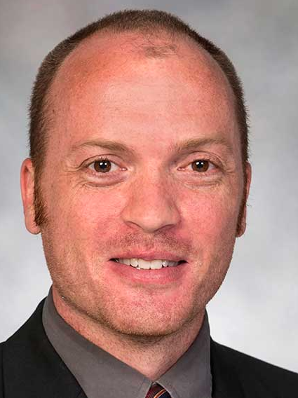 Jordan Miller合作论文数Department of Bioengineering, Rice University;3D Systems Corporation3Yi Chu合作论文数3John A. Waldhausen合作论文数3
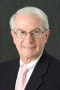 Donald Heistad合作论文数Department of Internal Medicine, Carver College of Medicine, University of Iowa;Division of Cardiology, University of Iowa3Jeffrey E. Everett合作论文数2
 Robert Weiss合作论文数Department of Human Genetics, School of Medicine, The University of Utah2
 Peña Silva Ricardo A合作论文数From the Departments of Internal Medicine (R.A.P.S., I.J.M., University of Iowa;Facultad de Medicina, Universidad de los Andes;Max-Delbrück Center for Molecular Medicine;and Department of Physiology and Biophysics, Federal University of Minas Gerais;University of Iowa, Federal University of Minas Gerais2Kristine M. Serrano合作论文数2Scott D. Niles合作论文数2Donald D. Lund合作论文数2MICHAEL R. PETRACEK合作论文数1WILLIAM W. ANGELL合作论文数1Tl Fudge合作论文数1
 Peter R. Vale合作论文数1Deepak M. Gangahar合作论文数1WILLIAM S. PIERCE合作论文数1Douglas Murphy合作论文数1Ensley Rd合作论文数1Christopher J Berry合作论文数1adriana banarova合作论文数1A. DeLucia合作论文数1Ws Pierce合作论文数1WA GAY合作论文数1Robert D. Dowling合作论文数1Ga Prophet合作论文数1Db Campbell合作论文数1Sl Selinger合作论文数1Alan F Ross合作论文数Dept Anesthesia, Univ Iowa1Donachy J H合作论文数1Landis D L合作论文数1Izakovic M合作论文数1
 Jeffrey Michael Isner合作论文数St. Elizabeth's Medical Center1Donald B. Doty合作论文数1
 Richard A. Schatz合作论文数Ralph H. Johnson Veterans Affairs Medical Center1
 John J. Lopez合作论文数1Timothy D. Henry合作论文数1Marie M. Gleason合作论文数1Keith B. Allen合作论文数1Joseph Van Camp合作论文数1Mark A. Grise合作论文数1
 Gerson Rosenberg合作论文数Department of Biomedical Engineering, College of Engineering, The Pennsylvania State University;Department of Surgery, The Pennsylvania State University;The Materials Research Institute, The Pennsylvania State University1F. David Fortuin合作论文数1John D. Marks合作论文数1
 Stephen G Young合作论文数School of Medicine, University of California, Los Angeles;Ronald Reagan UCLA Medical Center1Shreekanth V. Karwande合作论文数1John P. Reilly合作论文数1
 Gary L. Schaer合作论文数Med Ctr, Rush Univ1M. Elizabeth H. Hammond合作论文数1




