Abstract Background A comparison of diagnostic performance comparing AI-QCTISCHEMIA, CT-FFR, and physician visual interpretation on the prediction of invasive adenosine FFR have not been evaluated. Furthermore, the coronary plaque characteristics impacting these tests have not been assessed. Methods In a single center, 43-month retrospective review of 442 patients referred for CCTA and CT-FFR, 44 patients with CT-FFR had 54 vessels assessed using intracoronary adenosine FFR within 60 days. A comparison of the diagnostic performance among these three techniques for the prediction of FFR ≤ 0.80 was reported. Results The mean age of the study population was 65 years, 76.9% were male, and the median CAC was 623. When analyzing the per vessel ischemia prediction, AI-QCTISCHEMIA had greater specificity, PPV, diagnostic accuracy, and AUC vs. CT- FFR and physician visual interpretation CAD-RADS. The AUC for AI-QCTISCHEMIA was 0.91 vs. 0.76 for CT-FFR and 0.62 for CADRADS ≥3. Plaque characteristics that were different in false positive vs true positive cases for AI-QCTISCHEMIA was max stenosis diameter (50 vs 70%, p = 0.03); for CT-FFR were maximum stenosis diameter (40 vs 70%, p < 0.001), total noncalcified plaque (9 vs 13%, p = 0.02); and for physician visual interpretation CADRADS ≥3 were total noncalcified plaque (8 vs 12%, p = 0.01), lumen volume (681 vs 510mm3, p = 0.03), maximum stenosis diameter (40 vs 60%, p < 0.001), total plaque (19 vs 33%, p = 0.006, total calcified plaque (11 vs 22%, p = 0.008). Conclusion Regarding per-vessel prediction of FFR ≤ 0.8, AI-QCTISCHEMIA revealed greater specificity, PPV, accuracy, and AUC vs. CT-FFR and physician visual interpretation CADRADS ≥3.
Aims:A comparison of diagnostic performance comparing AI-QCTISCHEMIA, coronary computed tomography angiography using fractional flow reserve (CT-FFR), and physician visual interpretation on the prediction of invasive adenosine FFR have not been evaluated. Furthermore, the coronary plaque characteristics impacting these tests have not been assessed. Methods and results:In a single centre, 43-month retrospective review of 442 patients referred for coronary computed tomography angiography and CT-FFR, 44 patients with CT-FFR had 54 vessels assessed using intracoronary adenosine FFR within 60 days. A comparison of the diagnostic performance among these three techniques for the prediction of FFR ≤ 0.80 was reported. The mean age of the study population was 65 years, 76.9% were male, and the median coronary artery calcium was 654. When analysing the per-vessel ischaemia prediction, AI-QCTISCHEMIA had greater specificity, positive predictive value (PPV), diagnostic accuracy, and area under the curve (AUC) vs. CT-FFR and physician visual interpretation CAD-RADS. The AUC for AI-QCTISCHEMIA was 0.91 vs. 0.76 for CT-FFR and 0.62 for CAD-RADS ≥ 3. Plaque characteristics that were different in false positive vs. true positive cases for AI-QCTISCHEMIA were max stenosis diameter % (54% vs. 67%, P < 0.01); for CT-FFR were maximum stenosis diameter % (40% vs. 65%, P < 0.001), total non-calcified plaque (9% vs. 13%, P < 0.01); and for physician visual interpretation CAD-RADS ≥ 3 were total non-calcified plaque (8% vs. 12%, P < 0.01), lumen volume (681 vs. 510 mm3, P = 0.02), maximum stenosis diameter % (40% vs. 62%, P < 0.001), total plaque (19% vs. 33%, P = 0.002), and total calcified plaque (11% vs. 22%, P = 0.003). Conclusion:Regarding per-vessel prediction of FFR ≤ 0.8, AI-QCTISCHEMIA revealed greater specificity, PPV, accuracy, and AUC vs. CT-FFR and physician visual interpretation CAD-RADS ≥ 3.
Eight years after the first coronary angioplasty was described by Andreas Grüntzig in 1978,1Grüntzig A. Transluminal dilatation of coronary-artery stenosis.Lancet. 1978; 311: 263Abstract Scopus (1072) Google Scholar the first series of coronary stent implantations in humans was described by Puel et al.2Puel J Joffre F Rousseau H et al.Endo-prothèses coronariennes autoexpansives dans la prévention des resteénoses après angioplastie transluminale.Arch Mal Coeur. 1987; 8: 1311-1312Google Scholar In parallel, a team based in Texas developed a miniaturized balloon-expandable stent,2Puel J Joffre F Rousseau H et al.Endo-prothèses coronariennes autoexpansives dans la prévention des resteénoses après angioplastie transluminale.Arch Mal Coeur. 1987; 8: 1311-1312Google Scholar which eventually became the first Food and Drug Administration (FDA)-approved coronary stent in 1994 (Fig. 1). Since then, the Palmaz-Schatz stent has been implanted in close to 100 million patients. More than 30 years later, and after iterative improvements of their designs, more than 2 million coronary stents are now implanted globally each year. For this special edition of the Canadian Journal of Cardiology, Dr Richard A. Schatz provides a firsthand account of how his innovation transitioned from a simple idea to one of medicine's most commonly implanted permanent devices in history.Can you tell us about the professional background you had at the time you developed the first balloon-expandable stent approved by the FDA?Dr Richard A. Schatz: It was in 1985, when I met Dr Palmaz in San Antonio, Texas. I was an active duty major cardiologist in the army Medical Corps at a big teaching hospital, called Brooke Army Medical Center. Our meeting was pure fate, as he was at the University of Texas as the head of Interventional Radiology. We were introduced by a mutual friend, who thought we had a lot in common and should work together. I was doing some animal work at a local research centre, looking at laser angioplasty in baboon hearts. He had been working for 7 years on his idea of a balloon-expandable stent in animal models only, which was far away from human trials.How did you and Dr Palmaz think of the idea that eventually became the Palmaz-Schatz stent?R.A.S.: It was all Dr Palmaz's idea. He saw Grüntzig give his first talk on percutaneous transluminal angioplasty in femoral arteries in humans in 1978 and immediately thought it would collapse over time. So he thought of a balloon-expandable cage to prop open the artery. He studied the various metal materials and settled on 316L stainless steel, then built his own prototypes from round wire; he started placing them in animal models as early as 1980, when he moved to San Antonio. When I met him, I had the idea of making them more flexible, so they would work better in small arteries, such as coronary arteries, in which tortuosity was a problem. These ideas later became the 984, 332, and 417 patents.What are the major hurdles you encountered during the development of your invention?R.A.S.: First, it was funding. None of the major players believed in our work. Once we found private funding, the real work lay ahead. Getting the studies done was easy, but convincing the world and the FDA was very difficult. It took a lot of perseverance and luck to finally get it approved.What advices would you provide early career cardiologists interested in pursuing a career dedicated to medical innovations?R.A.S.: First, have confidence in your own ability to innovate. It's not that hard. Every time you are in the lab and you see a problem or a hiccup, ask yourself, "Why did this happen, and how can I find a better way to do things?" As a young cardiologist in the lab, you are smarter than any engineer in the world for identifying problems and finding a solution. Second, every time a new product comes out, tear it apart, and look for ways to make it better. The first prototypes are usually very crude and have lots of room for improvement; the more you use a new product, you will see its shortcomings. Be creative, and look for solutions.You reported the implantation of balloon-expandable stents in dogs' coronary arteries for the first time 33 years ago. How do you picture the field of interventional cardiology 33 years from now?R.A.S.: I think there will be huge advances in genetic manipulation to prevent coronary artery disease from forming. There will be a role for devices, but we might see hybrid valves, for instance, with tissue engineered from stem cells. Very likely, we will see better medicines that can slow the process down of coronary artery disease, so plaques never rupture. I've always thought that genes and stem cells work well in selected cases, so I think something new will appear to reverse congestive heart failure and grow blood vessels in refractory cases to relieve angina. Cardiology is the most exciting field to be in, and we should all be very proud of our rich history in getting to where we are today.Funding SourcesDr Marquis-Gravel is supported by the Fonds de Recherche du Québec – Santé (FRQS).This article was published as part of a supplement supported by the Montreal Heart Institute Interventional Cardiology Division and the Chaire Yves Des Groseillers et André Bérard de Cardiologie Interventionnelle de l'Université de Montréal (Yves Des Groseillers and André Bérard Chair in Interventional Cardiology at the University of Montreal).DisclosuresThe author has no conflicts of interest to disclose. Eight years after the first coronary angioplasty was described by Andreas Grüntzig in 1978,1Grüntzig A. Transluminal dilatation of coronary-artery stenosis.Lancet. 1978; 311: 263Abstract Scopus (1072) Google Scholar the first series of coronary stent implantations in humans was described by Puel et al.2Puel J Joffre F Rousseau H et al.Endo-prothèses coronariennes autoexpansives dans la prévention des resteénoses après angioplastie transluminale.Arch Mal Coeur. 1987; 8: 1311-1312Google Scholar In parallel, a team based in Texas developed a miniaturized balloon-expandable stent,2Puel J Joffre F Rousseau H et al.Endo-prothèses coronariennes autoexpansives dans la prévention des resteénoses après angioplastie transluminale.Arch Mal Coeur. 1987; 8: 1311-1312Google Scholar which eventually became the first Food and Drug Administration (FDA)-approved coronary stent in 1994 (Fig. 1). Since then, the Palmaz-Schatz stent has been implanted in close to 100 million patients. More than 30 years later, and after iterative improvements of their designs, more than 2 million coronary stents are now implanted globally each year. For this special edition of the Canadian Journal of Cardiology, Dr Richard A. Schatz provides a firsthand account of how his innovation transitioned from a simple idea to one of medicine's most commonly implanted permanent devices in history. Can you tell us about the professional background you had at the time you developed the first balloon-expandable stent approved by the FDA? Dr Richard A. Schatz: It was in 1985, when I met Dr Palmaz in San Antonio, Texas. I was an active duty major cardiologist in the army Medical Corps at a big teaching hospital, called Brooke Army Medical Center. Our meeting was pure fate, as he was at the University of Texas as the head of Interventional Radiology. We were introduced by a mutual friend, who thought we had a lot in common and should work together. I was doing some animal work at a local research centre, looking at laser angioplasty in baboon hearts. He had been working for 7 years on his idea of a balloon-expandable stent in animal models only, which was far away from human trials. How did you and Dr Palmaz think of the idea that eventually became the Palmaz-Schatz stent? R.A.S.: It was all Dr Palmaz's idea. He saw Grüntzig give his first talk on percutaneous transluminal angioplasty in femoral arteries in humans in 1978 and immediately thought it would collapse over time. So he thought of a balloon-expandable cage to prop open the artery. He studied the various metal materials and settled on 316L stainless steel, then built his own prototypes from round wire; he started placing them in animal models as early as 1980, when he moved to San Antonio. When I met him, I had the idea of making them more flexible, so they would work better in small arteries, such as coronary arteries, in which tortuosity was a problem. These ideas later became the 984, 332, and 417 patents. What are the major hurdles you encountered during the development of your invention? R.A.S.: First, it was funding. None of the major players believed in our work. Once we found private funding, the real work lay ahead. Getting the studies done was easy, but convincing the world and the FDA was very difficult. It took a lot of perseverance and luck to finally get it approved. What advices would you provide early career cardiologists interested in pursuing a career dedicated to medical innovations? R.A.S.: First, have confidence in your own ability to innovate. It's not that hard. Every time you are in the lab and you see a problem or a hiccup, ask yourself, "Why did this happen, and how can I find a better way to do things?" As a young cardiologist in the lab, you are smarter than any engineer in the world for identifying problems and finding a solution. Second, every time a new product comes out, tear it apart, and look for ways to make it better. The first prototypes are usually very crude and have lots of room for improvement; the more you use a new product, you will see its shortcomings. Be creative, and look for solutions. You reported the implantation of balloon-expandable stents in dogs' coronary arteries for the first time 33 years ago. How do you picture the field of interventional cardiology 33 years from now? R.A.S.: I think there will be huge advances in genetic manipulation to prevent coronary artery disease from forming. There will be a role for devices, but we might see hybrid valves, for instance, with tissue engineered from stem cells. Very likely, we will see better medicines that can slow the process down of coronary artery disease, so plaques never rupture. I've always thought that genes and stem cells work well in selected cases, so I think something new will appear to reverse congestive heart failure and grow blood vessels in refractory cases to relieve angina. Cardiology is the most exciting field to be in, and we should all be very proud of our rich history in getting to where we are today. Funding SourcesDr Marquis-Gravel is supported by the Fonds de Recherche du Québec – Santé (FRQS).This article was published as part of a supplement supported by the Montreal Heart Institute Interventional Cardiology Division and the Chaire Yves Des Groseillers et André Bérard de Cardiologie Interventionnelle de l'Université de Montréal (Yves Des Groseillers and André Bérard Chair in Interventional Cardiology at the University of Montreal). Dr Marquis-Gravel is supported by the Fonds de Recherche du Québec – Santé (FRQS).
Background Most cell therapy trials failed to show an improvement in global left ventricular (LV) function measures after myocardial infarction (MI). Myocardial segments are heterogeneously impacted by MI. Global LV function indices are not able to detect the small treatment effects on segmental myocardial function which may have prognostic implications for cardiac events. We aimed to test the efficacy of allogeneic cardiosphere-derived cells (CDCs) for improving regional myocardial function and contractility.Methods In this exploratory analysis of a randomised clinical trial, 142 patients with post-MI with LVEF <45% and 15% or greater LV scar size were randomised in 2:1 ratio to receive intracoronary infusion of allogenic CDCs or placebo, respectively. Change in segmental myocardial circumferential strain (Ecc) by MRI from baseline to 6 months was compared between CDCs and placebo groups.Results In total, 124 patients completed the 6-month follow-up (mean (SD) age 54.3 (10.8) and 108 (87.1%) men). Segmental Ecc improvement was significantly greater in patients receiving CDC (−0.5% (4.0)) compared with placebo (0.2% (3.7), p=0.05). The greatest benefit for improvement in segmental Ecc was observed in segments containing scar tissue (change in segmental Ecc of −0.7% (3.5) in patients receiving CDC vs 0.04% (3.7) in the placebo group, p=0.04).Conclusions In patients with post-MI LV dysfunction, CDC administration resulted in improved segmental myocardial function. Our findings highlight the importance of segmental myocardial function indices as an endpoint in future clinical trials of patients with post-MI.Trial registration number NCT01458405.
Introduction: Higher levels of low-density lipoprotein (LDL) are associated with an increased incidence of cardiovascular events. Little is known about the impact of lipid profile on the severity and extent of disease. This study aims to characterize the lipid profile’s ability to predict the number of and total length of coronary artery stents. Hypothesis: In patients undergoing percutaneous coronary intervention, the lipid panel is predictive of the total length and number of coronary stents deployed. Methods: We retrospectively identified unique, consecutive patients undergoing percutaneous coronary intervention at a single center from April 2018 to March 2021. Inclusion criteria included the availability of a lipid panel in the electronic medical record. Associations between lipid levels with stent number or stent length were tested by univariable linear regression. Results: For a single coronary angiogram procedure, 1547 unique patients had a lipid panel in the EMR within 2 weeks of the procedure with a median of 2 stents deployed with median total stent length of 38 mm. High-density lipoprotein (HDL), LDL, but not total cholesterol, triglycerides (TG), were predictive of stent length (HDL inversely, p =0.004; LDL p =0.01) and number of stents (HDL inversely, p =0.008; LDL p =0.04). Conclusions: These results indicate that the lipid panel has implications for the severity and extent of coronary artery disease found during coronary angiography, reflected in the number of and total length of coronary artery stents.
Abstract Background Regenerative therapies offer new approaches to improve cardiac function after acute ST-elevation myocardial infarction (STEMI). Mobilization of stem cells and homing within the infarcted area have been identified as the key mechanisms for successful treatment. Application of granulocyte-colony stimulating factor (G-CSF) is the least invasive way to mobilize stem cells while DDP4-inhibitor facilitates homing via stromal cell-derived factor 1 alpha (SDF-1α). Dutogliptin, a novel DPP4 inhibitor, combined with stem cell mobilization using G-CSF significantly improved survival and reduced infarct size in a murine model. Purpose We initiated a phase II, multicenter, randomized, placebo-controlled efficacy and safety study (N=140) analyzing the effect of combined application of G-CSF and dutogliptin, a small molecule DPP-IV-inhibitor for subcutaneous use after acute myocardial infarction. Methods The primary objective of the study is to evaluate the safety and tolerability of dutogliptin (14 days) in combination with filgrastim (5 days) in patients with STEMI (EF <45%) following percutaneous coronary intervention (PCI). Preliminary efficacy will be analyzed using cardiac magnetic resonance imaging (cMRI) to detect >3.8% improvement in left ventricular ejection fraction (LV-EF). 140 subjects will be randomized to filgrastim plus dutogliptin or matching placebos. Results Baseline characteristics of the first 26 patients randomized (24 treated) in this trial reveal a majority of male patients (70.8%) and a medium age of 58.4 years (37 to 84). During the 2-week active treatment period, 35 adverse events occurred in 13 patients, with 4 rated as serious (hospitalization due to pneumonia N=3, hospitalization due to acute myocardial infarction N=1), and 1 adverse event was rated as severe (fatal pneumonia), 9 moderate, and 25 as mild. 6 adverse events were considered possibly related to the study medication, including cases of increased hepatic enzymes (N=3), nausea (N=1), subcutaneous node/suffusion (N=1) and syncope (N=1). Conclusions Our data demonstrate that the combined application of dutogliptin and G-CSF appears to be safe on the short term and feasible after acute myocardial infarction and may represent a new therapeutic option in future. Funding Acknowledgement Type of funding source: Other. Main funding source(s): This research is funded by the sponsor RECARDIO, Inc., 1 Market Street San Francisco, CA 94150, USA. RECARDIO Inc. is funding the complete study. The Scientific Board of RECARDIO designed the study. Data Collection is at the participating sites. Interpretation of the data by the Scientific Board and Manuscript written by the authors and approved by the Sponsor
Primary percutaneous coronary intervention (PCI) is now the recommended reperfusion technique for patients with acute ST-segment elevation myocardial infarction. However, despite early reperfusion in the majority of patients, PCI does not achieve effective myocardial reperfusion in a significant proportion of patients due to the prevalence of coronary microvascular obstruction. The amount of infarcted myocardium has been considered to be a reliable indicator of major adverse cardiovascular events and resultant adverse left ventricular remodeling. The purpose of this paper is to review the clinical benefits of supersaturated oxygen therapy following PCI for ST-segment elevation myocardial infarction.
Introduction: Cell therapy failed to improve global LV ejection fraction (LVEF) in most trials of post-MI LV dysfunction. LVEF does not consider the effect of cell therapy on different segments of the myocardium, which may be heterogeneous across different myocardial regions in patients with regional MI. Hypothesis: Allogeneic cardiosphere-derived cells (CDCs) improve segmental (but not global) myocardial function indexed as circumferential strain by MRI. Methods: In this randomized double-blind trial, 142 post-MI patients with LVEF<45% and 15% or greater LV scar size were randomized in 2:1 ratio to receive intracoronary infusion of allogenic CDCs or placebo, respectively. Change in segmental myocardial circumferential strain (Ecc) by MRI from baseline to 6-month was compared between CDCs and placebo groups. Results: In total, 124 patients completed the 6-month follow-up (mean (SD) age 54.3(10.8) and 108 (87.1%) males). Segmental Ecc improvement was significantly greater in patients receiving CDC (-0.5%(4.0)) compared to placebo (0.2%(3.7), p=0.05). The greatest benefit for improvement in segmental Ecc was observed in segments containing scar tissue (change in segmental Ecc of -0.7% (3.5) in patients receiving CDC vs. 0.04% (3.7) in the placebo group, p=0.04). The beneficial effect of CDCs for improving segmental Ecc was greater in patients with LV ejection fraction<41%, LV scar size>18.8% or LV end-diastolic volume index>100 [mixed effect regression coefficients of -0.92(p=0.02),-0.78(p=0.03), and -1.16(p=0.004) respectively versus -0.58(p=0.05) for the entire cohort]. Conclusions: In patients with post-MI LV dysfunction, allogeneic CDC administration resulted in improved segmental myocardial function. This CDC induced improvement in segmental myocardial function was greater in patients with severe LV dysfunction, dilated LV and greater infarct size. (clinicaltrials.gov Identifier: NCT01458405).
Myocardial free wall rupture is a rare but usually fatal complication of acute myocardial infarction (MI) especially if it occurs out of hospital and occurs in 2-4% of patients who suffer from acute MI. Rapid diagnosis is essential but not always easy as diagnostic tests may be inconclusive. In this case report authors examine a rare and unique patient survival after left ventricular free wall rupture following MI. The patient developed chest pain and hypotension in the hospital and was taken directly to the catheterization laboratory where a diagnostic angiogram showed a high-grade occlusion of a very small marginal branch, fluoroscopy demonstrated a large pericardial effusion, which was drained then auto transfused back to the patient using a femoral vein sheath. Rapid diagnostic testing including transesophageal echocardiography with Definity, transthoracic echocardiography, aortography and left ventriculography were all negative for dissection and rupture. Despite the negative diagnostic test, a high index of suspicion for rupture led to urgent surgical exploration where a large 4-cm hole was found in the lateral wall. Repair was successful and the patient left the hospital about several weeks later.
AIMS:Cardiosphere-derived cells (CDCs) are cardiac progenitor cells that exhibit disease-modifying bioactivity in various models of cardiomyopathy and in previous clinical studies of acute myocardial infarction (MI), dilated cardiomyopathy, and Duchenne muscular dystrophy. The aim of the study was to assess the safety and efficacy of intracoronary administration of allogeneic CDCs in the multicentre, randomized, double-blinded, placebo-controlled, intracoronary ALLogeneic heart STem cells to Achieve myocardial Regeneration (ALLSTAR) trial. METHODS AND RESULTS:We enrolled patients 4 weeks to 12 months after MI, with left ventricular ejection fraction (LVEF) ≤45% and LV scar size ≥15% of LV mass by magnetic resonance imaging (MRI). A pre-specified interim analysis was performed when 6-month MRI data were available. The trial was subsequently stopped due to the low probability of detecting a significant treatment effect of CDCs based on the primary endpoint. Patients were randomly allocated in a 2:1 ratio to receive CDCs or placebo in the infarct-related artery by stop-flow technique. The primary safety endpoint was the occurrence, during 1-month post-intracoronary infusion, of acute myocarditis attributable to allogeneic CDCs, ventricular tachycardia- or ventricular fibrillation-related death, sudden unexpected death, or a major adverse cardiac event (death or hospitalization for heart failure or non-fatal MI or need for left ventricular assist device or heart transplant). The primary efficacy endpoint was the relative percentage change in infarct size at 12 months post-infusion as assessed by contrast-enhanced cardiac MRI. We randomly allocated 142 eligible patients of whom 134 were treated (90 to the CDC group and 44 to the placebo group). The mean baseline LVEF was 40% and the mean scar size was 22% of LV mass. No primary safety endpoint events occurred. There was no difference in the percentage change from baseline in scar size (P = 0.51) between CDCs and placebo groups at 6 months. Compared with placebo, there were significant reductions in LV end-diastolic volume (P = 0.02), LV end-systolic volume (P = 0.02), and N-terminal pro b-type natriuretic peptide (NT-proBNP) (P = 0.02) at 6 months in CDC-treated patients. CONCLUSION:Intracoronary infusion of allogeneic CDCs in patients with post-MI LV dysfunction was safe but did not reduce scar size relative to placebo at 6 months. Nevertheless, the reductions in LV volumes and NT-proBNP reveal disease-modifying bioactivity of CDCs. TRIAL REGISTRATION:Clinicaltrials.gov identifier: NCT01458405.
Background Regenerative therapies offer new approaches to improve cardiac function after acute ST-elevation myocardial infarction (STEMI). Previous trials using bone marrow cells, selected stem cell populations, or cardiac stem cell progenitors require invasive procedures and had so far inconclusive results. A less invasive approach utilizes granulocyte-colony stimulating factor (G-CSF) to mobilize stem cells to circulating blood and induce neovascularization and differentiation into endothelial cells and cardiomyocytes. Stromal cell-derived factor 1 alpha (SDF-1α) is an important chemokine for initiating stem cell migration and homing to ischemic myocardium. SDF-1α concentrations can be increased by inhibition of CD26/DPP4. Dutogliptin, a novel DPP4 inhibitor, combined with stem cell mobilization using G-CSF significantly improved survival and reduced infarct size in a murine model. Methods We test the safety and tolerability and efficacy of dutogliptin in combination with filgrastim (G-CSF) in patients with STEMI (EF < 45%) following percutaneous coronary intervention (PCI). Preliminary efficacy will be analyzed using cardiac magnetic resonance imaging (cMRI) to detect > 3.8% improvement in left ventricular ejection fraction (LV-EF) compared to placebo. One hundred forty subjects will be randomized to filgrastim plus dutogliptin or matching placebos. Discussion The REC-DUT-002 trial is the first to evaluate dutogliptin in combination with G-CSF in patients with STEMI. Results will lay the foundation for an appropriately powered cardiovascular outcome trial to test the efficacy of this combined pharmacological strategy. Trial registration EudraCT no.: 2018-000916-75 . Registered on 7 June 2018. IND number: 123717
Aims:Autologous CD34+ (auto-CD34+) cells represent an attractive option for the treatment of refractory angina. Three double-blinded randomized trials (n = 304) compared intramyocardial (IM) auto-CD34+ cells with IM placebo injections to affect total exercise time (TET), angina frequency (AF), and major adverse cardiac events (MACE). Patient-level data were pooled from the Phase I, Phase II ACT-34, ACT-34 extension, and Phase III RENEW trials to determine the efficacy and safety of auto-CD34+ cells.Methods and results:Treatment effects for TET were analysed using an analysis of covariance mixed-effects model and for AF using Poisson regression in a log linear model with repeated measures. The Kaplan-Meier rate estimates for MACE were compared using the log-rank test. Autologous CD34+ cell therapy improved TET by 46.6 s [3 months, 95% confidence interval (CI) 13.0 s-80.3 s; P = 0.007], 49.5 s (6 months, 95% CI 9.3-89.7; P = 0.016), and 44.7 s (12 months, 95% CI - 2.7 s-92.1 s; P = 0.065). The relative frequency of angina was 0.78 (95% CI 0.63-0.98; P = 0.032), 0.66 (0.48-0.91; P = 0.012), and 0.58 (0.38-0.88; P = 0.011) at 3-, 6- and 12-months in auto-CD34+ compared with placebo patients. Results remained concordant when analysed by treatment received and when confined to the Phase III dose of 1 × 10⁵ cells/kg. Autologous CD34 + cell therapy significantly decreased mortality (12.1% vs. 2.5%; P = 0.0025) and numerically reduced MACE (38.9% vs. 30.0; P = 0.14) at 24 months.Conclusion:Treatment with auto-CD34+ cells resulted in clinically meaningful durable improvements in TET and AF at 3-, 6- and 12-months, as well as a reduction in 24-month mortality in this patient-level meta-analysis.
Objective To assess safety and feasibility of autologous adipose‐derived regenerative cells (ADRCs), for treatment of chronic ischemic cardiomyopathy patients. Background Preclinical and early clinical trials suggest ADRCs have excellent potential for ischemic conditions. Methods The Athena program consisted of two parallel, prospective, randomized (2:1, active: placebo), double‐blind trials assessing intramyocardial (IM) ADRC delivery [40‐million, n = 28 (ATHENA) and 80‐million (ATHENA II) cells, n = 3]). Patients with an EF ≥20% but ≤45%, multivessel coronary artery disease (CAD) not amenable to revascularization, inducible ischemia, and symptoms of either angina (CCS II–IV) or heart failure (NYHA Class II–III) on maximal medical therapy were enrolled. All patients underwent fat harvest procedure (≤450 mL adipose), on‐site cell processing (Celution® System, Cytori Therapeutics), electromechanical mapping, and IM delivery of ADRCs or placebo. Results Enrollment was terminated prematurely due to non‐ADRC‐related adverse events and subsequent prolonged enrollment time. Thirty‐one patients (17‐ADRCs, 14‐placebo) mean age 65 ± 8 years, baseline LVEF(%) 31.1 ± 8.7 (ADRC), 31.8 ± 7.7 (placebo) were enrolled. Change in V0 2 max favored ADRCs (+45.4 ± 222 vs. −9.5 ± 137 mL/min) but there was no difference in left ventricular function or volumes. At 12‐months, heart failure hospitalizations occurred in 2/17 (11.7%) [ADRC] and 3/14 (21.4%) [placebo]. Differences in NYHA and CCS classes favored ADRCs at 12‐months with significant improvement in MLHFQ (−21.6 + 13.9 vs. −5.5 + 23.8, P = 0.038). Conclusions A small volume fat harvest, automated local processing, and IM delivery of autologous ADRCs is feasible with suggestion of benefit in “no option” CAD patients. Although the sample size is limited, the findings support feasibility and scalability for treatment of ischemic cardiomyopathy with ADRCs. © 2016 Wiley Periodicals, Inc.
A pproximately 1 in 5 patients beyond the age of 45 years who experience acute myocardial infarction (MI) will be dead within a year. Although the incidence of ST-segment–elevation myocardial infarction (STEMI) has declined and survival after STEMI has improved, the prognosis remains poor for those with residual left ventricular (LV) dysfunction after STEMI. Objective: To evaluate the safety and bioactivity of autologous CD34+ cell (CLBS10) intracoronary infusion in patients with left ventricular dysfunction post STEMI. (ejection fraction≤48%) ≥4 days poststent were eligible for enrollment. Subjects (N=161) underwent mini bone marrow harvest and were randomized 1:1 to receive (1) autologous CD34+ cells (minimum 10 mol/L±20% cells; N=78) or (2) diluent alone (N=83), via intracoronary infusion. The primary safety end point was adverse events, serious adverse events, and major adverse cardiac event. The primary efficacy end point was change in resting myocardial perfusion over 6 months. No differences in myocardial perfusion or adverse events were observed between the control and treatment groups, although increased perfusion was observed within each group from baseline to 6 months (P<0.001). In secondary analyses, when adjusted for time of ischemia, a consistently favorable cell dose–dependent effect was observed in the change in left ventricular ejection fraction and infarct size, and the duration of time subjects was alive and out of hospital (P=0.05). At 1 year, 3.6% (N=3) and 0% deaths were observed in the control and treatment group, respectively.
OBJECTIVES:To assess subjects' perception of healthcare costs and physician reimbursement. BACKGROUND:The lack of transparency in healthcare reimbursement leaves patients and physicians unaware of the distribution of health care dollars. METHODS:Anonymous survey-based study by means of convenience sampling. Participants were asked to estimate the total hospital cost and physician fee for one of the six medical procedures (n = 250). RESULTS:On the average for all 6 procedures, patients estimated the total cost was $36,177, ∼1,540% more than the actual Medicare rate of $7,333. Similarly, patients estimated the physician fee was $7,694, 1,474% more the actual Medicare rate of $589. CONCLUSION:Patients' perception of the total cost and physician fee are significantly higher than Medicare rates for all 6 procedures. This lack of insight may have widespread negative implications on the patient-physician relationship, on political trends to reduce physician reimbursement, and on a physician's desire to continue practicing medicine.
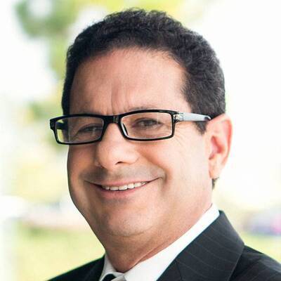 Paul Teirstein合作论文数Scripps Clinic;Prebys Cardiovascular Institute33
 Timothy D. Henry合作论文数30
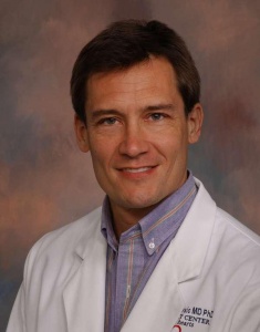 Thomas Joseph Povsic合作论文数Department of Medicine, School of Medicine, Duke University School of Medicine19
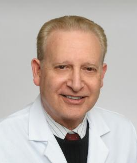 Martin Leon合作论文数Center for Interventional Vascular Therapy, Columbia University Medical Center19
 Matthew Price合作论文数Cardiac Catheterization Laboratory, Division of Cardiovascular Diseases, Scripps Clinic;Scripps Translational Science Institute18
 Douglas Losordo合作论文数Longeveron LLC;Cadrenal Therapeutics, Inc15
 Jay Traverse合作论文数14
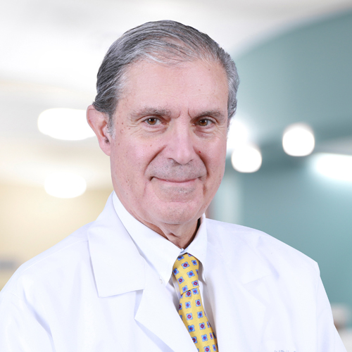 Dean J. Kereiakes合作论文数The Christ Hospital Heart and Vascular Institute, The Christ Hospital Health Network;The Christ Hospital Research Institute, The Christ Hospital Health Network;The Ohio State University;Medpace14F. David Fortuin合作论文数13
 Donald Baim合作论文数Baim Institute for Clinical Research10Curtiss T. Stinis合作论文数10
 Gary L. Schaer合作论文数Med Ctr, Rush Univ9
 David L Fischman合作论文数From Florida Heart Institute9candice e junge合作论文数8
 Robert J Russo合作论文数8Sheldon Goldberg合作论文数8
 Christopher J. White合作论文数The John Ochsner Cardiovascular Institute, Ochsner Clinic Foundation8
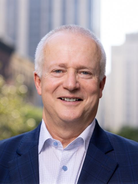 Robert A. Harrington合作论文数Weill Cornell Medicine;Cornell University8
 Warren Sherman合作论文数7
 Charles J Davidson合作论文数Department of Medicine, Feinberg School of Medicine, Northwestern University7Marbán Linda合作论文数7
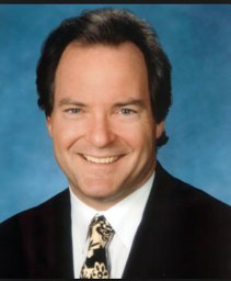 Richard R. Heuser合作论文数Arizona Heart Institute7
 Rajendra Makkar合作论文数Smidt Heart Institute, Cedars-Sinai Medical Center7
 Rachel Smith合作论文数7Ashkan Akhavein Mohammadi合作论文数6
 Eduardo Marbán合作论文数Department of physiology, University of California, Los Angeles6Mendelsohn Farrell O合作论文数6Zhang Qing合作论文数6Story Kenneth合作论文数6Phillip P. Toskes合作论文数6
 Jeffrey J. Popma合作论文数Medtronic6
 Theodore A Bass合作论文数6
 Douglas W. Losordo合作论文数Medicine at Northwestern University Feinberg School of Medicine6Steve S. Lee合作论文数Department of Psychology, University of California, Los Angeles6Stephen Steuterman合作论文数6Raghava R. Gollapudi合作论文数6Shirish Jani合作论文数6Vincent Massullo合作论文数6
 Shing-Chiu Wong合作论文数Greenberg Division of Cardiology, Department of Medicine, Weill Cornell Medical College, Cornell University;Cardiac Catheterization Laboratories, NewYork-Presbyterian Hospital6
 Michael P. Savage合作论文数Cardiac Catheterization Laboratory, Jefferson Medical College, Thomas Jefferson University;Cardiac Catheterization Laboratory, Thomas Jefferson University Hospital6
 Baharak Moshiree合作论文数6
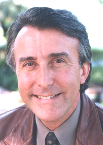 Julio César Palmaz合作论文数Advanced Bio Prosthetic Surfaces;University of Texas Health and Science Center at San Antonio6R Rake合作论文数6Martin L. Charlat合作论文数5Emem Udoh合作论文数5Andrea S Hunt合作论文数5Rajbir Sidhu合作论文数5George Wesbey合作论文数5




