Computational biomechanics increasingly requires models that combine mechanics, transport, chemistry, and biological regulation across different spatial and temporal scales. The FEBio simulation software provides extensive open-source capabilities for modeling these processes using monolithic approaches. However, assembling independently developed physics models into reproducible coupled workflows remains challenging. Existing approaches often require custom scripts or external software pipelines, which can limit model reuse and complicate development. We present FUSE, the FEBio Unified Simulation and Exchange framework, a partitioned coupling plugin that enables separately defined FEBio models to communicate through structured field exchange. FUSE is designed for problems that are best solved independently, particularly when fast mechanical responses influence slower biological or chemical evolution. The framework uses a time-decoupled strategy in which a primary model advances on the longer time scale, while one or more secondary models are repeatedly initialized, supplied with updated fields, solved over shorter time horizons, with results returned to the primary model. Field exchange utilizes existing FEBio data maps, output fields, and user-specified filters, allowing coupled workflows to be constructed without modifying the underlying solvers. The framework was able to reproduce reference coupled solutions while handling bidirectional transfer, spatial field mapping, and filtered exchange of model variables. Example applications demonstrated coupling between mechanical loading and chemical degradation in injured cartilage and interaction between biological tissue formation and mechanical feedback during bone healing. By separating coupling logic from physics implementation, FUSE provides a practical mechanism for building maintainable multiphysics workflows within FEBio.
Cartilage damage under loading is significantly affected by viscoelasticity. This study validates a finite deformation, nonlinear viscoelastic constitutive model for the collagen matrix of immature bovine articular cartilage, using reactive viscoelasticity. To examine the flow-independent viscoelasticity of cartilage collagen, tissue samples underwent proteoglycan (PG) digestion, losing more than 98% of their initial PG content to increase their hydraulic permeability. To verify that this PG-digestion eliminated flow-dependent viscoelasticity, PG-depleted samples were subjected to a gravitational permeation experiment, demonstrating that their hydraulic permeability, k=268 ± 152 mm4/N⋅s (n = 8), was five orders of magnitude greater than reported for untreated cartilage, confirming negligible flow-dependent viscoelasticity. Digested cartilage plugs were then subjected to unconfined compression stress relaxation (four consecutive ramp-hold profiles, each increasing the compressive strain by 10%) to fit the load response and extract material properties (RMSEfit=1.86 ± 0.61 kPa, n = 8). Successful curve-fitting served as a necessary condition for validating the model. Then, a separate unconfined compression stress-relaxation test was performed on the same samples, to 40% compressive strain at the same ramp rate. The model was able to faithfully predict this experimental response using fitted material properties (RMSEpred=3.95 ± 1.33 kPa, with 0≤ stresses ≤ 155 ± 37 kPa), providing a sufficient condition for validation in unconfined compression stress-relaxation. A computational model then showed that flow-independent viscoelasticity of cartilage collagen can enhance the stress response by ∼15% at fast strain rates, over flow-dependent effects. However, we estimate from prior studies that flow-independent viscoelasticity may enhance the stress response of cartilage by up to 200%, implying that PGs probably contribute significantly to the tissue's flow-independent viscoelasticity.
Computational fluid dynamics (CFD) is commonly used to investigate hemodynamics in the cardiovascular system, particularly in regions prone to cardiovascular disease, such as the carotid artery bifurcation. Despite its potential, significant variability exists across different computational approaches, highlighting the need for systematic solver comparisons. This study provides a comprehensive evaluation of three open-source finite element method (FEM) solvers—SimVascular, FEBio, and FEniCS Oasis—for simulating blood flow in a subject-specific carotid artery model. We conducted a rigorous comparison using a model derived from 4D phase-contrast magnetic resonance imaging (4D Flow MRI), examining solver performance across multiple mesh resolutions. This analysis focused on key hemodynamic metrics, including velocity fields, time-averaged wall shear stress (TAWSS), oscillatory shear index (OSI), and wall shear stress (WSS) topology. By maintaining identical meshes, boundary conditions, and postprocessing methods, we isolated solver-specific characteristics while focusing on high-resolution mesh refinements. All solvers demonstrated similar capability in representing the 4D-Flow MRI data. Notably, all solvers consistently identified critical hemodynamic regions, such as flow disturbance zones in the carotid sinus. Mesh convergence analysis showed the ability of all solvers to achieve converged predictions at relatively lower mesh resolutions. The computational time was also compared across the three solvers. While demonstrating the capabilities of each solver in predicting physiologically relevant hemodynamic patterns, our study underscores the utility of open-source solvers for high-fidelity hemodynamic predictions.
Patient-specific finite element models of hip cartilage and labrum (chondrolabral) mechanics have improved the understanding of form-function relationships underpinning hip osteoarthritis. While these models often assume the pelvis and femur are rigid bodies to reduce development and computational time, contact stresses may be overestimated when the pelvis and femur cannot deform. Recent advancements in element formulations, constitutive models, and integration of patient-specific boundary/loading conditions warrant a re-examination of this rigid body assumption. We assessed the influence of material representations for the pelvis and femur on finite element predictions of chondrolabral mechanics during level walking and squatting. Four material representations were evaluated: (1) rigid bodies, (2) deformable-inhomogeneous with CT-derived material properties, (3) deformable-two-material with distinct trabecular and cortical bone materials, and (4) deformable-one-material model. Patient-specific kinematics and joint reaction forces for level walking and squatting were derived from motion capture and musculoskeletal models, respectively. During level walking, there were no significant differences between bone material representations for maximum cartilage contact pressure, contact area, shear stress, or first principal strain. During squatting, rigid and one-material models produced significantly higher maximum contact pressures, while other metrics remained unaffected. Rigid models required significantly less computational runtime and memory than deformable models. Our findings indicate rigid bone assumptions suffice for level walking, improving efficiency in large cohort studies, but deformable bone models are likely warranted for activities that produce larger bone deformations such as squatting. This study provides guidance for selecting bone material representations that balance accuracy and computational efficiency in hip biomechanical analyses.
Purpose Transcatheter edge-to-edge repair (TEER) and annuloplasty devices are increasingly used to treat mitral valve regurgitation, yet their mechanical effects and interactions remain poorly understood. This study aimed to establish an open-source finite element modeling (FEM) framework for simulating patient-specific mitral valve repairs and to evaluate how TEER, annuloplasty, and combined strategies influence leaflet coaptation and valve mechanics. A central objective was to demonstrate how such simulations may support surgical planning by identifying optimal interventions. Methods A patient-specific mitral valve model was reconstructed using SlicerHeart and 3D Slicer. Four G4 MitraClip geometries were modeled and deployed in FEBio to capture leaflet grasp and subsequent clip-leaflet motion under physiologic pressurization. CardioBand annuloplasty was simulated by reducing annular circumference via displacement-controlled boundary conditions, and Mitralign suture annuloplasty was modeled using discrete nodal constraints. Simulations were performed for prolapse and dilated annulus cases, comparing repairs individually and in combination. Valve competence (regurgitant orifice area, ROA), coaptation/contact area (CA), and leaflet stress and strain distributions were quantified. Results In the prolapse anatomy, the simulations showed that while TEER restored coaptation, it also increased the stresses on the leaflets, whereas band and suture annuloplasty generated distinct morphologies with lower stresses. In the dilation anatomy, TEER alone left residual regurgitation, and annuloplasty improved the leaflet closure. Quantitatively, the model found that combined TEER + band annuloplasty yielded the smallest ROA (0.06 cm(2)), the largest CA (2.57cm(2) ), and reduced stresses relative to TEER alone. Conclusion This study establishes a reproducible, open-source FEM framework for simulating transcatheter TEER and annuloplasty repairs. The framework enables quantitative evaluation of the mechanical impact of different transcatheter valve repair strategies, offers a foundation for extending virtual repair analyses to additional valve geometries, and supports the broader goal of incorporating virtual repair into procedure planning.
Valvular heart disease is prevalent and a major contributor to heart failure. Valve leaflet strain is a promising metric for evaluating the mechanics underlying the initiation and progression of valvular pathology. However, generalizable methods for noninvasively quantifying valvular strain from clinically acquired patient images remain limited. This study aims to develop a robust feature tracking framework that enables accurate shape matching across variable valve morphologies and quantification of in vivo atrioventricular leaflet strain from three-dimensional echocardiographic (3DE) images in pediatric and adult patients. We developed a geometric feature tracking framework to quantify in vivo leaflet strain from 3DE images and to assess anatomical deformation across the cardiac cycle. Our approach integrates a cohort-derived geometric reference atlas to establish geometric correspondence and introduces a novel distance-weighted coherent point drift algorithm within a Gaussian mixture model framework for non-rigid registration. We evaluated performance against a finite element benchmark model and compared the approach with conventional point-based tracking methods. The framework was applied to pediatric and adult patient datasets (N = 31) to assess robustness across variable valve morphologies. The proposed method demonstrated greater accuracy in quantifying anatomical alignment and leaflet strain than conventional point-based approaches. Validation against the finite element benchmark confirmed improved strain estimation. The framework achieved reliable inter-phase tracking of valve deformation across diverse morphologies in pediatric and adult patients. Analysis identified a consistent distribution pattern of the 1^st principal strain associated with leaflet billow (prolapse). This feature tracking framework provides a generalizable method for noninvasive quantification of atrioventricular valve leaflet strain from clinical 3DE images. Characterization of biomechanical strain patterns may improve prognostic assessment and support longitudinal evaluation of valvular heart disease. Further investigation of the biomechanical signatures of heart valve disease has the potential to enhance prognostic assessment and longitudinal evaluation of valvular heart disease.
Fluid–structure interaction (FSI) simulation of biological systems presents significant computational challenges, particularly for applications involving large structural deformations and contact mechanics, such as heart valve dynamics. Traditional arbitrary Lagrangian–Eulerian methods encounter fundamental difficulties with such problems due to mesh distortion, motivating immersed techniques. This work presents a novel open-source immersed FSI framework that strategically couples two mature finite element libraries: MFEM, a GPU-ready and scalable library with state-of-the-art parallel performance developed at Lawrence Livermore National Laboratory, and FEBio, a nonlinear finite element solver with sophisticated solid mechanics capabilities designed for biomechanics applications developed at the University of Utah and Columbia University. This coupling creates a unique synergy wherein the fluid solver leverages MFEM’s state-of-the-art parallel performance, while the immersed solid exploits FEBio’s comprehensive suite of hyperelastic and viscoelastic constitutive models and advanced solid mechanics modeling targeted for biomechanics applications. FSI coupling is achieved using a fictitious domain/distributed Lagrange multiplier methodology with variational multiscale stabilization for enhanced accuracy on under-resolved grids expected with unfitted meshes used in immersed FSI. A fully implicit, monolithic scheme provides robust coupling for strongly coupled fluid-solid interactions characteristic of cardiovascular applications. The framework’s modular architecture facilitates straightforward extension to additional physics and element technologies. Several test problems are considered to demonstrate the capabilities of the proposed framework, including a three-dimensional semilunar heart valve simulation. This platform addresses a critical need for open-source immersed FSI software combining advanced biomechanics modeling with high-performance computing infrastructure.
Subject-specific finite element (FE) modeling enables detailed evaluation of cartilage and labral contact mechanics in the hip; however, the computational demands of this approach limit its use in large cohorts. Soft tissue overlap (STO) modeling is a simplified alternative that estimates strain from geometric overlap between articulating surfaces; however, its predictive accuracy remains unclear. The objective of this study was to evaluate the accuracy of STO models for predicting acetabular cartilage and labral strains during simulated walking by direct comparison with subject-specific FE models. Eight individuals with radiographically normal hips and eight patients with cam-type femoroacetabular impingement syndrome were evaluated using subject-specific anatomy, kinematics, and joint reaction forces. STO-predicted strains were compared with FE-predicted compressive strains using correspondence-based Network Analysis and Bland-Altman analyses at heel-strike and heel-off. No group-dependent differences in agreement were observed, so the results for the two cohorts were pooled. STO models captured the general spatial patterns and locations of peak strain in both cartilage and labrum. However, significant differences between STO and FE predictions were present across 1% to 75% of the gait cycle, particularly during higher magnitudes of joint loading. STO systematically underpredicted strain at higher magnitudes, with disagreement increasing as strain magnitude increased. Discrepancies were most pronounced in the anterosuperior cartilage and labrum near heel-off, where FE models predicted higher, more localized strains. These findings indicate that while STO modeling provides qualitative insight into strain patterns and contact locations, its ability to quantify strain magnitude, particularly under higher loading conditions and in the labrum, is limited.
In this study, we address fundamental theoretical considerations that should guide biomedical engineers in the assessment of patient-specific risk of tissue failure, or the assessment of other material properties needed for patient-specific computational modeling, based on noninvasive imaging modalities. Upon reviewing theoretical concepts of mechanics, the primary conclusion is that patient-specific material properties, such as measures of tissue failure, cannot be observed directly, because material properties are dependent on nonobservable functions of state. However, since functions of state may be formulated to depend on observable state variables, and since noninvasive imaging may be used to assess such variables, it behooves investigators to find strong correlations in vitro between the material property of interest and relevant observable state variables, such as measures of tissue morphology, transport characteristics, and composition. Once such univariate or multivariate correlations have been established experimentally in vitro, the next challenge is to relate imaging-based observable measures, acquired noninvasively (e.g., in vivo), to relevant material properties such as failure criteria. The uncertainty associated with these observation-derived material properties is, at best, equal to the uncertainty of the in vitro correlation.
Many approaches have been used to model chordae tendineae geometries in finite element simulations of atrioventricular heart valves. Unfortunately, current "functional" chordae tendineae geometries lack fidelity that would be helpful when informing clinical decisions. The objectives of this work are (i) to improve synthetic chordae tendineae geometry fidelity to consider branching and (ii) to define how the chordae tendineae geometry affects finite element simulations of valve closure. In this work, we develop an open-source method to construct synthetic chordae tendineae geometries in the SlicerHeart Extension of 3D Slicer. The generated geometries are then used in FEBio finite element simulations of atrioventricular valve function to evaluate how variations in chordae tendineae geometry influence valve behavior. Effects are evaluated using functional and mechanical metrics. Our findings demonstrated that altering the chordae tendineae geometry of a stereotypical mitral valve led to changes in clinically relevant valve metrics and valve mechanics. Specifically, cross sectional area had the most influence over valve closure metrics, followed by chordae tendineae density, length, radius and branches. We then used this information to showcase the flexibility of our new workflow by altering the chordae tendineae geometry of two additional geometries (mitral valve with annular dilation and tricuspid valve) to improve finite element predictions. This study presents a flexible, open-source method for generating synthetic chordae tendineae with realistic branching structures. Further, we establish relationships between the chordae tendineae geometry and valve functional/mechanical metrics. This research contribution helps enrich our open-source workflow and brings the finite element simulations closer to use in a patient-specific clinical setting.
Atherosclerotic plaque rupture is the precipitating event in most acute coronary syndromes. As rupture results from the material failure of arterial tissue under mechanical loading, in vivo image-based techniques that can accurately characterize arterial material stiffness offer potential in risk-stratifying lesions. This study developed and validated a novel magnetic resonance (MR) image-based computational framework to evaluate the material stiffness of vascular tissue. Porcine carotid arteries (n=4) were subjected to biaxial mechanical testing, followed by MR image acquisition under controlled loading. Best-fit material parameters for an anisotropic material model were estimated via regression analysis on the biaxial data. A deformable image registration technique, termed Hyperelastic Warping, was utilized to derive strain fields from the MR images and integrated with an inverse parameter estimation algorithm to identify the parameters for the same constitutive model. Experimentally- and Warping-estimated material stiffness values (tangent moduli) were not significantly different at physiologic lumen pressures of 80 (0.36±0.15 and 0.48±0.20 MPa; p=0.14) and 120 mmHg (0.64±0.27 and 0.73±0.36 MPa; p=0.60). The Warping-directed inverse modeling framework identified subtle, but observable variations in material stiffness within a sample and accurately illustrated the physical influence of loading conditions on those properties. Collectively, these results demonstrated the robustness of an innovative approach to characterize non-linear, hyperelastic behaviors of arterial tissue and quantify material stiffness directly from image data.
Computer simulation of "virtual interventions" may inform optimal valve repair for a given patient prior to intervention. However, the paucity of noninvasive methods to determine in vivo mechanical parameters of valves limits the accuracy of computer prediction and their clinical application. To address this, we propose a noninvasive method for determining elastic parameters of valve tissue using physics-informed neural networks. In this work, we demonstrated its application to the tricuspid valve of a child. We first tracked valve displacements from open to closed frames within a 3D echocardiogram time sequence using image registration. Physics-informed neural networks were subsequently applied to estimate the nonlinear mechanical properties from first principles and reference displacements. The simulated model using these patient-specific parameters closely aligned with the reference image segmentation, achieving a mean symmetric distance of less than 1 mm. Our approach doubled the accuracy of the simulated model compared to the generic parameters reported in the literature.
Biological tissues and biomaterials routinely feature a fibrous microstructure that contributes to physical and mechanical properties while influencing cellular guidance, organization and extracellular matrix (ECM) production. Specialized three-dimensional (3D) imaging techniques can visualize fibrillar structure and orientation, and previously we developed a nonparametric approach to extract orientation distribution functions (ODFs) directly from 3D image data [1]. In this work, we expanded our previous approach to provide a complete algorithmic and software framework to characterize inhomogeneous ODFs in image data and use ODFs to model the physics of materials with the finite element method. We characterized inhomogeneity using image subdomains and specialized interpolation methods, and we developed methods to incorporate ODFs directly into constitutive models. To facilitate its adoption by the biomechanics and biophysics communities, we developed a unified software framework in FEBio Studio (www.febio.org). This included new interpolation methods to spatially map the ODFs onto finite element meshes and an approach to downsample ODFs for efficient numerical calculations. The software provides the option to fit ODFs to parametric distributions, and scalar metrics provide means to assess goodness of fit. We evaluated the utility and accuracy of the algorithms and implementation using representative 3D image datasets. Our results demonstrated that utilizing the true measured ODFs provide a more accurate and spatially resolved representation of fiber ODFs and the resulting predicted mechanical response when compared with parametric approaches to approximating the true ODFs. This research provides a powerful, interactive software framework to extract and represent the inhomogeneous anisotropic characteristics of fibrous tissues directly from image data, and to incorporate them into biomechanics and biophysics simulations using the finite element method. Statement of Significance Biological tissues and biomaterials routinely feature a fibrous microstructure that contributes to physical and mechanical properties while influencing cellular guidance, organization and extracellular matrix (ECM) production. In this study, we developed a complete algorithmic and software framework to characterize inhomogeneous orientation distribution functions (ODFs) directly from biomedical image data and apply the ODFs to model the physics of biological materials. We characterized inhomogeneity using image subdomains and specialized interpolation methods, and we developed methods to incorporate ODFs directly into constitutive models. We developed a unified software framework in FEBio Studio (www.febio.org) to accommodate its adoption by the biomechanics and biophysics communities. The result is a powerful, interactive software framework to extract and represent inhomogeneous, anisotropic characteristics directly from image data, and incorporate them into biomechanics and biophysics simulations.
Multiscale coupling between cell-scale biology and tissue-scale mechanics is a promising approach for modeling disease growth. In such models, tissue-level growth and remodeling (G&R) are driven by cell-level signaling pathways and systems biology models, where each model operates at different scales. Herein, we generate multiscale G&R models to capture the associated multiscale connections. At the cell-scale, we consider systems biology models in the form of systems of ordinary differential equations (ODEs) and partial differential equations (PDEs) representing the reactions between the biochemicals causing the growth based on mass-action or logic-based Hill-type kinetics. At the tissue-scale, we employ kinematic growth in continuum frameworks. Two illustrative test problems (a tissue graft and aneurysm growth) are examined with various chemical signaling networks, boundary conditions, and mechano-chemical coupling strategies. We extend two open-source software frameworks-febio and fenics-to disseminate examples of multiscale growth and remodeling simulations. One-way and two-way coupling between the systems biology and the growth models are compared and the effect of biochemical diffusivity and ODE versus PDE-based systems biology modeling on the G&R results are studied. The results show that growth patterns emerge from reactions between biochemicals, the choice between ODEs and PDEs systems biology modeling, and the coupling strategy. Cross-verification confirms that results for febio and fenics are nearly identical. We hope that these open-source tools will support reproducibility and education within the biomechanics community.
Rehabilitation can help promote functional restoration following surgical reconstruction of severe bone injuries, yet prescribed regimens are often conservative due to limited understanding of their impact on healing. This study examined rodent physical activity parameters, including wheel running duration, distance, bout frequency, bout duration, velocity, and rest time, assessing their combined impact on bone healing in 2 and 3 mm segmental defects. Artifical intelligence (AI)-based genetic programming generated high-accuracy nonlinear models, revealing a “goldilocks” phenomenon: some rehabilitation stimulates bone formation, but excessive activity is detrimental. Subject-specific finite element modeling showed that local defect compressive strains decreased only in injuries that achieved union and that the correlation between strain and healing evolved over time. These findings highlight the dynamic healing process, necessitating a subject-specific approach. While bone healing is often thought to be primarily driven by achieving optimal strain magnitudes, our results suggest a more complex reality. Bone healing depends not only on optimal strain magnitude but also on balancing activity and rest, which shifts with injury severity and healing progression. Overall, effective rehabilitation must consider injury stabilization, severity, and healing status while ensuring adequate rest to promote optimal bone regeneration.
Valvular heart disease is prevalent and a major contributor to heart failure. Valve leaflet strain is a promising metric for evaluating the mechanics underlying the initiation and progression of valvular pathology. However, robust and generalizable methods for noninvasively quantifying valvular strain from clinically acquired patient images remain limited. In this work, we present a novel feature-tracking framework for quantifying leaflet strain in atrioventricular valves using 3D echocardiographic images of pediatric and adult patients. Our method demonstrated superior accuracy in the assessment of anatomical deformation and strain of heart valves compared to other point-based approaches, as verified against a finite element benchmark. Further, our approach can robustly track inter-phase deformation of valves across highly variable morphologies without parameter tuning. Our analysis revealed that a median and interquartile range of the 1st principal strain greater than 0.5 is associated with leaflet billow (prolapse). Further investigation of the biomechanical signatures of heart valve disease has the potential to enhance prognostic assessment and longitudinal evaluation of valvular disease.
Background - Pulmonary insufficiency is a consequence of transannular patch repair in Tetralogy of Fallot (ToF), leading to late morbidity and mortality. Transcatheter native outflow tract pulmonary valve replacement (TPVR) has become common, but assessment of patient candidacy and selection of the optimal device remains challenging. We demonstrate an integrated open-source workflow for simulation of TPVR in image-derived models to inform device selection. Methods - Machine learning-based segmentation of CT scans was implemented to define the right ventricular outflow tract (RVOT). A custom workflow for device positioning and pre-compression was implemented in SlicerHeart. Resulting geometries were exported to FEBio for simulation. Visualization of results and quantification were performed using custom metrics implemented in SlicerHeart and FEBio. Results - RVOT model creation and device placement could be completed in under 1 minute. Virtual device placement using FE simulations visually mimicked actual device placement and allowed quantification of vessel strain, stress, and contact area. Regions of higher strain and stress were observed at the proximal and distal end locations of the TPVs where the devices impinge the RVOT wall. No other consistent trends were observed across simulations. The observed variability in mechanical metrics across RVOTS, stents, and locations in the RVOT highlights that no single device performs optimally in all anatomies, thereby reinforcing the need for simulation-based patient-specific assessment. Conclusions - This study demonstrates the feasibility of a novel open-source workflow for the rapid simulation of TPVR which with further refinement may inform assessment of patient candidacy and optimal device selection.
Longitudinal estimation of local strain distributions within the regenerative niche of segmental femoral fractures is important for understanding mechanobiology principles for bone healing to design more effective rehabilitation regimens and mitigate nonunion complications. Finite element (FE) modeling is the standard for investigating these biomechanical parameters, yet most existing models lack clinical relevance due to their use of generic data and computational inefficiency. This study developed a subject-specific FE workflow aimed at accurate biomechanical predictions based on subject-specific data while addressing the limitations of previous approaches. For the experimental study, near-critical-sized segmental bone defects were created in the femurs of Wistar rats and stabilized with internal fixators before rehabilitation. Subject-specific geometries of the defect were generated from in vivo micro-CT scans, which were also used to assign material coefficients. Generalized geometries of the cortical and trabecular bone and fixator were integrated to increase computational efficiency. In addition, axial strain data from strain gauges on the fixators were used to define subject-specific boundary conditions, enabling a longitudinal study of the healing process. Sensitivity analyses revealed that incorporating subject-specific boundary conditions significantly enhanced model accuracy, a factor often overlooked in conventional approaches. The workflow was used to build six defect models to approximate compressive strains within the defect and the joint contact force. Strain distributions correlated with experimentally observed mineralization and better predicted functional bone bridging (union) compared to bone volume metrics. This efficient workflow facilitates the assessment of local biomechanics during bone healing and highlights their influence on adaptive regeneration. Further, the findings support the potential application of the subject-specific modeling workflow to guide clinical decision-making and improve therapeutic outcomes for treating bone fractures.
Computer simulation of "virtual interventions" may inform optimal valve repair for a given patient prior to intervention. However, the paucity of noninvasive methods to determine in vivo mechanical parameters of valves limits the accuracy of computer prediction and their clinical application. To address this, we propose ADEPT: A noninvasive method for Determining Elastic Parameters of valve Tissue. In this work, we demonstrated its application to the tricuspid valve of a child. We first tracked valve displacements from open to closed frames within a 3D echocardiogram time sequence using image registration. Physics-informed neural networks were subsequently applied to estimate the nonlinear mechanical properties from first principles and reference displacements. The simulated model using these patient-specific parameters closely aligned with the reference image segmentation, achieving a mean symmetric distance of less than 1 mm. Our approach doubled the accuracy of the simulated model compared to the generic parameters reported in the literature.
Clinical failure rates associated with in-stent restenosis are difficult to predict and manage, particularly at the patient-specific level. Studies have linked biomechanical factors to focal disease development and progression, suggesting that physics-based simulations using finite element (FE) approaches hold potential to mitigate stent failure rates. However, insufficient validation to assess the accuracy of model predictions limit model credibility for clinical translation. Herein, we established a computational framework to validate vascular stent deployment by integrating robust simulation and rigorous experimental approaches. Experimental testing characterized the transient deformation of a commercially available balloon-expandable stent system, and high-resolution image data were post-processed to create a representative FE model. Non-linear material behaviors and physical boundary conditions were varied to create mixed-fidelity models that assessed the effects of modeling assumptions on stent deformation metrics. Qualitative comparisons of stent deployment stages showed that high-fidelity FE models captured the characteristic burst opening of the stent edges, followed by the central stent region. Quantitative metrics determined from pressure–diameter curves showed strong agreement, with root mean square error and concordance correlation coefficient values for the proximal, central, and distal diameters ranging from 0.31 mm and 0.96, respectively (lowest fidelity) to 0.21 mm and 0.99 (highest fidelity). Analysis of higher-order metrics (i.e., dog-boning, foreshortening) further demonstrated strong agreement. This framework successfully established a validation plan for vascular stent deployment, analyzed errors in model development, and demonstrated the utility of quantitative assessments, potentially improving the translatability of in silico tools and reducing device failure rates.
 Steve Maas合作论文数Laboratory of Biomedical Physics, University of Antwerp72
 Benjamin J. Ellis合作论文数Department of Bioengineering;University of Utah;Department of Bioengineering, University of Utah54
 Gerard A. Ateshian合作论文数Musculoskeletal Biomechanics Laboratory, Department of Mechanical Engineering, The Fu Foundation School of Engineering and Applied Science, Columbia University in the City of New York42
 James B. Hoying合作论文数Health Sciences Center, University of Arizona25Christopher L. Peters合作论文数23
 Andrew J Anderson合作论文数Department of Optometry and Vision Sciences, Melbourne School of Health Sciences, Faculty of Medicine, Dentistry and Health Sciences, The University of Melbourne21Clayton J. Underwood合作论文数20Shawn P. Reese合作论文数19
 Krishnan Laxminarayanan合作论文数Parker H Petit Inst Bioengn & Biosci, Georgia Inst Technol17
 Corinne Henak合作论文数Department of Biomedical Engineering, College of Engineering, University of Wisconsin-Madison16
 Grant T. Gullberg合作论文数Department of Radiology and Biomedical Imaging, University of California San Francisco16James E. Guilkey合作论文数Los Alamos Natl. Laboratory, University of Utah15
 Steven LaBelle合作论文数Sci Comp & Imaging Inst, Univ Utah14
 Richard Debski合作论文数Department of Bioengineering, Swanson School of Engineering, University of Pittsburgh14
 Henninger Heath B合作论文数Orthopaedic Research Laboratory, University of Utah13
 Lowell Taylor Edgar合作论文数Fujitsu Research12
 Michael D Harris合作论文数Department of Bioengineering &Scientific Computing and Imaging Institute, University of Utah;Department of Bioengineering & Scientific Computing and Imaging Institute, University of Utah12
 Alexander Veress合作论文数Department of Bioengineering, College of Engineering & UW Medicine, University of Washington;Department of Mechanical Engineering, University of Washington11
 Robert Guldberg合作论文数College of Arts and Sciences, University of Oregon;Phil and Penny Knight Campus for Accelerating Scientific Impact, University of Oregon11
 Richard Rabbitt合作论文数Department of Biomedical Engineering, College of Engineering, University of Utah11
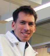 Trevor J. Lujan合作论文数Department of Bioengineering;University of Utah;Department of Bioengineering, University of Utah11
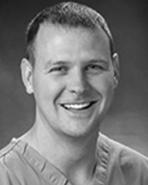 Matthew A. Jolley合作论文数Department of Anesthesia and Critical Care, Children’s Hospital of Philadelphia10
 Jared Zitnay合作论文数10
 John C Gardiner合作论文数Department of Bioengineering;University of Utah;Department of Bioengineering, University of Utah10
 Patrick J. McMahon合作论文数McGowan Institute for Regenerative Medicine, University of Pittsburgh;Department of Bioengineering, University of Pittsburgh;UPMC Department of Orthopedics10
 Timmins Lucas H合作论文数Wallace H. Coulter Department of Biomedical Engineering, Georgia Institute of Technology and Emory University9
 Andrew E. Anderson合作论文数Department of Bioengineering;Scientific Computing and Imaging Institute;University of Utah;Scientific Computing and Imaging Institute, University of Utah9
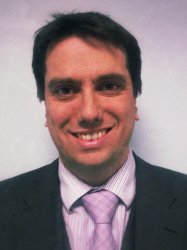 Christopher Peters合作论文数Surgery, Department of Surgery and Cancer, Faculty of Medicine, Imperial College London8
 Nick Willett合作论文数8AI Veress合作论文数7Jay J Shim合作论文数7
 Adam Rauff合作论文数The University of Utah7Allen H Lin合作论文数7
 Michael Yu合作论文数7
 Ross Whitaker合作论文数School of Computing, University of Utah;Scientific Computing and Imaging Institute, University of Utah7
 Susan M. Moore合作论文数Department of Psychology, Melbourne State College Australia7
 Urs Utzinger合作论文数University of Texas;Biomedical Engineering Program;Biomedical Engineering Program, University of Texas7
 Christine L Abraham合作论文数6
 Anton Bowden合作论文数Exponent, Philadelphia, PA, USA6
 Keat Ghee Ong合作论文数Dept Biomed Engn, Michigan Technol Univ6Sly Woo合作论文数5
 Christian Herz合作论文数Department of Anesthesiology and Critical Care Medicine (H.H.N., M.F., Children's Hospital of Philadelphia5
 Devin W Laurence合作论文数School of Aerospace and Mechanical Engineering, The University of Oklahoma5
 Penny R Atkins合作论文数Orthopaedics, University of Utah5
 James C. McWilliams合作论文数Institute of the Environment & Sustainability, University of California, Los Angeles;Department of Atmospheric and Oceanic Sciences, University of California, Los Angeles;Nanjing University of Information Science & Technology5
 Maak Travis G合作论文数Hospital for Special Surgery, New York, NY 10021, USA. maakt@hss.edu5Drury Nicholas J合作论文数5
 Ashley L Kapron合作论文数5
 Stephen K Aoki合作论文数5




