Case report of a phytobezoar causing gastric outlet obstruction as a late complication after biliopancreatic diversion with duodenal switch, review of the literature and discussion of bezoars as a differential diagnosis in patients after bariatric surgery presenting with signs of intestinal obstruction.
En tenant compte de plusieurs nouvelles etudes portant sur le developpement et le traitement de la maladie diverticulaire, cet article apporte un eclairage critique sur les traitements standard de reference.
Mit Rucksicht auf mehrere neue Studien zur Entstehung und Behandlung der Divertikelkrankheit werden in diesem Artikel die traditionellen Behandlungsstandards kritisch beleuchtet.
Background: There is still limited data on the safety of propofol sedation during ERCP, where deep sedation is mandatory.Objective: We asked if non-anesthetist sedation with propofol is safe for ERCP. Setting: A hospital-based gastroenterology center in Switzerland. Patients and Design: Between November 1997 and October 2007, all ERCP procedures were prospectively assessed regarding patient characteristics, American Society of Anesthesiologists (ASA) status, dosage of propofol, fall of oxygen saturation, need to increase nasal oxygen administration above 2 L/min, need for assisted ventilation, need for endotracheal intubation, feasibility of the ERCP procedure, sedation-related adverse events or death. Intervention: ERCP procedures were performed under deep sedation with propofol, administered by the endoscopy team consisting of 1 physician endoscopist, 1 endoscopy assistant and 1 radiology assistant. Patient monitoring consisted of pulse oxymetry, breathing frequency, electrocardiogram and clinical assessment. Results: During the 10 years period, 1305 ERCP procedures (9 % diagnostic, 91 % therapeutic) were performed (mean patient age 64 years, range 16-98 years). In 36 ERCP procedures (2.7 %, all patients ASA III and IV), sedation was administered by an anesthesiology team. In the remaining 1269 ERCP procedures (97.3 %), propofol sedation was administered by the endoscopy team. During ERCP, patients received a mean propofol dose of 459 mg (range 50 -1990 mg) in addition to 50 mg of meperidine. Oxygen saturation fell below 90% (lowest 70%) in 92 patients (7%), normalizing within 30 seconds by stimulating the patient and increasing the nasal oxygen flow to 4 to 10 L/min. 4 patients (0.3%) required mask ventilation for less than 30 seconds. No endotracheal intubation was necessary, no death occurred. 12 ERCP procedures (0.9%) had to be discontinued due to increasing hypoxemia, danger of aspiration or insufficient sedation level. 6 of these patients underwent successful ERCP when sedation was applied by an anesthesiology team, the other 6 patients underwent surgery. Conclusions: The vast majority of ERCP procedures can safely be performed with propofol sedation administered by an endoscopy team consisting of 1 physician endoscopist, 1 endoscopy assistant and 1 radiology assistant. Only a small minority of patients (usually ASA III and IV patients) need anesthetist sedation for ERCP.
Background: Propofol has been shown to be safe for nonanesthetist use during GI endoscopy. However, published studies involved propofol administration by an additional nurse or used specialized patient monitoring or were carried out in tertiary hospitals.Objective: Considering the downward pressure on reimbursement for endoscopic procedures, we asked how much staff and monitoring is necessary for safe use of propofol.Setting: Two private gastroenterology practices.Patients and Design: A total of 27,061 endoscopic procedures (14,856 EGDs and 12,205 colonoscopies) were prospectively assessed regarding patient characteristics, American Society of Anesthesiologists (ASA) status, dosage of propofol, fall of oxygen saturation below 90%, need to increase nasal oxygen administration above 2 L/min, and need for assisted ventilation.Intervention: Propofol was administered by the endoscopy nurse supervised by the endoscopist. Patient monitoring consisted of only pulse oximetry and clinical assessment.Results: The mean propofol dose for EGD was 161 mg (range 50-650 mg). During colonoscopy patients received a mean propofol dose of 116 mg (30-500 mg) in addition to 25 mg of meperidine. Oxygen saturation fell below 90% (lowest 74%) in 623 procedures (2.3%), normalizing within less than 30 seconds by stimulating the patient and increasing the nasal oxygen flow to 4 to 10 L/min. Six patients (ASA 111) required mask ventilation for less than 30 seconds. No endotracheal intubation was necessary.Limitations: There was no further follow-up regarding adverse events after patient discharge from the endoscopy unit.Conclusions: An endoscopy team, consisting of I physician endoscopist and I endoscopy nurse, can safely administer propofol sedation for GI endoscopy in a practice setting without additional staff or specialized monitoring.
Die medizinischen Titel der Preistrager entsprechen dem Stand zur Zeit der jeweiligen Preisverleihung. Die 21 mit einem * gekennzeichneten Preistrager haben in der Zwischenzeit den Titel eines Professors erhalten. Daraus kann man erkennen, dass der Wissenschaftliche Ausschuss der Schweizerischen Herzstiftung weit vorausschauend dem Potenzial und der Qualitat der Kandidaten entsprechend den Preis zugesprochen hat.
Introduction: Preoperative chemoradiotherapy is generally recommended for locally advanced esophageal cancer (clinical stage T3 or T4 or nodal positive disease) but not for early cancer (clinical stage T0 to T2, N0). EUS has been described as the most accurate method to distinguish between early and locally advanced stage in several studies. Recently however, the high accuracy of EUS (90% or higher) was questioned by some investigators. This raises the issue whether the results of studies focused on EUS accuracy may be directly translated into daily clinical practice. Aim & Methods: The aim of this retrospective analysis was to assess the accuracy of preoperative EUS to distinguish between early and locally advanced esophageal cancer in daily clinical practice outside a study setting. EUS was performed by several investigators, including trainees in one university hospital. For this purpose, EUS reports and patient files (medical and surgical) including histological reports of 300 consecutive pts with esophageal tumors were reviewed. In pts with adenocarcinoma or squamous cell cancer and surgical resection without previous radio-/chemotherapy, EUS tumor staging was compared with histological diagnosis. Results: Out of the 300 consecutive pts with esophageal tumor and EUS 102 pts had esophageal surgery after EUS-staging without any radio-/chemotherapy. In 93 pts oesophageal cancer was confirmed, whereas 9 had other tumors. The mean age was 65 years (range 27-89), sex ratio female:male was 1:3.2. To distinguish between early and late tumor stage, the accuracy was 85%. The sensitivity and specificity for early cancer was 59%, and 93%, respectively. The diagnostic accuracy for local tumor spread was 90%, 90%, 68%, 69%, 89% for pT0, pT1, pT2, pT3 and pT4 lesions, respectively. The overall accuracy for T-stage was 74%. For pN-positive staging the accuracy of EUS was 73%. Conclusion: In daily clinical practice, the accuracy of EUS in assessing esophageal tumor staging is lower than in specific studies focusing on EUS accuracy. Mainly early esophageal cancer stages were overstaged. Thus, the implementation of recommendations for diagnostic work-up of esophageal cancer patients resulting from highly specific studies should consider the appropriate clinical setting.
Heartburn is the cardinal symptom of GERD.In patients with non-erosive reflux disease (NERD), the relief of symptoms with acid suppression is less reliable than in patients with erosive GERD.A study comparing rabeprazole sodium 10 and 20 mg tablets once daily vs. placebo was recently completed in NERD patients evaluating GERD symptoms including heartburn over a 4-week period.The response to the first 2 doses was evaluated.METHODS: This was a double blind placebo controlled trial in patients with endoscopically confirmed NERD.Patients were eligible if they reported at least moderate heartburn for at least 5 periods (2: 3 day time periods and?: 1 nighttime period) during the second week of a 2-week, single blind, run-in on placebo.Baseline symptoms were assessed as the last observation before the first dose of the double blind phase.The diary captured assessments for daytime and nighttime heartburn.RESULTS: A total of 203 subjects enrolled in the study.Age and gender distribution was similar in all 3 groups.Intent-to-treat analysis includes 199 evaluable subjects.Symptoms were rated on a 5-point Likert scale from 0 (no symptoms) to 4 (severe symptoms).The mean heartburn score response to the first 2 doses for both daytime and nighttime are displayed in the tables below.No significant difference between rabeprazole 10 vs. rabeprazole 20 mg was observed.CONCLUSION: In NERD patients, rabeprazole 10 and 20 mg provide significant symptom improvement for both daytime and nighttime heartburn.This fast symptom relief is seen on the first day with the first dose.
Objective: In healthy subjects and patients with bleeding peptic ulcers, ranitidine and omeprazole, given parenterally, achieve high intragastric pH values on the first day of therapy. However, data on the antisecretory effect beyond the first 24 h is scanty. In addition, the superiority of either infusion or injection of omeprazole remains unproven. Thus, we have compared the antisecretory effect of high dose omeprazole and ranitidine infusion and injection over the critical first 72 h. Methods: A total of 34 healthy volunteers were randomized into a double-blind crossover 72 h intragastric pH-metry study (data compared: median pH, percentage of time with pH >4 and pH >6). Omeprazole-infusion: initial bolus of 80 mg + 8 mg/h; omeprazole-injection: initial bolus of 80 mg + 40 mg/6 h; Ranitidine-infusion: initial bolus of 50 mg + 0.25 mg/kg/h; ranitidine-injection: 100 mg/6 h. Results: Omeprazole-infusion versus ranitidine-infusion: on day 1: median pH 6.1vs5.1 (p= 0.01) and 95%vs70% was pH >4 (p< 0.01); on day 2: median pH 6.2vs3.2 (p< 0.01); and 100%vs38% was pH >4 (p< 0.01); on day 3: median pH 6.3vs2.7 (p< 0.01); 100%vs26% was pH >4 (p< 0.01). Injections of both drugs were significantly less effective than the infusions on day 1. Thereafter, omeprazole injection was almost as effective as omeprazole infusion, whereas ranitidine injection and infusion were equally effective. Conclusion: Our study shows, for the first time, that omeprazole infusion was significantly superior to all other regimens by having a high median pH >6 on each day. The tolerance effect of ranitidine, however, led to a rapid loss of antisecretory activity on days 2 and 3, rendering it inappropriate for situations in which high intragastric pH-levels appear to be essential.
A careful patient history is the first step of an examination which may lead to the diagnosis of gastro(o)esophageal reflux disease (GERD). Heartburn and regurgitation have a high specificity in GERD. Less typical symptoms of GERD are epigastric pain, dysphagia, odynophagia, pharyngitis, reflux-induced pulmonal symptoms or intermittent chest pain. If alarm symptoms are lacking, the diagnosis of GERD is highly likely if symptoms improve during antisecretory drug treatment. In all other cases an endoscopic investigation of the upper gastrointestinal tract should be performed. If endoscopy reveals oesophagitis, specific treatment can be started. Based on endoscopy, patients with complications can be identified and included into surveillance programs. This is especially important for patients with endobrachyoesophagus (Barrett's oesophagus) where the original squamous epithelial cell lining is replaced by a metaplastic columnar-type epithelium with malignant potential. In the absence of oesophagitis, GERD can be reliably detected or excluded by ambulatory 24-hour pH-metry. Combination of pH-metry with manometry allows to detect motility disorders of the oesophagus which may be due to GERD or to other diseases, causing secondary reflux oesophagitis. Further diagnostic tools are radiology, scintigraphy, provocation tests and endosonography which are indicated only in selected cases? particularly within clinical trials.
We investigated the effect of oral anticoagulation on thrombogenesis induced by the subendothelium of rabbit aorta. Eighteen healthy volunteers underwent a 2-week treatment with the oral coumarin preparation phenprocoumon to a target international normalized ratio (INR) of 5. By using an ex-vivo perfusion chamber system, the interaction between flowing blood and exposed subendothelium was measured at low (50 sec-1) and high (650 sec-1) wall shear rates. The low shear rate simulated blood flow in venous vessels and the high shear rate simulated blood flow in arterial vessels. Deposition of fibrin, platelets, and platelet thrombi on subendothelium was quantified by morphometric and immunologic techniques. Fibrin deposition prevailed at the low shear rate (183 +/- 57 ng/mm2 vs 46 +/- 16 ng/mm2, low vs high shear rate; mean +/- SEM; p less than 0.01). In contrast, the interaction of platelets with subendothelium was more intense at high shear rates when compared with low shear rates, as indicated by higher platelet adhesion (44% +/- 2% vs 9% +/- 1% coverage of subendothelium with platelets, p less than 0.001) and platelet thrombus volumes (3.6 +/- 0.4 microns 3/microns 2 vs 1.3 +/- 0.3 microns 3/microns 2, p less than 0.001). Fibrin deposition on subendothelium was substantially reduced even at a low intensity of anticoagulation (reduction by 60% at INR 2.1 and by 75% at INR 3.7) and was abolished after high coumarin doses (INR 5.2). In contrast, a significant inhibition of platelet thrombus formation could be achieved only by high doses of phenprocoumon (INR 5.2). Our data indicate that relatively low doses of oral anticoagulants (INR between 2 and 3.5) substantially inhibit the fibrin formation on subendothelium prevailing at venous shear conditions, whereas a high intensity of anticoagulation (INR 4 to 5) is necessary to inhibit the formation of platelet-rich thrombi prevailing at arterial shear conditions.
We assessed the effect of hypoxia/reoxygenation on 14C-albumin flux across endothelial monolayers. Cultured bovine pulmonary artery endoehtlial cells were grown to confluence on nitrocellulose filters (pore size 12 μm). The endothelialized filters were mounted in Ussing-type chambers which were filled with cell culture medium (M 199). Equimolar amounts (33 nM) of 14C-labeled and unlabeled albumin were added to the “hot” and “cold” chambers, respectively. The monolayers were then exposed to successive periods (90 min) of normoxia (pO2 20 mmHg), and reoxygenation (pO2 145 mmHg). A gas bubbling system was used to control media pO2 and to ensure adequate mixing. Four aliquots of culture media were taken during each period in order to calculate 14C-albumin permeability across the endothelialized filter. In some experiments, either the xanthine oxidase inhibitor, oxypurinol (10 μM), or superoxide dismutase (600 U/mL), was added to the media immediately prior to the experiments. As compared to the normoxic control period, albumin permeability was 1.5 times higher during hypoxia (p<0.01) and 2.3 times higher during reoxygenation (p<0.01). The reoxygenation-induced increase in albumin permeability was prevented by either oxypurinol or superoxide dismutase. These data indicate that xanthine oxidase-derived oxygen radicals contribute to the hypoxia/reoxygenation-induced endothelial cell dysfunction. The altered endothelial barrier function induced by hypoxia/reoxygenation is consistent with the microvascular dysfunction observed following reperfusion of ischemic tissues.
The aim of this study was to assess the role of neutrophilic elastase in anoxia-reoxygenation-induced, neutrophil-mediated injury to microvascular endothelium. Cultured bovine microvascular endothelial cells were grown to confluence and labeled with 51Cr. The endothelial cells were exposed to a 30-min period of anoxia and subsequently reoxygenated. Endothelial cell injury, quantitated as 51Cr release and cell detachment, was determined 8 h after reoxygenation. Addition of neutrophils upon reoxygenation enhanced the anoxia-reoxygenation-induced increase in 51Cr release and cell detachment. The neutrophil-mediated injury was associated with elastase release from the neutrophils. Four agents were used to inhibit neutrophilic elastase activity: Eglin C, methoxysuccunyl-Ala2-Pro-Val-CH2Cl, L658,758, and a monoclonal antibody against neutrophilic elastase. All elastase inhibitors attenuated the neutrophil-mediated endothelial cell detachment but not 51Cr release. Addition of purified human neutrophilic elastase, at a level that mimicked the release from neutrophils, increased cell detachment in endothelial cells exposed to anoxia-reoxygenation but did not affect 51Cr release. Our results indicate that elastase plays an important role in anoxia-reoxygenation-induced, neutrophil-mediated endothelial cell dysfunction.
The effect of heparin on thrombogenesis induced by the subendothelium of rabbit aorta was investigated in 24 healthy volunteers after intravenous injection of different doses (0, 1000, 2500, and 5000 IU). By using an ex vivo perfusion chamber system, the interaction between flowing blood and exposed subendothelium was measured at low (50 s-1), intermediate (650 s-1), and high (2600 s-1) wall shear rates. The low shear rate simulated blood flow in venous, the intermediate shear rate in arterial, and the high shear rate in small or stenosed arterial vessels. Deposition of fibrin, platelets, and platelet thrombi on vascular subendothelium (SE) was quantified by morphometrical and immunological techniques. Fibrin deposition prevailed at low shear rates and was only minimal at high shear rates (30 +/- 1% vs. 1 +/- 0.4% coverage of SE with fibrin, means +/- SEM, p less than 0.001). In contrast, the interaction of platelets with SE was more intense at high compared to low shear rates, as indicated by higher platelet adhesion (54 +/- 5% vs. 4 +/- 1% coverage of SE with platelets, p less than 0.001) and platelet thrombus volumes (4.8 +/- 1.3 vs. 0.5 +/- 0.1 microns 3/microns 2, p less than 0.001). Fibrin deposition on SE was inhibited by heparin in a dose-dependent manner and was abolished after high doses. In addition, high doses of heparin reduced the height and volume of platelet thrombi at low and intermediate wall shear rates, but no effect was found at the high shear rate. Our data show that heparin inhibits the formation of both fibrin and platelet thrombi on vascular subendothelium. The lack of effect of heparin on platelet thrombus formation at high shear rates indicates that thrombin modulates the growth rate and/or stability of platelet thrombi at low and intermediate shear rates, whereas additional factors may control platelet thrombus growth and stability at high shear conditions.
The objective of this study was to determine whether superoxide mediates the leukocyte-endothelial cell interactions elicited by reperfusion (reoxygenation) of ischemic (hypoxic) tissues. Mesenteric and intestinal blood flows were reduced to 20% of control for 1 h, followed by 1 h of reperfusion. Sixty minutes after reperfusion, red blood cell velocity (Vr), leukocyte rolling velocity (Vw), and the number of adherent leukocytes were measured in mesenteric venules. Then, either human superoxide dismutase (hSOD), hydrogen peroxide-inactivated hSOD, or MoAb IB4 (a monoclonal antibody against the leukocyte adhesion molecule CD18) was injected intravenously. Ten minutes later, repeat measurements were obtained and compared with pretreatment values. hSOD attenuated reperfusion-induced neutrophil adherence and increased Vw/Vr, an index of the fracture stress between leukocytes and endothelium. Peroxide-inactivated hSOD did not alter any parameter. MoAb IB4 attenuated reperfusion-induced adherence but did not alter Vw/Vr. In a correlate study, cultured bovine microvascular endothelium was exposed to 30 min of anoxia, followed by 60 min of reoxygenation. Cat neutrophils were added during reoxygenation. Reoxygenation-induced leukocyte adherence was attenuated by either hSOD or MoAb IB4 but not by inactivated hSOD. Adherence of phorbol 12-myristate 13-acetate-activated cat neutrophils to plastic was unaffected by hSOD or inactive hSOD, yet MoAb IB4 virtually abolished the response. These results indicate that superoxide mediates reperfusion-induced leukocyte adherence and that endothelial cells are required for this superoxide-mediated adherence.
Oxygen-derived free radicals have been implicated as a mediator of the microvascular and parenchymal cell injury associated with reperfusion of ischemic tissues. Xanthine oxidase and neutrophilic NADPH oxidase are commonly invoked to explain reperfusion-induced production of oxygen radicals. The strengths and weaknesses of evidence used to suggest the involvement of both sources are discussed. Evidence is also presented which suggests that xanthine oxidase and neutrophils are redundant yet interactive mechanisms that play an important role in reperfusion injury.
 A HAEBERLI合作论文数7HR BAUMGARTNER合作论文数6
 D. Neil Granger合作论文数Department of Molecular and Cellular Physiology, School of Graduate Studies, Louisiana State University Health Sciences Center in Shreveport6PW STRAUB合作论文数5
 Peter Netzer合作论文数5Pr Kvietys合作论文数4Christophe Petrig合作论文数3Fred Halter合作论文数3Thomas Bombeli合作论文数3HJ SCHRAG合作论文数2P. Aeberhard合作论文数2S. Navarro合作论文数2M. Prados合作论文数2T. Obeid合作论文数2Jan Schmielau合作论文数2ME SCHELLING合作论文数2Erminia Caldiron合作论文数2J. Triller合作论文数2Christian Roeder合作论文数2André Gut合作论文数2Hans Weidenbach合作论文数2Alain Vandermeeren合作论文数2Enrique Domínguez-Muñoz合作论文数2Guido Adler合作论文数2Nicholas A. Wright合作论文数2Gisli H. Sigurdsson合作论文数2Jean-Marc Dumonceau合作论文数2Ch. Becker合作论文数2Peter Malfertheiner合作论文数2Jacques Devière合作论文数2PC Bornman合作论文数2Günter Klöppel合作论文数2Roland M. Schmid合作论文数2HU Baer合作论文数2Sabine Beeler合作论文数2David L. Can-Locke合作论文数2Rudolf W. Ammann合作论文数2Helmut Friess合作论文数2Myriam Delhaye合作论文数2P. Vock合作论文数2C Gaia合作论文数2Hj Granger合作论文数2
 Nicholas Robert Lemoine (尼古拉斯·罗伯特·莱蒙)合作论文数Zhengzhou University2A Schmassmann合作论文数2
 Leslie I. Gold合作论文数Department of Pathology, Grossman School of Medicine, New York University2
 Matthias Ebert合作论文数School of Translational Medicine, Medical Faculty Mannheim, Heidelberg University2
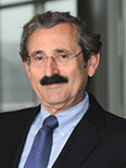 Murray Korc合作论文数School of Medicine, Indiana University2
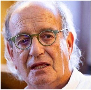 Claudio Bassi合作论文数Department of General and Pancreatic Surgery, Verona University2Dale E. Bockman合作论文数2




