OBJECTIVE:Chiari malformation type I (CM-I) is traditionally conceptualized as a structural disorder of the posterior cranial fossa characterized by cerebellar tonsillar herniation. However, increasing clinical and experimental evidence suggests that CM-I may be associated with distributed neurocognitive and affective sequelae consistent with cerebellar network dysfunction. METHODS:The authors conducted a PRISMA-compliant systematic review of the MEDLINE and Embase databases from database inception through November 2025 to identify studies reporting standardized neuropsychological outcomes in pediatric and adult patients with CM-I. Given substantial heterogeneity in study design, cognitive instruments, and outcome reporting, findings were synthesized using a structured narrative approach. Methodological quality was assessed using an adapted Newcastle-Ottawa Scale and mapped to Agency for Healthcare Research and Quality criteria. RESULTS:Thirty-four studies comprising 2113 individuals with CM-I met inclusion criteria. Across pediatric and adult cohorts, selective cognitive vulnerabilities were most consistently observed in complex attention, executive function, visuospatial processing, learning and memory, language, and higher-order social cognition. These profiles closely resembled features of cerebellar cognitive affective syndrome. Cognitive deficits varied by developmental stage and were modulated by chronic pain, psychiatric comorbidity, and neurodevelopmental factors. Advanced neuroimaging studies demonstrated widespread disruption of cerebello-thalamo-cortical and cortico-ponto-cerebellar networks. Reported effects of posterior fossa decompression on neurocognitive outcomes were heterogeneous and inconsistent across domains and study designs. CONCLUSIONS:The available evidence supports reconceptualizing CM-I as a heterogeneous, large-scale brain network disorder with clinically meaningful cognitive and affective consequences in a subset of patients. Integration of domain-specific neuropsychological assessment into routine clinical evaluation may improve prognostication and guide future mechanism-based interventions.
Chiari malformation type I (CM1) is traditionally viewed as a structural disorder of the posterior fossa characterized by cerebellar tonsillar herniation. However, growing evidence indicates that CM1 is frequently accompanied by disturbances in cognition, emotion, and behavior that cannot be fully explained by pain, hydrocephalus, or radiographic severity alone. Advances in cerebellar neuroscience highlight the cerebellum’s critical role in higher-order cognition and affect through distributed cerebrocerebellar networks. Here, we synthesize neuropsychological, neuroimaging, developmental, and clinical evidence to reframe CM1 as a disorder of cerebellar cognitive affective dysfunction. Across pediatric and adult populations, CM1 is associated with domain-specific inefficiencies in attention, executive control, working memory, visuospatial processing, language fluency, and social cognition, closely paralleling the cerebellar cognitive affective syndrome (CCAS). Network-level imaging studies further demonstrate disruption of cortico-ponto-cerebellar and cerebello-thalamo-cortical connectivity, supporting a distributed rather than focal model of disease. We highlight the modulatory roles of development and chronic pain, critically evaluate cognitive outcomes after posterior fossa decompression, and discuss implications for assessment and future research. Recognizing CM1 as a disorder of cerebellar network integration has important consequences for clinical care and mechanistic investigation.
Consciousness is a fundamental component of cognition1, but the degree to which higher-order pattern recognition relies on it remains disputed2,3. Here we demonstrate the persistence of oddball discrimination, semantic processing and online prediction in individuals under general-anaesthesia-induced loss of consciousness4,5. Using high-density Neuropixels microelectrodes6 to record both single-unit and local-field-potential neural activity in the human hippocampus while playing a series of tones to anaesthetized patients, we found that hippocampal neurons and local oscillations retained some detection of oddball tones. This effect size grew over the course of the experiment (around 10 min), demonstrating representational plasticity. A biologically plausible recurrent neural network model showed that learning and oddball representation are an emergent property of flexible tone discrimination. Moreover, when we played language stimuli, single units and local field potentials carried information about the semantic and grammatical features of natural speech, even predicting semantic information about upcoming words. Together these results indicate that in the hippocampus, which is anatomically and functionally distant from primary sensory cortices7, complex processing of sensory stimuli occurs even in the unconscious state. In the hippocampus, complex processing of sensory stimuli occurs even in the unconscious state.
Abstract Objective Expanded indications, diagnostic tools, and treatment options have transformed the landscape of modern pediatric epilepsy surgery. Published real‐world experiences from large surgical cohorts are still needed. To close this gap, we evaluated access, indications, treatment, and outcomes in a contemporary pediatric epilepsy surgery program. Methods We evaluated data from 100 consecutive diagnostic and therapeutic procedures in 62 pediatric and young adult patients. Data collected included demographics, diagnostics, procedures, 12‐month seizure and medication outcomes, and adverse events. Primary outcome for patients who underwent treatment with the goal of cure or resection, along with the intent of palliation, was 12‐month postoperative Engel/ILAE scores. For those who underwent RNS‐implant (alone or in combination with a second procedure) with the goal of seizure reduction, the primary outcome was proportion seizure reduction in the prior 28 days at 12 months postoperatively. Results Patients largely matched state and regional demographics. Epilepsy types included unifocal (n = 33, 53.2%), multifocal (n = 15, 24.2%), generalized (n = 12, 19.4%), and combined (n = 2, 3.2%). Of the 100 procedures, 36.0% were diagnostic SEEG (n = 35) and 64.0% were treatment procedures (n = 58; note: n = 4 pending). Among patients who underwent surgical treatment with the goal of cure (n = 33), a 12‐month Engel I/ILAE I or III outcome was achieved in 81.8% (n = 27). Among patients who underwent RNS implantation, 79.0% were responders (>50% reduction) and 38.0% were super‐responders (>90% reduction), with a median seizure reduction of 78% at 12 months. Three treatment procedures (4.7%, n = 3 patients) had a surgical complication, none permanent. Significance Contemporary pediatric epilepsy surgery, utilizing modern diagnostic and surgical techniques, including off‐label use of RNS, provides safe, effective, accessible, and equitable treatment to children across a broad range of indications, many of whom, historically, have not been considered viable surgical candidates. Plain Language Summary “In this article by McLaren et al, 100 consecutive procedures were examined from a modern pediatric epilepsy surgery program. By utilizing advanced diagnostic and surgical techniques, they've shown that pediatric epilepsy surgery can be safe, effective, and accessible across diverse conditions and demographics. Notably, 82% of patients who underwent surgery aimed at curing their condition achieved seizure‐freedom in 12 months and 79% of patients with Responsive Neurostimulation (RNS) implants experienced significant seizure reduction.”
As implantable brain-computer interfaces (iBCIs) for communication and movement transition from cutting-edge research to clinical practice, a standardized approach will be required to reliably plan neurosurgeries involving complex microelectrode arrays and other neural sensors. Here, through our BrainGate study experiences, we present a replicable methodology, using open-source tools, to create interactive, personalized, 3-dimensional, virtual and physical, functional mapping models to guide iBCI surgical planning and provide intra-operative imaging displays. ### Competing Interest Statement The MGH Translational Research Center has a clinical research support agreement (CRSA) with Ability Neurotech. Axoft, Neuralink, Neurobionics, Paradromics, Precision Neuro, Synchron, and Reach Neuro, for which LRH and SSC provide consultative input. LRH is a non-compensated member of the Board of Directors of a nonprofit assistive communication device technology foundation (Speak Your Mind Foundation). Mass General Brigham (MGB) is convening the Implantable Brain-Computer Interface Collaborative Community (iBCI-CC); charitable gift agreements to MGB, including those received to date from Paradromics, Synchron, Precision Neuro, Neuralink, and Blackrock Neurotech, support the iBCI-CC, for which LRH provides effort. SSC has stock/stock options in Beacon Biosignals. DBR has no relevant financial disclosures. JMH is a consultant for Neuralink and Paradromics, is a shareholder in Maplight Therapeutics and Enspire DBS, and is a co-founder and shareholder in Re-EmergeDBS. He is also an inventor on intellectual property licensed by Stanford University to Blackrock Neurotech and Neuralink. SDS is an inventor on intellectual property licensed by Stanford University to Blackrock Neurotech and Neuralink Corp. He has patent applications related to speech BCI owned by the Regents of the University of California. Stavisky is an advisor to Sonera. DMB is a surgical consultant for Paradromics Inc. EYC is a consultant at Neuralink Corp. All other authors have no competing interests. ### Clinical Trial NCT00912041 ### Funding Statement The investigators would like to thank the participants, their families, and care partners for their extraordinary dedication to this research. The investigators would also like to thank the collaborators on the scientific research teams at Massachusetts General Hospital, Brown University, Providence VA Medical Center, Stanford, UC Davis, and Emory. CAUTION: Investigational Device. Limited by Federal Law to Investigational Use. The content is solely the responsibility of the authors and does not necessarily represent the official views of the National Institutes of Health, or the Department of Veterans Affairs, or the United States Government. The funders had no role in the study design, data collection and interpretation, or the decision to submit the work for publication. The work described in this manuscript was supported by: NIH-National Institute on Deafness and Other Communication Disorders (NIDCD) from U01DC017844 (LRH), R01DC014034 (JMH), and NIH-National Institute of Neurologic Disorders and Stroke (NINDS) from UH2NS095548 (LRH) and U01NS098968 (SSC) and R01NS134410 (ACP), Searle Scholars (SDS), Burroughs Wellcome Fund Career Award at the Scientific Interface (SDS), CDMRP ALS Pilot Clinical Trial Award (AL220043) from the Office of the Assistant Secretary of Defense for Health Affairs (SDS), the Wu Tsai Neurosciences Institute at Stanford (JMH), and Larry and Pamela Garlick (JMH). ### Author Declarations I confirm all relevant ethical guidelines have been followed, and any necessary IRB and/or ethics committee approvals have been obtained. Yes The details of the IRB/oversight body that provided approval or exemption for the research described are given below: The IRB of the Massachusetts General Hospital gave ethical approval for this work. I confirm that all necessary patient/participant consent has been obtained and the appropriate institutional forms have been archived, and that any patient/participant/sample identifiers included were not known to anyone (e.g., hospital staff, patients or participants themselves) outside the research group so cannot be used to identify individuals. Yes I understand that all clinical trials and any other prospective interventional studies must be registered with an ICMJE-approved registry, such as ClinicalTrials.gov. I confirm that any such study reported in the manuscript has been registered and the trial registration ID is provided (note: if posting a prospective study registered retrospectively, please provide a statement in the trial ID field explaining why the study was not registered in advance). Yes I have followed all appropriate research reporting guidelines, such as any relevant EQUATOR Network research reporting checklist(s) and other pertinent material, if applicable. Yes The code for reproducing the figures is made available at https://github.com/Center-For-Neurotechnology/BrainInterface3D. The data required to reproduce the visualizations in this study are publicly available on FigShare. The dataset contains neuroimaging data recorded from participants, including 3D models of the brain and skull.
Chiari malformation (CM) types 1 and 2 are common congenital hindbrain disorders characterized by varying degrees of cerebellar herniation and association with neural tube defects. Knowledge of CM pathogenesis has thus far relied on preclinical animal models. Here, we provide a comprehensive review of animal models (genetic, teratogen-induced, surgical, and spontaneous) of CM1 and CM2, highlighting their construct validity, pathophysiological insights, and translational relevance. Genetic models exhibit defects in cranial base development, cerebrospinal fluid (CSF) flow, and neural tube closure. Teratogenic and surgical models have been informative for understanding the consequences of CSF leakage and validating prenatal treatments, especially in fetal sheep and chick embryos. Spontaneous models in dogs, cattle, and primates have offered complementary insights but the heterogeneity arising from species-specific differences obscures further understanding. Despite these advances, translational challenges persist, including species-specific differences in skull anatomy, CSF physiology, and symptomatology. We highlight the need for a patient-first approach, emphasizing the translational importance of large-scale gene discovery research in human patients to inform and prioritize dowstream functional investigations in animal models to enhance mechanistic understanding and support the development of etiology-specific therapies for CM.
BACKGROUND AND OBJECTIVES:Minimal exposure to neurosurgery in standard medical school curricula alongside a growing need for neurosurgical care necessitate early exposure programs that promote medical student retention in neurosurgery. Here, we evaluate preclinical students' perceptions on a one-day, resident-designed introductory neurosurgical course. METHODS:Course curriculum involved hands-on and discussion-based elements split into three stations: (1) suturing/general information; (2) introduction to drilling/LPs/EVDs; and (3) case/clinical skills review. Anonymous online post-course surveys were administered immediately after the course and one year after the course following completion of clerkships. RESULTS:Ten medical students participated in the course, 70 % of which indicated desire to pursue neurosurgery at course onset. Nine students responded to an immediate post-survey, which revealed that, in students with a desire to pursue neurosurgery (n = 6) versus those undifferentiated in specialty interest (n = 3): the program was rated as having high versus moderate impacts on readiness and enthusiasm for surgical clerkships (readiness: avg 4 vs 3.67; enthusiasm: avg 4.3 vs 3.67), on fostering a sense of belonging within neurosurgery (avg 4.67 vs 3.33), and on clarifying specialty interests (avg 4.3 vs 3.3). At 1-year, 75 % of participants (n = 6), including 100 % of students (n = 4) from minority backgrounds, noted a persistent desire to pursue neurosurgery. In participants with a continued interest in neurosurgery versus those planning to pursue different fields, the course was rated as having high versus moderate impacts on preparedness for surgery clerkship experiences (average 4.17 vs 3.5) and moderate impact on participant desire to pursue neurosurgery (3.67 vs 3). CONCLUSIONS:Significant enthusiasm and sense of belonging fostered by the course, particularly amongst students with prior interests in neurosurgery, support that early specialty-specific exposure may promote retention of students in neurosurgery. High rates of persistent engagement in neurosurgery amongst minority students at 1-year post-course suggest that such courses may help to build a more representative neurosurgical workforce.
Chiari malformation type I (CM-I) and hydrocephalus often occur together, but their connection can involve a variety of different and overlapping pathophysiological mechanisms. These include posterior fossa hypoplasia, cerebrospinal fluid (CSF) flow obstruction at the foramen magnum, venous outflow restriction, craniospinal pressure dissociation, and genetically mediated brain overgrowth. Such mechanisms often converge in complex developmental disorders, most notably syndromic craniosynostoses where premature suture fusion restricts posterior fossa expansion and perturbs venous and CSF dynamics, driving hindbrain herniation and ventriculomegaly. Concurrent CM-I and hydrocephalus may be best categorized into one of three mechanistic patterns: 1) hydrocephalus with secondary Chiari-like tonsillar descent (acquired CM-I); 2) CM-I with secondary hydrocephalus; or 3) simultaneous/complex presentations driven by shared developmental anomalies. Recognizing these distinctions is crucial for guiding treatment selection, as clinical management often relies on identifying the primary pathological driver, with CSF diversion (ventriculoperitoneal shunt [VPS] or endoscopic third ventriculostomy [ETV]) used for hydrocephalus-driven cases, posterior fossa decompression (PFD) for CM-I-driven cases, and multidisciplinary staged interventions reserved for complex or syndromic cases. Future directions include genetic stratification, advanced CSF dynamics imaging, computational biomechanical modeling, and the integration of multimodal data to individualize intervention timing and techniques. A mechanism-based framework, rather than a purely anatomical classification, may enhance diagnostic accuracy and improve surgical outcomes in CM-I with hydrocephalus.
OBJECTIVE:Chiari malformation type 1 (CM1) is the most common neurological disorder of the craniocervical junction. CM1 is characterized by cerebellar tonsillar herniation below the foramen magnum, causing CSF obstruction and neural compression. Patients with CM1 suffer from highly variable symptoms, progression, comorbidities, and outcomes, partly due to poor understanding of CM1 pathogenesis. In this paper, the authors present the only familial CM1 cohort study to statistically assess intrafamilial clinical phenotypes to date. METHODS:Comprehensive medical, surgical, and family histories and neuroimaging were collected from families with multiple CM1-affected family members. Univariate analysis was performed for each symptom, comorbidity, or surgery to compare observed versus expected frequencies of affected patients with another CM1-affected family member with the same clinical characteristic. RESULTS:Twenty-four new familial CM1 cases (totaling 57 patients with CM1) are presented. Intrafamilial similarities were identified for age of onset, symptoms such as headaches (p = 0.007) and neck pain (p = 0.018), neurological comorbidities such as syringomyelia (p = 0.003) and hydrocephalus (p = 0.0001), neurodevelopmental conditions such as dyslexia (p < 0.0001), and connective tissue disorders (CTDs) such as Ehlers-Danlos syndrome (p < 0.0001). CONCLUSIONS:These results suggest that CM1 and its associated clinical phenotypes, including age of onset, clinical symptoms, neurological comorbidities, neurodevelopmental conditions, and CTDs, are genetically influenced. Whole-exome sequencing of CM1 patient-parent trios has the potential to identify the genetic determinants of CM1, with implications for neurosurgical management.
Consciousness is a fundamental component of cognition, 1 but the degree to which higher-order perception relies on it remains disputed. 2,3 Here we demonstrate the persistence of learning, semantic processing, and online prediction in individuals under general anesthesia-induced loss of consciousness. 4,5 Using high-density Neuropixels microelectrodes 6 to record neural activity in the human hippocampus while playing a series of tones to anesthetized patients, we found that hippocampal neurons could reliably detect oddball tones. This effect size grew over the course of the experiment (∼10 minutes), consistent with learning effects. A biologically plausible recurrent neural network model showed that learning and oddball representation are an emergent property of flexible tone discrimination. Last, when we played language stimuli, single units and ensembles carried information about the semantic and grammatical features of natural speech, even predicting semantic information about upcoming words. Together these results indicate that in the hippocampus, which is anatomically and functionally distant from primary sensory cortices, 7 complex processing of sensory stimuli occurs even in the unconscious state.
Background: Calvarial venous malformations (VMs) are rare and genetically understudied. While somatic TEK receptor tyrosine kinase (TEK) mutations drive sporadic VMs, their role in scalp-calvarial VMs is unknown. We report the first pediatric case of a calvarial VM with a pathogenic somatic TEK mutation and its molecular implications. Methods: A 16-year-old female with a symptomatic parietal scalp VM underwent neurosurgical resection. Exome sequencing was performed on both lesional and blood DNA. Single-cell RNA sequencing (scRNA-seq) data from normal brain vasculature were analyzed for TEK expression and pathway enrichment. Results: A novel somatic TEK L914F mutation (chr9:27212760-C-T [GRCh38]), absent in germline DNA and population databases, was identified and predicted to be deleterious (CADD: 24). scRNA-seq data analysis revealed TEK enrichment in endothelial cells, particularly in fetal and arterial subtypes, and implicated angiogenesis and PI3K/Rho signaling as potential downstream phenotypic and molecular consequences. Conclusions: This first pediatric scalp VM with a somatic TEK L914F mutation expands the phenotypes associated with TEK-related vascular anomalies. These findings emphasize the role of somatic TEK mutation in diverse VMs and support genetic testing in sporadic cases. Further studies are needed to define therapeutic targets.
High-density microelectrode arrays have opened new possibilities for systems neuroscience, but brain motion relative to the array poses challenges for downstream analyses. We introduce DREDge (Decentralized Registration of Electrophysiology Data), a robust algorithm for the registration of noisy, nonstationary extracellular electrophysiology recordings. In addition to estimating motion from action potential data, DREDge enables automated, high-temporal-resolution motion tracking in local field potential data. In human intraoperative recordings, DREDge's local field potential-based tracking reliably recovered evoked potentials and single-unit spike sorting. In recordings of deep probe insertions in nonhuman primates, DREDge tracked motion across centimeters of tissue and several brain regions while mapping single-unit electrophysiological features. DREDge reliably improved motion correction in acute mouse recordings, especially in those made with a recent ultrahigh-density probe. Applying DREDge to recordings from chronic implantations in mice yielded stable motion tracking despite changes in neural activity between experimental sessions. These advances enable automated, scalable registration of electrophysiological data across species, probes and drift types, providing a foundation for downstream analyses of these rich datasets.
Humans are capable of generating extraordinarily diverse articulatory movement combinations to produce meaningful speech. This ability to orchestrate specific phonetic sequences, and their syllabification and inflection over subsecond timescales allows us to produce thousands of word sounds and is a core component of language 1 , 2 . The fundamental cellular units and constructs by which we plan and produce words during speech, however, remain largely unknown. Here, using acute ultrahigh-density Neuropixels recordings capable of sampling across the cortical column in humans, we discover neurons in the language-dominant prefrontal cortex that encoded detailed information about the phonetic arrangement and composition of planned words during the production of natural speech. These neurons represented the specific order and structure of articulatory events before utterance and reflected the segmentation of phonetic sequences into distinct syllables. They also accurately predicted the phonetic, syllabic and morphological components of upcoming words and showed a temporally ordered dynamic. Collectively, we show how these mixtures of cells are broadly organized along the cortical column and how their activity patterns transition from articulation planning to production. We also demonstrate how these cells reliably track the detailed composition of consonant and vowel sounds during perception and how they distinguish processes specifically related to speaking from those related to listening. Together, these findings reveal a remarkably structured organization and encoding cascade of phonetic representations by prefrontal neurons in humans and demonstrate a cellular process that can support the production of speech.
From sequences of speech sounds1,2 or letters3, humans can extract rich and nuanced meaning through language. This capacity is essential for human communication. Yet, despite a growing understanding of the brain areas that support linguistic and semantic processing4-12, the derivation of linguistic meaning in neural tissue at the cellular level and over the timescale of action potentials remains largely unknown. Here we recorded from single cells in the left language-dominant prefrontal cortex as participants listened to semantically diverse sentences and naturalistic stories. By tracking their activities during natural speech processing, we discover a fine-scale cortical representation of semantic information by individual neurons. These neurons responded selectively to specific word meanings and reliably distinguished words from nonwords. Moreover, rather than responding to the words as fixed memory representations, their activities were highly dynamic, reflecting the words' meanings based on their specific sentence contexts and independent of their phonetic form. Collectively, we show how these cell ensembles accurately predicted the broad semantic categories of the words as they were heard in real time during speech and how they tracked the sentences in which they appeared. We also show how they encoded the hierarchical structure of these meaning representations and how these representations mapped onto the cell population. Together, these findings reveal a finely detailed cortical organization of semantic representations at the neuron scale in humans and begin to illuminate the cellular-level processing of meaning during language comprehension.
Objective: Extraoperative electrical cortical stimulation (ECS) facilitates defining the seizure onset zone (SOZ) and eloquent cortex. The clinical relevance of stimulation-induced afterdischarges (ADs) is not well defined. Methods: Fifty-five patients who underwent intracranial electroencephalogram evaluations with ECS were retrospectively identified. ADs were identified in these recordings and categorized by pattern, location, and association with stimulation-induced seizures. Results: ADs were generated in 1774/9285 (19%) trials. Rhythmic spikes and irregular ADs within the stimulated bipolar contact pair were predictive of location within the SOZ compared to non-epilepto genic/non-irritative cortex (rhythmic spikes OR 2.24, p = 0.0098; irregular OR 1.39; p = 0.013). ADs immediately preceding stimulated seizures occurred at lower stimulation intensity thresholds compared to other stimulations (mean 2.94 +/- 0.28 mA vs. 4.16 +/- 0.05 mA respectively; p = 0.0068). Conclusions: Changes in AD properties can provide clinically relevant data in extraoperative stimulation mapping. Significance: Although not exclusive to the SOZ, the generation of rhythmic spikes may suggest that a stimulation location is within the SOZ, while decreased stimulation intensity thresholds eliciting ADs may alert clinicians to a heightened probability of seizure generation with subsequent stimulation. (c) 2024 International Federation of Clinical Neurophysiology. Published by Elsevier B.V. All rights reserved.
INTRODUCTION: The basic functional unit of the mammalian neocortex is the cortical column. It consists of a vast diversity of circuit elements and interconnected layers supporting a range of computations for cognitive functions. While much progress has been made in our understanding of its functional architecture in animal models, its role in complex, uniquely human cognitive processes remain unknown. METHODS: We utilized a new columnar recording approach in humans participating of cognitive tasks, while undergoing awake neurosurgical procedures. The novelty of this approach hinged on utilizing state-of-the-art Neuropixels probes for our recordings, a silicon-based electrophysiology recording electrodes with high channel count and recording site density. This technology allows us to measure neural activity with single cell resolution throughout the neocortical column and its layers, and at a scale that is far beyond the capabilities of current clinically-approved devices. The participants performed a naturalistic visual priming-based sentence production task that provided them with pictorial representations of events that had to be articulated in specific form and order. RESULTS: We observed distinct spatiotemporal activity dynamics throughout the cortical column in the language-dominant prefrontal cortex that encoded a preparatory mode for speech generation, as well as visual perception and sentence construction processing stages during natural speech. Furthermore, we observed the emergence of this columnar activity pattern with optimal visual inspection of presented images (by high resolution eye and pupil tracking) and associated improved performance during the task. CONCLUSIONS: These results highlight a functional organization of the cortical column in the human prefrontal cortex subserving sensory-motor transformations for visual conscious perception and speech production, with profound implications for future brain machine interfaces that aim to restore complex human cogntive functions.
Chiari malformation type 1 (CM1) is the most common structural brain disorder involving the craniocervical junction, characterized by caudal displacement of the cerebellar tonsils below the foramen magnum into the spinal canal. Despite the heterogeneity of CM1, its poorly understood patho-etiology has led to a 'one-size-fits-all' surgical approach, with predictably high rates of morbidity and treatment failure. In this review we present multiplex CM1 families, associated Mendelian syndromes, and candidate genes from recent whole exome sequencing (WES) and other genetic studies that suggest a significant genetic contribution from inherited and de novo germline variants impacting transcription regulation, craniovertebral osteogenesis, and embryonic developmental signaling. We suggest that more extensive WES may identify clinically relevant, genetically defined CM1 subtypes distinguished by unique neuroradiographic and neurophysiological endophenotypes.
High-density microelectrode arrays (MEAs) have opened new possibilities for systems neuroscience in human and non-human animals, but brain tissue motion relative to the array poses a challenge for downstream analyses, particularly in human recordings. We introduce DREDge (Decentralized Registration of Electrophysiology Data), a robust algorithm which is well suited for the registration of noisy, nonstationary extracellular electrophysiology recordings. In addition to estimating motion from spikes in the action potential (AP) frequency band, DREDge enables automated tracking of motion at high temporal resolution in the local field potential (LFP) frequency band. In human intraoperative recordings, which often feature fast (period <1s) motion, DREDge correction in the LFP band enabled reliable recovery of evoked potentials, and significantly reduced single-unit spike shape variability and spike sorting error. Applying DREDge to recordings made during deep probe insertions in nonhuman primates demonstrated the possibility of tracking probe motion of centimeters across several brain regions while simultaneously mapping single unit electrophysiological features. DREDge reliably delivered improved motion correction in acute mouse recordings, especially in those made with an recent ultra-high density probe. We also implemented a procedure for applying DREDge to recordings made across tens of days in chronic implantations in mice, reliably yielding stable motion tracking despite changes in neural activity across experimental sessions. Together, these advances enable automated, scalable registration of electrophysiological data across multiple species, probe types, and drift cases, providing a stable foundation for downstream scientific analyses of these rich datasets.
 Angelique C Paulk合作论文数11
 Domokos Meszéna合作论文数8
 Ziv Williams合作论文数8
 Sydney Cash合作论文数Division of Epilepsy, Department of Neurology, Mass General Brigham;Department of Neurology, Massachusetts General Hospital;Harvard Medical School, Harvard University7
 William E. Butler合作论文数Massachusetts General Hospital6Yoav Kfir合作论文数5
 Irene Caprara合作论文数5
 Kristopher Kahle合作论文数Department of Neurosurgery, School of Medicine, Yale University;Division of Pediatric Neurosurgery, Yale University5Brian Coughlin合作论文数4Charlie Windolf合作论文数4
 Adam J Kundishora合作论文数Dept Neurosurg, Yale Univ4
 Erdem Varol合作论文数Columbia University4
 Bernardo Rudy合作论文数Department of Anesthesiology, Perioperative Care, and Pain Medicine, Grossman School of Medicine, New York University;Department of Neuroscience and Physiology, Grossman School of Medicine, New York University4
 Khanna Arjun合作论文数French Family Science Center, Duke University4
 Hardstone Richard合作论文数Center for Neurogenomics and Cognitive Research, VU University Amsterdam Amsterdam;Neuroscience Campus Amsterdam;Center for Neurogenomics and Cognitive Research, VU University Amsterdam Amsterdam4
 Eric Trautmann合作论文数Dartmouth Thayer School of Engineering4
 Nicole R Provenza合作论文数Department of Neurosurgery, Baylor College of Medicine3
 Julien Boussard合作论文数Stanford university3
 Sarah R Heilbronner合作论文数Department of Neuroscience, University of Minnesota3
 Garrett Banks合作论文数From the Department of Neurological Surgery, Columbia University Medical Center;Weill Cornell Medical College, Cornell University;Department of Neurology, Columbia University Medical Center;Columbia University Medical Center, Columbia University Medical Center3Tremblay Robin合作论文数3
 Andrew Hale合作论文数From the Divisions of Cardiovascular and Network Medicine and.3
 Seth Leo Alper合作论文数Molecular and Vascular Medicine Unit, Beth Israel Deaconess Medical Center3
 Christopher Stapleton合作论文数Wang Ambulatory Care Center, Massachusetts General Hospital;Harvard Medical School3
 Duy Phan Q合作论文数Department of Neuroscience, The Ohio State University College of Medicine3Despenza Tyrone合作论文数Department of Physiology and Neurobiology, Geisel School of Medicine at Dartmouth Lebanon3
 Rungratsameetaweemana Nuttida合作论文数Neurosciences Graduate Program, University of California3
 Nanda Pranav合作论文数Department of Psychiatry, Beth Israel Deaconess Medical Center;College of Physicians &, Columbia University;Surgeons, New York, New York.;College of Physicians & Surgeons, Columbia University3
 Liam Paninski合作论文数Zuckerman Institute, Columbia University;Department of Statistics, Columbia University;Kavli Institute for Brain Science, Columbia University3
 Mark Richardson合作论文数Centre for Developmental Neurobiology, School of Neuroscience, Institute of Psychiatry, Psychology & Neuroscience, King’s College London3Carol Nelson-Williams合作论文数3
 Sameer Anil Sheth合作论文数Baylor College of Medicine3Jordan R Shaker合作论文数2Martina L Mustroph合作论文数2Jack Ocken合作论文数2Anna Lebedeva合作论文数UCL Queen Square Institute of Neurology, University College London2
 Jeffrey Schweitzer合作论文数2
 Edward Chang (张复伦)合作论文数Weill Institute for Neurosciences, University of California, San Francisco;Department of Neurological Surgery, School of Medicine, University of California, San Francisco2Han Yu合作论文数2
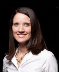 Anne Churchland合作论文数Department of Neurobiology, University of California, Los Angeles2
 Moreno-De-Luca Andres合作论文数Genomic Medicine Institute, Geisinger Health System2
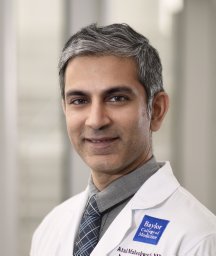 Atul Maheshwari合作论文数Department of Neurology, Massachusetts General Hospital;Harvard Medical School;Harvard Medical School, Massachusetts General Hospital2Chung Jason E合作论文数2
 Kristin Sellers合作论文数Department of Psychiatry, University of North Carolina at Chapel Hill;Neurobiology Curriculum, University of North Carolina at Chapel Hill;University of North Carolina at Chapel Hill, University of North Carolina at Chapel Hill2
 Jin Sheng Chih合作论文数Department of Psychiatry, Washington University School of Medicine2
 Alica Goldman合作论文数Baylor College of Medicine, Baylor College of Medicine2
 Joao Couto合作论文数Theoretical Neurobiology and Neuroengineering Laboratory, University of Antwerp;Neuro-Electronics Research Flanders (NERF),;Theoretical Neurobiology and Neuroengineering Laboratory, University of Antwerp2
 Duo Xu合作论文数2
 Zhiwen Ye合作论文数2




