Supplementary Figure 9 from Immortalization of Human Bronchial Epithelial Cells in the Absence of Viral Oncoproteins
PDF File - 35K, Annexin V staining by flow cytometry 24 hours after addition of 1nM docetaxel to MDA-MB-231 cells.
Supplementary Figures 1-5. Supplementary Fig. 1. Analysis of single-strand G-overhang by native and denatured DNA gel electrophoresis. Supplementary Fig. 2. Telomere FISH analysis of chromosomal aberrations induced by Ssb1 depletion in mouse and human cells at metaphase. Supplementary Fig. 3. Analysis of hTERT and hSsb1 co-dependent association with telomeres by ChIP. Supplementary Fig. 4. Analysis of hSsb1 interaction with TRF1 and TRF2 by co-immunoprecipitation and ChIP analysis of their influence on hSsb1 recruitment to telomeres. Supplementary Fig. 5. Measurement of in vitro telomerase activity (TRAP assay) in control and hSsb1 depleted cells.
PDF File - 2103K, Normalization of expression array datasets. Five expression arrays were normalized and pooled (see Methods).
PDF File - 86K, List of cell lines (from Neve et al. 2006, see text) with clinical annotations for analysis of KIF14 and TLN1 expression within each clinical subtype of breast cancer.
PDF File - 852K, Annotation of all shRNA clones used in the current study from Open Biosystems (Lafayette, CO) CSHL Hs shRNAmir 6.13 library.
Supplementary Data from The Telomerase Antagonist, Imetelstat, Efficiently Targets Glioblastoma Tumor-Initiating Cells Leading to Decreased Proliferation and Tumor Growth
Supplementary Figures 1-5 from The Telomerase Inhibitor Imetelstat Depletes Cancer Stem Cells in Breast and Pancreatic Cancer Cell Lines
PDF File - 77K, Descriptive statistics of IC50 determinations of MDA-MB-231 cells to docetaxel.
Supplementary Figures 1-8, Tables 1-5, Methods from Functional Parsing of Driver Mutations in the Colorectal Cancer Genome Reveals Numerous Suppressors of Anchorage-Independent Growth
PDF file - 1617KB, Figure S1. Malignant Characteristics of Invasive Adenocarcinoma in K-rasLA1 Mice; Figure S2. Radiation Effects on the Incidence of Various Endpoints in K-rasLA1 Mice; Figure S3. Comparative Genomic Analyses and Classifier Isolation from Irradiated Versus Unirradiated Control K-rasLA1 Mice; Figure S4. Comparative Genomic Analysis of Whole Lungs Reveals Unique Gene Classifiers Capable of Specifying Individual Experimental Cohorts; Figure S5. Only "Fractionated" Classifier Demonstrates Clinical Relevance for Lung Cancer Patient Survival; Figure S6. "Fractionated" Classifier Capable of Predicting Overall Survival in Patients with Breast, but not Lung Squamous Cell Cancer; Supplementary Figure S7. Cox Regression Analysis Exposes 6 Genes Within "Fractionated" Classifier Which Retain Predictive Capacity; Table S1. Logistic Regression Analysis of Unirradiated K-rasLA1 Mice for Gender and Strain Effects on Various Endpoints; Table S2. Logistic Regression Analysis for Gender and Strain Effects on Various Endpoints Controlling for Experiment; Table S3. Logistic Regression Analysis of Radiation Effects on the Incidence of Invasive Adenocarcinoma Controlling for Gender and Strain; Table S4. Multivariate Cox Analysis for Gender and Strain Effects on Overall Survival Controlling for Experiment; Table S5. Multivariate Cox Analysis of Radiation Effects on Overall Survival Controlling for Gender and Strain Effects; Table S6. IPA Network Annotations Associated with Corresponding Gene Lists.
Supplementary Figures 1-8 from Immortalization of Human Bronchial Epithelial Cells in the Absence of Viral Oncoproteins
Forkhead transcription factors (TFs) often dimerize outside their extensive family, whereas bHLH transcription factors typically dimerize with E12/E47. Based on structural similarities, we predicted that a member of the former, Forkhead Box P1 (FOXP1), might heterodimerize with a member of the latter, MYOD1 (MyoD). Data shown here support this hypothesis and further demonstrate the specificity of this forkhead/myogenic interaction among other myogenic regulatory factors. We found that FOXP1-MyoD heterodimerization compromises the ability of MyoD to bind to E-boxes and to transactivate E box- containing promoters. We observed that FOXP1 is required for the full ability of MyoD to convert fibroblasts into myotubules. We provide a model in which FOXP1 displaces ID and E12/E47 to repress MyoD during the proliferative phase of myoblast differentiation. These data identify FOXP1 as a hitherto unsuspected transcriptional repressor of MyoD. We suggest that isolation of paired E-box and forkhead sites within 1 turn helical spacings provides potential for cooperative interactions among heretofore distinct classes of transcription factors.
Aberrant expression of DUX4, a gene unique to humans and primates, causes Facioscapulohumeral Muscular Dystrophy-1 (FSHD), yet the pathogenic mechanism is unknown. As transgenic overexpression models have largely failed to replicate the genetic changes seen in FSHD, many studies of endogenously expressed DUX4 have been limited to patient biopsies and myogenic cell cultures, which never fully differentiate into mature muscle fibers. We have developed a method to xenograft immortalized human muscle precursor cells from patients with FSHD and first-degree relative controls into the tibialis anterior muscle compartment of immunodeficient mice, generating human muscle xenografts. We report that FSHD cells mature into organized and innervated human muscle fibers with minimal contamination of murine myonuclei. They also reconstitute the satellite cell niche within the xenografts. FSHD xenografts express DUX4 and DUX4 downstream targets, retain the 4q35 epigenetic signature of their original donors, and express a novel protein biomarker of FSHD, SLC34A2. Ours is the first scalable, mature in vivo human model of FSHD. It should be useful for studies of the pathogenic mechanism of the disease as well as for testing therapeutic strategies targeting DUX4 expression.
Alternative lengthening of telomeres (ALT) is a telomerase-independent telomere maintenance mechanism that occurs in a subset of cancers. One of the hallmarks of ALT cancer is the excessively clustered telomeres in promyelocytic leukemia (PML) bodies, represented as large bright telomere foci. Here, we present a model system that generates telomere clustering in nuclear polySUMO (small ubiquitin-like modification)/polySIM (SUMO-interacting motif) condensates, analogous to PML bodies, and thus artificially engineered ALT-associated PML body (APB)-like condensates in vivo. We observed that the ALT-like phenotypes (i.e., a small fraction of heterogeneous telomere lengths and formation of C circles) are rapidly induced by introducing the APB-like condensates together with BLM through its helicase domain, accompanied by ssDNA generation and RPA accumulation at telomeres. Moreover, these events lead to mitotic DNA synthesis (MiDAS) at telomeres mediated by RAD52 through its highly conserved N-terminal domain. We propose that the clustering of large amounts of telomeres in human cancers promotes ALT that is mediated by MiDAS, analogous to Saccharomyces cerevisiae type II ALT survivors.
Linear chromosome ends are capped by telomeres that have been previously reported to adopt a t-loop structure. The lack of simple methods for detecting t-loops has hindered progress in understanding the dynamics of t-loop formation and its function in protecting chromosome ends. Here, we employed a classical two-dimensional agarose gel method (2D gel method) to innovatively apply to t-loop detection. Briefly, restriction fragments of genomic DNA were separated in a 2D gel, and the telomere sequence was detected by in-gel hybridization with telomeric probe. Using this method, we found that t-loops are present throughout the cell cycle, and t-loop formation tightly couples to telomere replication. We also observed that t-loop abundance positively correlates with chromatin condensation, i.e. cells with less compact telomeric chromatin (ALT cells and trichostatin A (TSA)-treated HeLa cells) exhibited fewer t-loops. Moreover, we observed that telomere dysfunction-induced foci, ALT-associated promyelocytic leukemia bodies, and telomere sister chromatid exchanges are activated upon TSA-induced loss of t-loops. These findings confirm the importance of the t-loop in protecting linear chromosomes from damage or illegitimate recombination.
Summary: Keloids are benign fibroproliferative skin tumors that can cause disfigurement and disability. Although they frequently recur after excision or medical management and can affect 6 to 16 percent of African Americans, there is no gold standard therapy. Keloids are challenging to study because there are no animal or in vitro models of this disorder. This makes it very difficult to validate data from treated tissue samples or cells and develop targeted therapies for this disease. In this study, the authors demonstrate that intralesional 5-fluorouracil injection after keloid excision prevents recurrence for 2 years, with no reported adverse events. The authors analyze the expression of treated and untreated biopsy specimens of the same keloids in their native context to capture insights that may be missed by in vitro cell culture models and correct for intrakeloid variability. Random forest analysis of the microarray data dramatically increased the statistical power of the authors’ results, permitting hypothesis-free creation of a gene expression profile of 5-fluorouracil–treated keloids. Through this analysis, the authors found a set of genes, including YAP1 and CCL-2, whose expression changes predict 5-fluorouracil therapy status and include genes that have not previously been associated with keloid biology and are of unknown function. The authors further describe keloid heterogeneity for the first time using multidimensional analysis of their microarray results. The methods and tools the authors developed in this research may overcome some of the challenges in studying keloids and developing effective treatments for this disease. CLINICAL QUESTION/LEVEL OF EVIDENCE: Therapeutic, V.
 Jerry W. Shay合作论文数University of Texas Southwestern Medical Center298
 Kimberly Batten合作论文数25Crystal Cornelius合作论文数18Michael D. West合作论文数Department of Cell Biology and Neuroscience, The University of Texas Southwestern Medical Center18Sergei M. Gryaznov合作论文数17
 Sangbum Kim合作论文数Motor Behavior Laboratory, University of Florida16Guido Stadler合作论文数15
 Michel M Ouellette合作论文数Dept Biochem & Mol Biol, Univ Nebraska Med Ctr13
 Andrew Ludlow合作论文数School of Kinesiology, University of Michigan-Ann Arbor13Ugur Eskiocak合作论文数Southwestern Medical Center, University of Texas13Fasciani Gail合作论文数12walter d funk合作论文数11Stina M. Singel合作论文数11
 Zeliha Gunnur Dikmen合作论文数11Calvin B. Harley合作论文数11
 Tsung-Po Lai合作论文数UT Southwestern Med Ctr, Dept Cell Biol, Dallas, TX 75390 USA10Robin Jérôme D合作论文数10
 Yue Zou合作论文数James H. Quillen College of Medicine, East Tennessee State University10Mender Ilgen合作论文数Faculty of Medicine, Hacettepe University9Andres I. Roig合作论文数9SE Holt合作论文数8
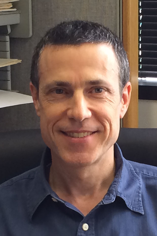 Luc Girard合作论文数Department of Hamon Center for Therapeutic Oncology Research, Medical School, University of Texas Southwestern Medical Center;Department of Pharmacology, University of Texas Southwestern Medical Center8
 Yong Zhao (赵勇)合作论文数School of Life Sciences, Sun Yat-sen University8
 Peter Ly合作论文数8
 Melville B Vaughan合作论文数Department of Biology, University of Central Oklahoma8
 Weihang Chai合作论文数Section of Microbiology, Cornell University8MA PIATYSZEK合作论文数7
 Ying Zou合作论文数Boston Univ, Sch Med, Ctr Human Genet, Boston, MA 02118 USA7
 Joseph A. Baur合作论文数Department of Physiology, Perelman School of Medicine, University of Pennsylvania;Center of Excellence in Environmental Toxicology, Perelman School of Medicine, University of Pennsylvania7
 Dara L. Aisner合作论文数Colorado Molecular Correlates Laboratory (CMOCO), Department of Pathology, University of Colorado Anschutz Medical Campus7
 Brody Holohan合作论文数Department of Cell Biology, UT Southwestern Medical Center7
 Lu Zhang合作论文数7
 Brittney-Shea Herbert合作论文数Indiana University School of Medicine7Rd Ramirez合作论文数6Bs Herbert合作论文数6Scott L Weinrich合作论文数6lance p ford合作论文数6Lawrence Lum合作论文数University of Texas Southwestern Medical Center6Tesmer Valerie M合作论文数6Aadil Kaisani合作论文数6
 Lawrence G. Lum合作论文数Detroit Medical Center, Wayne State University6
 Adi F. Gazdar合作论文数Department of Pathology, Medical Center, University of Texas Southwestern6Tracy T. Chow合作论文数6Ginelle C. Gellert合作论文数6
 Nam Kim合作论文数6Oliver Delgado合作论文数6Michael A White合作论文数5Cp Morales合作论文数5Hirotoshi Hoshiyama合作论文数5




