In advanced castration resistant prostate cancer (CRPC), mutations in the DNA damage response (DDR) gene ataxia telangiectasia mutated ( ATM ) are common. While poly(ADP-ribose) polymerase inhibitors are approved in this context, their clinical efficacy remains limited. Thus, there is a compelling need to identify alternative therapeutic avenues for ATM mutant prostate cancer patients. Here, we generated matched ATM-proficient and ATM-deficient CRPC lines to elucidate the impact of ATM loss on DDR in response to DNA damage via irradiation. Through unbiased phosphoproteomic screening, we unveiled that ATM-deficient CRPC lines maintain dependence on downstream ATM targets through activation of ATR and DNA-PKcs kinases. Dual inhibition of ATR and DNA-PKcs effectively inhibited downstream γH2AX foci formation in response to irradiation and radiosensitized ATM-deficient lines to a greater extent than either ATM-proficient controls or single drug treatment. Further, dual inhibition abrogated residual downstream ATM pathway signaling and impaired replication fork dynamics. To circumvent potential toxicity, we leveraged the RUVBL1/2 ATPase inhibitor Compound B, which leads to the degradation of both ATR and DNA-PKcs kinases. Compound B effectively radiosensitized ATM-deficient CRPC in vitro and in vivo , and impacted replication fork dynamics. Overall, dual targeting of both ATR and DNA-PKcs is necessary to block DDR in ATM-deficient CRPC, and Compound B could be utilized as a novel therapy in combination with irradiation in these patients.
The seamless transition through stages of pluripotency relies on a balance between transcription factor networks and epigenetic mechanisms. Here, we reveal the crucial role of the transgene activation suppressor (TASOR), a component of the human silencing hub (HUSH) complex, in maintaining cell viability during the transition from naive to primed pluripotency. TASOR loss in naive pluripotent stem cells (PSCs) triggers replication stress, disrupts H3K9me3 heterochromatin, and impairs silencing of LINE-1 (L1) transposable elements, with more severe effects in primed PSCs. Notably, the survival of Tasor knockout PSCs during this transition can be restored by inhibiting caspase or deleting the mitochondrial antiviral signaling protein (MAVS). This suggests that unscheduled L1 expression activates an innate immune response, leading to cell death specifically in cells exiting naive pluripotency. Our findings highlight the importance of epigenetic programs established in naive pluripotency for normal development.
Abstract Background: Mutations in DNA Damage Response (DDR) genes, including those resulting in loss of Ataxia-Telangiectasia, Mutated (ATM) expression are common in advanced prostate cancers (PC). Poly (ADP-ribose) polymerase (PARP) inhibitors are approved in this setting but demonstrate limited clinical efficacy in ATM-mutant PCs. In this project, we sought to define the impact of ATM loss on DDR pathways in PC, with the goal of identifying alternate therapeutic vulnerabilities. Methods: ATM-KO PC cell lines were generated via CRISPR-Cas9. ATM loss and attenuation of downstream kinase activity were confirmed via western blot. Phosphoproteomic evaluation in parental and ATM-KO cells was performed following ionizing radiation (IR). Clonogenic survival assays were performed after treatment with inhibitors and/or IR. Kinetics of DDR foci recruitment and resolution were interrogated with immunofluorescence (IF). DNA fiber assays were utilized to assess replication stress. Results: ATM-KO PC cells were able to effectively repair DNA damage following IR, as measured by resolution of yH2ax, 53BP1, and MDC1 foci. Phosphoproteomic studies indicated that ATM-KO cells maintained canonical DDR pathways through activation of ATR and DNA-PKcs kinases. Treatment of ATM-KO cells with either the ATR inhibitor VX970 or the DNA-PKcs inhibitor M3814 incrementally affected DDR in ATM-KO cells compared to ATM-proficient parental control cells. Importantly, combination treatment with VX970 and M3814 was needed to prevent downstream DDR foci recruitment in ATM-KO cells and was more effective in radiosensitizing ATM-KO PC than either parental control cells or single drug treatment. These data indicate that the activity of any one of the trinities of these kinases is sufficient to mediate DDR and that blockade of both ATR and DNA-PKcs is needed to effectively prevent DDR in ATM-KO PC. Further, DNA fiber assays showed significantly decreased fork length with dual treatment compared to control or single drug, indicating increased replication stress. We then leveraged a RUVBL1 ATPase inhibitor Compound B, which affects the expression of these three kinases. In ATM-KO PC cells, Compound B attenuated ATR and DNA-PKcs expression and radiosensitized the cells as shown by clonogenic survival assays. DNA fiber assays indicated that Compound B induced replication stress in ATM-KO PC cells to the same extent as combination therapy. In combination with IR, Compound B also significantly attenuated the growth of ATM-KO PC xenografts in vivo. Conclusions: Our unbiased phosphoproteomic approach indicated that ATR and DNA-PKCs mediate DDR following IR in ATM-KO PC cells, and that dual targeting of both ATR and DNA-PKcs is necessary to block DDR in ATM-KO PC cells. We have identified that Compound B effectively depletes ATR and DNA-PKcs, sensitizes to DNA damage, and induces replication stress in ATM-KO PCs. Our studies suggest that Compound B may be a novel therapeutic strategy in ATM-mutant PC. Citation Format: Mia E. Hofstad, Lan Yu, Andrea Woods, Zoi E. Sychev, Alice Mazzagatti, Xiaofang Huo, Ralf Kittler, Peter Ly, Justin M. Drake, Ganesh V. Raj. Deciphering DNA damage repair in ATM mutant prostate cancers [abstract]. In: Proceedings of the AACR Special Conference in Cancer Research: DNA Damage Repair: From Basic Science to Future Clinical Application; 2024 Jan 9-11; Washington, DC. Philadelphia (PA): AACR; Cancer Res 2024;84(1 Suppl):Abstract nr A020.
Mitotic errors generate micronuclei entrapping mis-segregated chromosomes, which are susceptible to catastrophic fragmentation through chromothripsis. The reassembly of fragmented chromosomes by error-prone DNA double-strand break (DSB) repair generates diverse genomic rearrangements associated with human diseases. How specific repair pathways recognize and process these lesions remains poorly understood. Here we use CRISPR/Cas9 to systematically inactivate distinct DSB repair pathways and interrogate the rearrangement landscape of fragmented chromosomes. Deletion of canonical non-homologous end joining (NHEJ) components substantially reduces complex rearrangements and shifts the rearrangement landscape toward simple alterations without the characteristic patterns of chromothripsis. Following reincorporation into the nucleus, fragmented chromosomes localize within sub-nuclear micronuclei bodies (MN bodies) and undergo ligation by NHEJ within a single cell cycle. In the absence of NHEJ, chromosome fragments are rarely engaged by alternative end-joining or recombination-based mechanisms, resulting in delayed repair kinetics, persistent 53BP1-labeled MN bodies, and cell cycle arrest. Thus, we provide evidence supporting NHEJ as the exclusive DSB repair pathway generating complex rearrangements from mitotic errors.
Chromosome-containing micronuclei are a hallmark of aggressive cancers. Micronuclei frequently undergo irreversible collapse, exposing their enclosed chromatin to the cytosol. Micronuclear rupture catalyzes chromosomal rearrangements, epigenetic abnormalities, and inflammation, yet mechanisms safeguarding micronuclear integrity are poorly understood. In this study, we found that mitochondria-derived reactive oxygen species (ROS) disrupt micronuclei by promoting a noncanonical function of charged multivesicular body protein 7 (CHMP7), a scaffolding protein for the membrane repair complex known as endosomal sorting complex required for transport III (ESCRT-III). ROS retained CHMP7 in micronuclei while disrupting its interaction with other ESCRT-III components. ROS-induced cysteine oxidation stimulated CHMP7 oligomerization and binding to the nuclear membrane protein LEMD2, disrupting micronuclear envelopes. Furthermore, this ROS-CHMP7 pathological axis engendered chromosome shattering known to result from micronuclear rupture. It also mediated micronuclear disintegrity under hypoxic conditions, linking tumor hypoxia with downstream processes driving cancer progression.
Chromosomal instability (CIN) generates micronuclei-aberrant extranuclear structures that catalyze the acquisition of complex chromosomal rearrangements present in cancer. Micronuclei are characterized by persistent DNA damage and catastrophic nuclear envelope collapse, which exposes DNA to the cytoplasm. We found that the autophagic receptor p62/SQSTM1 modulates micronuclear stability, influencing chromosome fragmentation and rearrangements. Mechanistically, proximity of micronuclei to mitochondria led to oxidation-driven homo-oligomerization of p62, limiting endosomal sorting complex required for transport (ESCRT)-dependent micronuclear envelope repair by triggering autophagic degradation. We also found that p62 levels correlate with increased chromothripsis across human cancer cell lines and with increased CIN in colorectal tumors. Thus, p62 acts as a regulator of micronuclei and may serve as a prognostic marker for tumors with high CIN.
Chromothripsis describes the catastrophic shattering of mis-segregated chromosomes trapped within micronuclei. Although micronuclei accumulate DNA double-strand breaks and replication defects throughout interphase, how chromosomes undergo shattering remains unresolved. Using CRISPR-Cas9 screens, we identify a non-canonical role of the Fanconi anemia (FA) pathway as a driver of chromothripsis. Inactivation of the FA pathway suppresses chromosome shattering during mitosis without impacting interphase-associated defects within micronuclei. Mono-ubiquitination of FANCI-FANCD2 by the FA core complex promotes its mitotic engagement with under-replicated micronuclear chromosomes. The structure-selective SLX4-XPF-ERCC1 endonuclease subsequently induces large-scale nucleolytic cleavage of persistent DNA replication intermediates, which stimulates POLD3-dependent mitotic DNA synthesis to prime shattered fragments for reassembly in the ensuing cell cycle. Notably, FA-pathway-induced chromothripsis generates complex genomic rearrangements and extrachromosomal DNA that confer acquired resistance to anti-cancer therapies. Our findings demonstrate how pathological activation of a central DNA repair mechanism paradoxically triggers cancer genome evolution through chromothripsis.
Abstract Chromosomal instability and changes in the epigenome are defining features in most metastatic cancers. Our research links chromosomal missegregation, their containment in micronuclei, and subsequent rupture of the micronuclear envelope to histone post-translational modification aberrations. This relationship was across all tested human and murine cancer cell lines, as well as non-transformed cells. Furthermore, we discovered that the observed changes in histone PTMs were caused by either micronuclear rupture or the persistence of histone PTM status during cell division. Moreover, we revealed substantial chromatin accessibility differences in micronuclei. Notably, there is a distinct bias in chromatin accessibility between promoter regions compared to distal and intergenic regions of the genome, which aligned well with observed changes in histone modifications. The induction of chromosomal instability leads to widespread, heterogeneous, and heritable abnormalities in the epigenetic landscape of cancer cells. Therefore, on top of genomic copy number changes, chromosomal instability causes epigenetic reprogramming, which adds another layer to cancer heterogeneity. Citation Format: Albert S. Agustinus, Duaa Al-Rawi, Bhargavi Dameracharla, Ramya Raviram, Bailey S. Jones, Stephanie Stransky, Lorenzo Scipioni, Jens Luebeck, Melody Di Bona, Danguole Norkunaite, Robert M. Myers, Mercedes Duran, Seongmin Choi, Britta Weigelt, Shira Yomtoubian, Andrew McPherson, Eleonore Toufektchan, Kristina Keuper, Paul S. Mischel, Vivek Mittal, Sohrab P. Shah, John Maciejowski, Zuzana Storchova, Enrico Gratton, Peter Ly, Dan Landau, Mathieu F. Bakhoum, Richard P. Koche, Simone Sidoli, Vineet Bafna, Yael David, Samuel F. Bakhoum. Chromosomal instability causes epigenetic aberrations in cancer [abstract]. In: Proceedings of the American Association for Cancer Research Annual Meeting 2024; Part 1 (Regular Abstracts); 2024 Apr 5-10; San Diego, CA. Philadelphia (PA): AACR; Cancer Res 2024;84(6_Suppl):Abstract nr 4293.
This abstract is being presented as a short talk in the scientific program. A full abstract is printed in the Proffered Abstracts section (PR012) of the Conference Program/Proceedings. Citation Format: Frank M. Mason, Anteneh T. Tebeje, Emily S. Kounlavong, Rashmi Dahiya, Logan Vlach, Tiffany Guess, Ruhee Dere, Ryoma Ohi, Peter Ly, Cheryl L. Walker, W. Kimryn Rathmell. SETD2 safeguards the genome against isochromosome formation [abstract]. In: Proceedings of the AACR Special Conference: Advances in Kidney Cancer Research; 2023 Jun 24-27; Austin, Texas. Philadelphia (PA): AACR; Cancer Res 2023;83(16 Suppl):Abstract nr B016.
Biomolecular condensates participate in the regulation of gene transcription, yet the relationship between nuclear condensation and transcriptional activation remains elusive. Here, we devised a biotinylated CRISPR-dCas9–based optogenetic method, light-activated macromolecular phase separation (LAMPS), to enable inducible formation, affinity purification, and multiomic dissection of nuclear condensates at the targeted genomic loci. LAMPS-induced condensation at enhancers and promoters activates endogenous gene transcription by chromatin reconfiguration, causing increased chromatin accessibility and de novo formation of long-range chromosomal loops. Proteomic profiling of light-induced condensates by dCas9-mediated affinity purification uncovers multivalent interaction-dependent remodeling of macromolecular composition, resulting in the selective enrichment of transcriptional coactivators and chromatin structure proteins. Our findings support a model whereby the formation of nuclear condensates at native genomic loci reconfigures chromatin architecture and multiprotein assemblies to modulate gene transcription. Hence, LAMPS facilitates mechanistic interrogation of the relationship between nuclear condensation, genome structure, and gene transcription in living cells.
Centromere identity is defined and maintained epigenetically by the presence of the histone variant CENP-A. How centromeric CENP-A position is specified and precisely maintained through DNA replication is not fully understood. The recently released Telomere-to-Telomere (T2T) genome assembly containing the first complete human centromere sequences provides a new resource for examining CENP-A position. Mapping CENP-A position in clones of the same cell line to the T2T assembly identified highly similar CENP-A position after multiple cell divisions. In contrast, centromeric CENP-A epialleles were evident at several centromeres of different human cell lines, demonstrating the location of CENP-A enrichment and the site of kinetochore recruitment vary among human cells. Across the cell cycle, CENP-A molecules deposited in G1 phase are maintained in their precise position through DNA replication. Thus, despite CENP-A dilution during DNA replication, CENP-A is precisely reloaded onto the same sequences within the daughter centromeres, maintaining unique centromere identity among human cells.
Isochromosomes are mirror-imaged chromosomes with simultaneous duplication and deletion of genetic material which may contain two centromeres to create isodicentric chromosomes. Although isochromosomes commonly occur in cancer and developmental disorders and promote genome instability, mechanisms that prevent isochromosomes are not well understood. We show here that the tumor suppressor and methyltransferase SETD2 is essential to prevent these errors. Using cellular and cytogenetic approaches, we demonstrate that loss of SETD2 or its epigenetic mark, histone H3 lysine 36 trimethylation (H3K36me3), results in the formation of isochromosomes as well as isodicentric and acentric chromosomes. These defects arise during DNA replication and are likely due to faulty homologous recombination by RAD52. These data provide a mechanism for isochromosome generation and demonstrate that SETD2 and H3K36me3 are essential to prevent the formation of this common mutable chromatin structure known to initiate a cascade of genomic instability in cancer.
Mitotic cell division is tightly monitored by checkpoints that safeguard the genome from instability. Failures in accurate chromosome segregation during mitosis can cause numerical aneuploidy, which was hypothesized by Theodor Boveri over a century ago to promote tumorigenesis. Recent interrogation of pan-cancer genomes has identified unexpected classes of chromosomal abnormalities, including complex rearrangements arising through chromothripsis. This process is driven by mitotic errors that generate abnormal nuclear structures that provoke extensive yet localized shattering of mis-segregated chromosomes. Here, we discuss emerging mechanisms underlying chromothripsis from micronuclei and chromatin bridges, as well as highlight how this mutational cascade converges on the DNA damage response. A fundamental understanding of these catastrophic processes will provide insight into how initial errors in mitosis can precipitate rapid cancer genome evolution.
Chromosomal instability (CIN) and epigenetic alterations are characteristics of advanced and metastatic cancers [1-4], yet whether they are mechanistically linked is unknown. Here we show that missegregation of mitotic chromosomes, their sequestration in micronuclei [5, 6], and subsequent micronuclear envelope rupture [7] profoundly disrupt normal histone post-translational modifications (PTMs), a phenomenon conserved across humans and mice as well as cancer and non-transformed cells. Some of the changes to histone PTMs occur due to micronuclear envelope rupture whereas others are inherited from mitotic abnormalities prior to micronucleus formation. Using orthogonal techniques, we show that micronuclei exhibit extensive differences in chromatin accessibility with a strong positional bias between promoters and distal or intergenic regions. Finally, we show that inducing CIN engenders widespread epigenetic dysregulation and that chromosomes which transit in micronuclei experience durable abnormalities in their accessibility long after they have been reincorporated into the primary nucleus. Thus, in addition to genomic copy number alterations, CIN can serve as a vehicle for epigenetic reprogramming and heterogeneity in cancer.
The loss of one copy of chromosome 3p represents an early truncal genetic event in the majority of clear cell renal cell carcinoma (ccRCC) tumors. Chromosome 3p loss can be initiated by chromothripsis, a process in which mis-segregated chromosomes entrapped within abnormal nuclear structures called micronuclei become pulverized into small genomic fragments. These fragments are then re-stitched together to form complex rearrangements that are accompanied by extensive segmental deletions that inactivate critical tumor suppressor genes located on chromosome 3p. Despite being a driver of ccRCC development, the mechanism(s) that initiate chromosome 3p chromothripsis followed by pressures that select for specific rearrangement patterns are poorly understood. To recapitulate early genetic events arising throughout ccRCC evolution, here we developed chromosome 3p-specific micronuclei models in non-transformed human renal proximal tubule epithelial cells (RPTECs) to interrogate recurrent patterns of chromosomal alterations that promote renal cell tumorigenesis. To do so, we used CRISPR/Cas9 to induce a DNA double-strand break on chromosome 3p, which generates chromosome 3p-specific micronuclei when the acentric arm is left unrepaired into the subsequent mitosis. DNA fluorescence in situ hybridization revealed that mis-segregation of chromosome 3p into micronuclei induces chromosome pulverization and rearrangements. Notably, these alterations are sufficient to provide a selective advantage by enabling anchorage-independent growth in vitro. Cytogenetic characterization of partially transformed RPTECs revealed enrichment of chromosome 3p rearrangements, which were gradually lost upon propagation as conventional monolayer cultures. These results provide mechanistic insight into the origins of chromosome 3p alterations in ccRCC and further highlight the role of selection pressure in shaping genome evolution. Citation Format: Rashmi Dahiya, Peter Ly. Modeling recurrent chromosomal alterations in renal cell carcinoma evolution [abstract]. In: Proceedings of the AACR Special Conference: Advances in Kidney Cancer Research; 2023 Jun 24-27; Austin, Texas. Philadelphia (PA): AACR; Cancer Res 2023;83(16 Suppl):Abstract nr A014.
Supplementary Figures 1-8, Tables 1-5, Methods from Functional Parsing of Driver Mutations in the Colorectal Cancer Genome Reveals Numerous Suppressors of Anchorage-Independent Growth
Complex genome rearrangements can be generated by the catastrophic pulverization of missegregated chromosomes trapped within micronuclei through a process known as chromothripsis1-5. As each chromosome contains a single centromere, it remains unclear how acentric fragments derived from shattered chromosomes are inherited between daughter cells during mitosis6. Here we tracked micronucleated chromosomes with live-cell imaging and show that acentric fragments cluster in close spatial proximity throughout mitosis for asymmetric inheritance by a single daughter cell. Mechanistically, the CIP2A-TOPBP1 complex prematurely associates with DNA lesions within ruptured micronuclei during interphase, which poises pulverized chromosomes for clustering upon mitotic entry. Inactivation of CIP2A-TOPBP1 caused acentric fragments to disperse throughout the mitotic cytoplasm, stochastically partition into the nucleus of both daughter cells and aberrantly misaccumulate as cytoplasmic DNA. Mitotic clustering facilitates the reassembly of acentric fragments into rearranged chromosomes lacking the extensive DNA copy-number losses that are characteristic of canonical chromothripsis. Comprehensive analysis of pan-cancer genomes revealed clusters of DNA copy-number-neutral rearrangements-termed balanced chromothripsis-across diverse tumour types resulting in the acquisition of known cancer driver events. Thus, distinct patterns of chromothripsis can be explained by the spatial clustering of pulverized chromosomes from micronuclei.
Chromosome mis-segregation during cell division occurs in diverse tumor types, promoting aneuploidy and intratumoral genetic heterogeneity. While the mechanisms that govern chromosome segregation are well-established, how changes in the activities of tumor suppressors or oncogenes drive mitotic errors remain poorly understood. Loss of the tumor suppressor SETD2, the methyltransferase responsible for tri-methylation of histone 3 lysine 36 (H3K36me3), correlates with clear cell renal cell carcinoma (ccRCC) tumors exhibiting extensive intratumoral heterogeneity. Yet, how loss of H3K36me3 or SETD2 promotes heterogeneity is unknown. Here, we show that loss of SETD2 promotes chromosome mis-segregation during mitosis and interphase DNA bridges driven by the formation of dicentric chromosomes. Cytogenetic analyses revealed that these dicentrics loss were largely comprised of mirror-imaged isodicentric chromosomes that contain two active centromeres. In addition to isodicentrics, cells lacking SETD2 or H36K36me3 generated iso- and acentric chromosomes, however loss of SETD2 protein additionally caused chromosome instability. These data directly link loss of a tumor suppressor to these mutable chromatin structures that initiate intratumor heterogeneity by promoting gross chromosomal rearrangements. Citation Format: Frank M. Mason, Anteneh T. Tebeje, Emily S. Kounlavong, Rashmi Dahiya, Logan Vlach, Tiffany Guess, Ruhee Dere, Ryoma Ohi, Peter Ly, Cheryl L. Walker, W. Kimryn Rathmell. SETD2 safeguards the genome against isochromosome formation [abstract]. In: Proceedings of the AACR Special Conference: Advances in Kidney Cancer Research; 2023 Jun 24-27; Austin, Texas. Philadelphia (PA): AACR; Cancer Res 2023;83(16 Suppl):Abstract nr PR012.
 Don W. Cleveland合作论文数Department of Cellular and Molecular Medicine, School of Medicine, University of California, San Diego;Division of Genomics and Precision Medicine, Department of Medicine, School of Medicine, University of California, San Diego;Laboratory of Cell Biology, Ludwig Cancer Research12
 Rashmi Dahiya合作论文数9
 Mazzagatti Alice合作论文数9
 Woodring Erik Wright合作论文数Department of Cell Biology, The University of Texas Southwestern Medical Center;Southland Financial Corporation8Qing Hu合作论文数7
 Samuel Bakhoum合作论文数Cell & Developmental Biology, Weill Cornell Graduate School of Medical Sciences, Cornell University7
 Jerry W. Shay合作论文数University of Texas Southwestern Medical Center7
 Sangbum Kim合作论文数Motor Behavior Laboratory, University of Florida7Ugur Eskiocak合作论文数Southwestern Medical Center, University of Texas7Crystal Cornelius合作论文数5
 Mercedes Duran合作论文数IBGM5Andres I. Roig合作论文数5Michael A White合作论文数4Anteneh T. Tebeje合作论文数4Logan Vlach合作论文数4Emily S. Kounlavong合作论文数4
 Justin Engel合作论文数4
 Tiffany Guess合作论文数Dept Pathol Microbiol & Immunol, VUMC4
 Dong Hyun Kim合作论文数Univ Calif San Diego, Ludwig Inst Canc Res, San Diego, CA 92093 USA4
 Di Bona Melody合作论文数Nanoscopy, Nanophysics, Istituto Italiano di Tecnologia, Genoa, Italy4
 Ryoma Ohi合作论文数Department of Cell & Developmental Biology, Medical School, University of Michigan4
 Yael Nechemia-Arbely合作论文数Goldyne Savad Institute of Gene Therapy, Hadassah Hebrew University Hospital4Mason Frank M合作论文数4
 W. Kimryn Rathmell合作论文数Solove Research Institute, The Ohio State University Wexner Medical Center;James Cancer Hospital, The Ohio State University Wexner Medical Center4
 Daniele Fachinetti合作论文数DSBB-Università degli Studi di Milano4
 Cheryl Walker合作论文数Center for Precision Environmental Health, Baylor College of Medicine;Dan L Duncan Comprehensive Cancer Center, Baylor College of Medicine4Kakajan Komurov合作论文数Champions Oncology, Inc.4
 Ruhee Dere合作论文数Institute of Biosciences and Technology, Texas A&M University System Health Science Center4Andrea Woods合作论文数3Zoi E Sychev合作论文数3Mia Hofstad合作论文数3Sebastian Biglione合作论文数3Alison Guyer合作论文数3
 Elizabeth Maurais合作论文数3Yu-Fen Lin合作论文数3
 Lorenzo Scipioni合作论文数LFD, Irvine, CA, USA3
 Jose Espejo Valle-Inclan合作论文数Wageningen Univ & Res, Lab Phytopathol, Droevendaalsesteeg 1, NL-6708 PB Wageningen, Netherlands3
 Albert S Agustinus合作论文数Chemical Biology Program, Memorial Sloan Kettering3Lan Yu合作论文数3Jacob T Sanders合作论文数Department of Biochemistry & Cellular and Molecular BiologyUniversity of TennesseeKnoxvilleTN37996United States3
 John Maciejowski合作论文数Molecular Biology, Weill Cornell Graduate School of Medical Sciences, Cornell University3
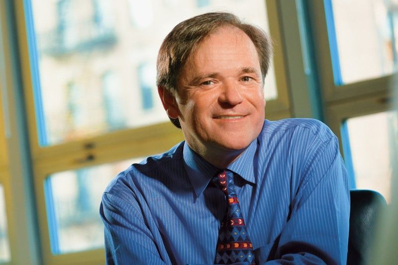 Simon Powell合作论文数Molecular Biology, Weill Cornell Graduate School of Medical Sciences, Cornell University3McMahon Moira A合作论文数3
 Shoshani Ofer合作论文数Ludwig Inst Canc Res, Univ Calif San Diego3
 Enrico Gratton合作论文数Laboratory for Fluorescence Dynamics, Department of Biomedical Engineering, Samueli School of Engineering, University of California, Irvine;Department of Physics, University of Illinois, Urbana-Champaign3
 Simone Sidoli合作论文数Department of Biochemistry and Molecular Biology, University of Southern Denmark3
 Isidro Cortes-Ciriano合作论文数Dept Biol Struct & Chim, Inst Pasteur3
 Nancy Y. Lee合作论文数Department of Radiation Oncology, Memorial Sloan-Kettering Cancer Center3
 Ralf Kittler合作论文数Max Planck;Institute of Molecular Cell Biology;Max Planck|Institute of Molecular Cell Biology3




