Abstract DNA double-strand break (DSB) repair can be mediated by non-homologous end joining (NHEJ) and homologous recombination (HR). Homologous recombination repair (HRR) is important because it accurately repairs DNA DSBs using a sister chromatid as a template, which is crucial for maintaining genome stability and preventing mutations. Deficiency in HRR makes cancer cells sensitive to DNA damage drugs. BRCA1/2 and RAD51 proteins play very important roles in HRR. PARP is a key enzyme in the repair of DNA single-strand breaks (SSB). Unrepaired SSBs will convert to DSBs in cells. Therefore, BRCA1/2 -mutated cancer cells are sensitive to PARP inhibitors. However, PARP inhibitor resistance develops quickly, mainly through mechanisms that restore homologous recombination repair. This can happen through secondary mutations that restore function in genes of BRCA1 or BRCA2, and overexpression of RAD51. RAD51 overexpression is reported in many cancers, such as breast, prostate, and glioblastoma, and has been involved in chemotherapy resistance. Inhibition of RAD51 creates an HR-deficient status, which can sensitize cancer cells to PARP inhibitor treatments. It has been reported that ISR (integrated stress response) leads to downregulation of RAD51. We reported before that PG3 treatment induced potent ISR. Hence, we hypothesized that PG3 can sensitize PRAP-resistant tumor cells (both BRCA1-mutant and wild-type) to PARP inhibitors by downregulating RAD51. The combination treatments of PG3 and Olaparib/Talazoparib showed synergistic inhibitory effects on triple-negative breast cancer cells, BRCA1-mutated SUM149 and MD-MB-436, and BRCA1-wildtype MD-MB-231 and MD-MB-468. Western blots showed that two BRCA1-mutated cell lines, SUM149 and MD-MB436, express very low levels of BRCA1 protein compared to wild-type cell lines MD-MB231and MD-MB468. On the other hand, SUM149 and MD-MB436 show much higher RAD51 expression than MD-MB231 and MD-MB468. PG3 downregulates RAD51 in both SUM149 and MD-MB231 cells, but not in MD-MB436 and MD-MB468 cells. Transcriptional factors c-Myc, E2F1, and FoxM1 regulate RAD51 gene expression. We found that PG3 induced downregulation of c-Myc, E2F1, and FoxM1 in both SUM149 and MB231 cells, but not in MB436 and MB468 cells. That is consistent with previous publications. PG3 also downregulates wild-type BRCA1 in MD-MB231 cells but not in MD-MB468 cells. We found that olaparib treatment induced upregulation of RAD51 in SUM149 cells and upregulation of both BRCA1 and RAD51 in MD-MB231 cells. PG3 blocked the olaparib-induced upregulation of RAD51 in SUM149cells, and the upregulation of BRCA1 and RAD51 in MD-MB231 cells. The combined treatment induced more DNA damage than olaparib or PG3 alone in SUM149 and MB231 cells, as indicated by increased γH2AX level. Citation Format: Xiaobing Tian, Wafik S. El-Deiry. A combination of PG3 and PARP inhibitors exhibits antitumor effects in both BRCA1-mutated and BRCA1-wild-type TNBC [abstract]. In: Proceedings of the American Association for Cancer Research Annual Meeting 2026; Part 1 (Regular Abstracts); 2026 Apr 17-22; San Diego, CA. Philadelphia (PA): AACR; Cancer Res 2026;86(7 Suppl):Abstract nr 1747.
Prostate cancer (PCa) is the most prevalent malignancy in American men excluding skin cancer. In metastatic disease, resistance to androgen pathway inhibitors (APIs) is a major obstacle to curative treatment. One such resistance mechanism is the histological transformation from adenocarcinoma to a neuroendocrine (NE) phenotype, driven by transcription factors like SOX2 and BRN2. Of note, SOX2 is typically overexpressed in PCa lacking functional Rb and p53 and can regulate cell cycle proteins including Cyclin D1. These findings warrant an investigation into a potential connection between SOX2, the cell cycle, and tumor suppressors to further establish molecular mechanisms of neuroendocrine differentiation (NED). We also plan to investigate the potential role of BRN2 in this process. Lentiviral Tet-On® systems were cloned and transduced into the PCa cell lines LNCaP (WT RB1, WT TP53) and DU145 (RB1 -/-, TP53 mutant) for the inducible expression of SOX2 (iSOX2) and BRN2 (iBRN2). In vitro growth assays were performed by plating 5, 000 cells/well in a 96 well plate and adding doxycycline the following day (DOX). Western blots were performed to determine protein expression. Cell cycle analysis was performed by PI staining and flow cytometry. Inducible models showed robust induction of SOX2 or BRN2 after 24h induction. At 4d, iBRN2 cell lines demonstrated NE marker upregulation, specifically synaptophysin. SOX2, but not BRN2, induction decreased Cyclin D1 expression irrespective of Rb and p53 status. Intriguingly, LNCaP iSOX2 cells were less viable following induction and had distinct morphological changes including the loss of their stellate shape. LNCaP iBRN2 cells had less profound, but apparent, changes in morphology. No significant morphological changes were identified in DU145. Cell cycle analysis found a greater percentage of LNCaP iSOX2 cells in the sub-G1 state and a lesser percentage in the G2/M phase versus the empty vector. These results suggest that SOX2 overexpression (OE) may only be tolerated in an Rb and/or p53 deficient setting. Further research will seek to determine if this phenomenon is due to SOX2-induced repression of Cyclin D1. Subsequent experiments will use RB1 and/orTP53 KO cell lines to generate new SOX2 and BRN2 inducible systems. Novel experiments will seek to determine if RB1 and/or TP53 KO increases tolerance to SOX2 OE. Connecting this knowledge of the SOX2 protein to the cell cycle and hormonal signaling in PCa may offer insight into the interplay between emerging cell cycle-targeting therapeutics and the NED process. Connor Purcell, Praveen R. Srinivasan, Shengliang Zhang, Maximilian Pinho-Schwermann, William J. MacDonald, Lanlan Zhou, Andrea Schmidt, Claire Lin, Xiaobing Tian, Tyler J. Roady, Vida Tajiknia, Viva Voong, Wafik El-Deiry. Tolerance of SOX2 and BRN2 in prostate cancer cell lines: effects on cell cycle and lineage transformation [abstract]. In: Proceedings of the American Association for Cancer Research Annual Meeting 2025; Part 1 (Regular Abstracts); 2025 Apr 25-30; Chicago, IL. Philadelphia (PA): AACR; Cancer Res 2025;85(8_Suppl_1):Abstract nr 1458.
More than 90% of head & neck (H&N) cancers are squamous cell carcinoma (HNSCC) that occur from the mucosal epithelial tissue of the oral cavity, oropharynx, and larynx. About 30∼40% of stage I or II HNC patients are curable and show improved survival rates after surgery or radiotherapy alone. However, over 60% of stage III or IV HNC patients require chemotherapy. Cisplatin is one of standard chemotherapy drugs for stage III and IV patients. In addition, the mutation frequency of the p53 gene in HNSCC is 65-85%. DNA damage following cisplatin treatment can activate apoptosis via p53 in p53 wild-type cancer cells. In p53-mutated/deleted cancer cells, DNA-damage drugs can lead to cell death through other mechanisms, such as integrated stress response (ISR). There multiple cisplatin-resistance mechanisms were reported. Nucleotide excision repair (NER) is known as the primary strategy for repair of cisplatin-induced DNA damage. Elevated ERCC1 and ERCC5 expression enhance NER and are associated with cisplatin resistance in HNSCC patients. It was reported that ATF4 downregulation promoted cisplatin resistance in p53 mutated and deleted gastric cancers. A small molecule PG3 treatment triggers ISR and leads to cell apoptosis through HRI/eIF2α/ATF4/PUMA pathway in p53-mutated and deleted colorectal cancer cell lines. We hypothesize that (1) combination treatment of PG3 with cisplatin can reduce side effects of cisplatin through reducing the dose of cisplatin, which can be achieved by enhanced induction of integrated stress response, (2) PG3 can sensitize cisplatin-resistant and p53-mutated H&N cancer cells to cisplatin through enhanced induction of integrated stress response. The combination treatment shows synergistic effects in p53-mutated FaDu and Cal27 H&N cancer cells. In combination therapy, use of PG3 allows use of a lower cisplatin dose to achieve a therapeutic benefit. The combined treatment enhances ISR induction, and the ISR contributes to cell apoptosis. We identified that cisplatin or the combination treatment activates the HRI/ATF4/NOXA pathway. NOXA mediates Mcl-1 degradation and is responsible for the combination treatment-induced apoptosis. Xiaobing Tian, Wafik S. El-Deiry. Combination treatment with cisplatin and PG3 for p53-mutated head & neck cancers [abstract]. In: Proceedings of the American Association for Cancer Research Annual Meeting 2025; Part 1 (Regular Abstracts); 2025 Apr 25-30; Chicago, IL. Philadelphia (PA): AACR; Cancer Res 2025;85(8_Suppl_1):Abstract nr 6928.
Apoptosis is a form of programmed cell death that is mediated by intrinsic and extrinsic pathways. Dysregulation of and resistance to cell death are hallmarks of cancer. For over three decades, the development of therapies to promote treatment of cancer by inducing various cell death modalities, including apoptosis, has been a main goal of clinical oncology. Apoptosis pathways also interact with other signaling mechanisms, such as the p53 signaling pathway and the integrated stress response (ISR) pathway. In addition to agents directly targeting the intrinsic and extrinsic pathway components, anticancer drugs that target the p53 and ISR signaling pathways are actively being developed. In this Review, we discuss selected and promising anticancer therapies in various stages of development, including drug targets, mechanisms, and resistance to related treatments, focusing especially on B cell lymphoma 2 (BCL-2) inhibitors, TRAIL analogues, DR5 antibodies, and strategies that target p53, mutant p53, and the ISR.
Abstract HRI was initially identified as a kinase essential for maintaining heme-globin balance within red blood cells as well as for controlling the ISR in response to oxidative stress. HRI also responds to a broad range of stresses such as osmotic stress, heat shock, proteasome inhibition. The recently discovered unexpected functions of HRI include innate immunity, translational control of immune evasion in cancer by upregulating PD-L1, proteostasis, mitochondrial stress, inhibition of histone H3 lysine 27 (H3K27) demethylase (KDM6A) and iron deficiency. Importantly, recent evaluation of patient data uncovered high expression of HRI mRNA in a subset of epithelial tumors versus normal tissues. Elevated expression of HRI protein in these tumor cells lead to cell death when BIRC6 ubiquitin complex is inhibited, which mediates degradation of HRI and is required for the survival of the tumors. These further broaden the importance of this member of the eIF2α kinase family as a cancer therapeutic strategy. Dordaviprone (ONC201), an imipridone small molecule, binds to and activates mitochondrial protease ClpP leading to integrated stress response (ISR) and ATF4 transcription factor activation. PG3, a prodigiosin analog, induces ATF4 and pro-apoptotic PUMA. Our data indicate that PG3 activates ATF4 through ISR via eIF2α kinase HRI. ALAS1 (5'-aminolevulinate synthase 1) catalyzes the first rate-limiting step in heme (Iron-protoporphyrin) biosynthesis. ONC201 treatment leads to potent downregulation and inhibition of ALAS1, indicating that ONC201 inhibits heme biosynthesis. It is well known that reduced heme results in activation of the HRI kinase. We show that an inhibitor of heme biosynthesis or knockdown of ALAS1 results in HRI activation, while silencing of HRI or knockout of HRI gene potently inhibit the eIF2α phosphorylation and upregulation of ATF4, CHOP and PUMA by PG3 and imipridones. Knockdown of ClpP rescues ONC201-induced downregulation of ALAS1 which blocks ONC201-induced upregulation of CHOP. Also, silencing of ClpP significantly reduced PARP cleavage in HCT116 p53−/- and MDA-MB-468 cancer cells. Our studies identify a novel link between ClpP activation induced by ONC201 treatment and ATF4 upregulation, via the ClpP/ALAS1/HRI/ATF4 pathway. However, PG3 treatment did not lead to degradation of ALAS1, indicating that PG3 does not activate ClpP. PG3 potently induced cell apoptosis through ISR via HRI/ATF4/PUMA pathway independent of ClpP. We are further investigating the targets of PG3 and the signaling pathway that leads to PG3-induced activation of HRI. Our results suggest that different small molecule inducers of the ISR such as ONC201 and PG3 can achieve an anti-tumor effect through different pathways converging on kinase HRI ultimately leading to ATF4 activation and tumor cell death. Citation Format: Xiaobing Tian, Praveen Srinivasan, Wafik S. El-Deiry. ClpP-dependent and -independent activation of HRI kinase by small molecules [abstract]. In: Proceedings of the American Association for Cancer Research Annual Meeting 2024; Part 1 (Regular Abstracts); 2024 Apr 5-10; San Diego, CA. Philadelphia (PA): AACR; Cancer Res 2024;84(6_Suppl):Abstract nr 610.
Abstract Diffuse midline gliomas (DMGs) are highly aggressive, high-grade gliomas which typically arise in children and young adults. With currently approved treatments the median survival from time of diagnosis for pediatric DMG is 8-10 months with only 10% of children living to 2 years post diagnosis. Despite being only 15% of the cases it makes up 40% of the pediatric brain cancer deaths making it the leading cause of death for all pediatric glioma cases. Two recent clinical studies, NCT03416530 and NCT03134131, have shown the clinical efficacy of Dordaviprone (ONC201/TIC10) for the treatment of DMG; increasing median survival to 22 months in patients following radiation treatment prior to recurrence. Imipridone ONC206, a chemical derivative of ONC201, is under clinical development for treatment of pediatric and adult patients with primary brain tumors (NCT04732065 and NCT04541082). To further improve treatment, we must continue to study how the tumor microenvironment impacts the efficacy of imipridones. One crucial aspect of all brain cancers is hypoxia, so the IC50s of the SU-DIGP-25, SU-DIPG-XIII and SU-DIPGIV pediatric DMG cell lines treated with ONC201 or ONC206 were measured using the CellTiter-Glo assay under conditions of hypoxia and normoxia. Imipridones mediate apoptosis through the upregulation of the TRAIL death receptor DR5 and the activation of the integrated stress response (ISR), so western blots were used to show changes in the ISR protein expression in ONC201 treated DMG cells at multiple different degrees of hypoxia. To understand how hypoxia inducible factors (HIFs) play a role in resistance and susceptibility to ONC201 or ONC206, HIF-1a, HIF-2a, and HIF-3a were knocked down showing altered ISR protein expression after treating with the imipridones under hypoxic and normoxic conditions. Citation Format: Tyler J. Roady, Nolan Stubbs, Josephine Chen, Yutong Xia, Ashley Sanchez Sevilla Uruchurtu, Xiaobing Tian, Lanlan Zhou, Wafik S. El-Deiry. Impact of hypoxia on the integrated stress response activated by imipridones ONC201 and ONC206 in pediatric diffuse midline glioma cells [abstract]. In: Proceedings of the American Association for Cancer Research Annual Meeting 2024; Part 1 (Regular Abstracts); 2024 Apr 5-10; San Diego, CA. Philadelphia (PA): AACR; Cancer Res 2024;84(6_Suppl):Abstract nr 3173.
S1: Raw bioluminescence data obtained from screens of kinase, protease, and redox inhibitors. S2: Bioluminescent and viability data from U251 DR5 Luciferase cells treated with an NH125 dilution series. S3: Representative images of T4213 neurospheres taken under 40X magnification demonstrate that T4213 enrich for CD133 (red) SOX2 (orange) and Nestin (green) (scale bar = 25 microns). S4: Representative phase contrast images of TMZ, and NH125 treated GSC taken under 10X magnification (scale bar = 100 microns). S5: Caspase 3/7 activity increases in NH125 treated GSC. GSC were incubated with either 0 M NH125 (0.1% DMSO) or 5 M NH125 for twenty-four hours followed by addition of Caspase 3/7 Glo. S6: NH125 leads to a dose dependent increase in markers of apoptosis. S7: GSC that acquire a differentiated morphology are less sensitive to NH125. S8: The ten most significant canonical pathways from analysis of the transcriptional data of NH125 treated U251 and NHA. S9: PEG-PCL-NH125 treatment of U251 leads to an increase in CHOP and DR5 expression in vitro. Table S1: Knockout of CHOP leads to an abrogation of NH125 mediated TRAIL synergy. Table S2: IC50 values from a panel of NH125 treated cells. Table S3: Addition of low dose TRAIL leads to increased synergy in glioma stem cells.
p53 is a transcription factor that regulates the expression of genes involved in tumor suppression. p53 mutations mediate tumorigenesis and occur in approximately 50% of human cancers. p53 regulates hundreds of target genes that induce various cell fates including apoptosis, cell cycle arrest, and DNA damage repair. p53 also plays an important role in anti-tumor immunity by regulating TRAIL, DR5, TLRs, Fas, PKR, ULBP1/2, and CCL2; T-cell inhibitory ligand PD-L1; pro-inflammatory cytokines; immune cell activation state; and antigen presentation. Genetic alteration of p53 can contribute to immune evasion by influencing immune cell recruitment to the tumor, cytokine secretion in the TME, and inflammatory signaling pathways. In some contexts, p53 mutations increase neoantigen load which improves response to immune checkpoint inhibition. Therapeutic restoration of mutated p53 can restore anti-cancer immune cell infiltration and ameliorate pro-tumor signaling to induce tumor regression. Indeed, there is clinical evidence to suggest that restoring p53 can induce an anti-cancer immune response in immunologically cold tumors. Clinical trials investigating the combination of p53-restoring compounds or p53-based vaccines with immunotherapy have demonstrated anti-tumor immune activation and tumor regression with heterogeneity across cancer type. In this Review, we discuss the impact of wild-type and mutant p53 on the anti-tumor immune response, outline clinical progress as far as activating p53 to induce an immune response across a variety of cancer types, and highlight open questions limiting effective clinical translation.
Supplementary Figures 1-2 from Tumor necrosis factor–related apoptosis-inducing ligand (TRAIL) and paclitaxel have cooperative in vivo effects against glioblastoma multiforme cells
KRAS mutation is found in 95% pancreatic ductal adenocarcinoma (PDAC). TP53 is altered in 70% of patients with PDAC that co-occur with KRAS mutations. Enhanced de novo cholesterol biosynthesis is a hallmark of cancer cells. P53 inhibits the mevalonate pathway to mediate tumor repression. p53 mutations drive de novo cholesterol pathway activation and are required for the proliferation of KRAS-mutant cancers. Inhibition of the mevalonate pathway leads to feedback activation of SREBP-2, which restores mevalonate pathway and leads to re-activation of KRAS and restoration of mutant p53 GOF. Imipridones induced ATF4 through the action of mitochondria protease ClpP. Simvastatin triggers ATF4 activation through inhibition of mevalonate pathway. We hypothesize that combined treatment with imipridones can overcome the resistance to simvastatin by two different mechanisms: (1) Imipridones inhibit oxidative phosphorylation through ClpP hyperactivation and result in AMPK activation. AMPK phosphorylates and inhibits SREBP-2 transcriptional function, which blocks the feedback activation of SREBP-2. (2) Enhanced ATF4 and CHOP induction through different pathways leads to cancer cell death. We also expect that the combined treatment through sustained induction of ATF4 and CHOP can sensitize cancer cells to imipridone treatment. Mouse and human PDAC cell lines (KPCY, HPAF-II, and PANC-1) with p53 and KRAS mutations were used to test the effects of combination treatment of simvastatin and imipridones. The combined treatments synergistically inhibited the proliferation of p53 and KRAS co-mutant cell lines. For example, the synergy scores of simvastatin and ONC201 combination are 75 (KPCY), 15 (HPAF-II), and 14 (PANC-1). Enhanced cell death is consistent with sustained and enhanced induction of ATF4 and CHOP compared to single drug treatment alone. Imipridones activated AMPK in the tested PDAC cell lines. Imipridones potently inhibit AKT and ERK1/2 activation at 72-hour time point, which might also contribute to block above mentioned feedback activation of KRAS. We also observed that cell lines with a structural mutant of p53 are more sensitive to the combination treatments than a cell line with a DNA-contact mutant of p53, which is consistent with the fact that simvastatin treatment leads to degradation of structural p53 mutants. Surprisingly, our data indicated that simvastatin potently inhibits kinase WEE1 and leads to CDK1 dephosphorylation and activation, which has not been reported before. Together, our data is consistent with publications that p53 mutated cancer cell lines are sensitive to statin treatments. In short, combination treatment may overcome simvastatin resistance and/or imipridone resistance by modulating ATF4/CHOP and SREBP-2 activity. Citation Format: Xiaobing Tian, Wafik S. Deiry. Targeting mutant p53 and KRAS for novel pancreatic cancer therapy by combination treatment of simvastatin and imipridones [abstract]. In: Proceedings of the American Association for Cancer Research Annual Meeting 2023; Part 1 (Regular and Invited Abstracts); 2023 Apr 14-19; Orlando, FL. Philadelphia (PA): AACR; Cancer Res 2023;83(7_Suppl):Abstract nr 6122.
Our Laboratory was established in 1994 at Univ. of Pennsylvania. Lab members demonstrated initial competencies by performing cell culture, western blots, immunofluorescence, and flow cytometry showing induction of p53/p21(WAF1) in cells treated with chemotherapy. Years later, our Laboratory of Translational Oncology & Experimental Cancer Therapeutics moved to Penn State Univ., Fox Chase Cancer Center/Temple Univ. and then Brown Univ. By 2020, with desire for inclusiveness (everyone succeeds), scientific rigor/reproducibility mandated by NIH, and as a training and mentoring activity (lab scientists/trainees/students mentoring others at High School level and beyond), we established a process for onboarding and training new cancer researchers. By Fall of 2022, there were 17 current Brown University undergraduate students (10 receiving research credit and 7 not receiving credit), HS students, 7 graduate students (PhD, masters, MD/PhD), and 6 medical students working with collaborating faculty at our laboratory at Brown’s Legorreta Cancer Center. After completion of biosafety training, and required trainings such as by IACUC, new lab members complete basic competencies in cell culture, cell viability, and western blot analysis that include technical, presentation quality output, and quantitative/statistical rigor to satisfy current standards for journal publication. For cell culture this includes pathogen free conditions, authentication, attention to details of routine procedures, documentation of morphology, freezing, thawing, passaging, seeding density, and managing cell populations to not run out of cells. Cell viability assessment includes attention to culture conditions, synergy analysis, data robustness, and presentation, and for western blots attention to quality of blots, protein quantification, loading, labeling, antibody specificity and sensitivity controls, presentation at 2022 standards, conventions for splicing, and issues with reproducibility including biological replicates, and generalizability. Additional and advanced competencies include RT-PCR, long-term colony assays, 3-D cultures (spheroids, organoids), transfection (overexpression, knockdown, CRISPR), co-culture and triculture with immune cells and fibroblasts, cytokine profiling, in vivo studies, in vivo imaging, immunohistochemistry, flow cytometric analysis, single cell techniques, viral infection, circulating tumor cell isolation, blood immune and cytokine analysis, and work with transgenic organoids and inducible cancer predisposing alleles. Modeling the tumor microenvironment, relevance to human cancer and translational directions are emphasized. Shared online lab resources, protocols, practices, videos, and manuscripts are available for lab members. The framework herein may be of interest to others involved in similar training programs. Citation Format: Wafik S. El-Deiry, Andrew George, Francesca Di Cristofano, Praveen Srinivasan, Lindsey Carlsen, Kelsey E. Huntington, Arielle De La Cruz, Leiqing Zhang, Marina Hahn, Shuai Zhao, Attila Seyhan, Bradley D. DeNardo, Aaron W. Maxwell, Dae Hee Kim, Alex Raufi, Hina Khan, Stephanie L. Graff, Don S. Dizon, Christopher Azzoli, Abbas E. Abbas, Roxanne Wood, Rishi R. Lulla, Howard P. Safran, Benedito A. Carneiro, Arunasalam Navaraj, Xiaobing Tian, Shengliang Zhang, Lanlan Zhou. Inclusive basic and advanced translational laboratory research competencies for research in cancer biology and therapeutics. [abstract]. In: Proceedings of the American Association for Cancer Research Annual Meeting 2023; Part 1 (Regular and Invited Abstracts); 2023 Apr 14-19; Orlando, FL. Philadelphia (PA): AACR; Cancer Res 2023;83(7_Suppl):Abstract nr 4185.
TP53 is a tumor suppressor gene that encodes a sequence-specific DNA-binding transcription factor activated by stressful stimuli; it upregulates target genes involved in growth suppression, cell death, DNA repair, metabolism, among others. TP53 is the most frequently mutated gene in tumors, with mutations not only leading to loss-of-function (LOF), but also gain-of-function (GOF) that promotes tumor progression, and metastasis. The tumor-specific status of mutant p53 protein has suggested it is a promising target for cancer therapy. We summarize the current progress of targeting wild-type and mutant p53 for cancer therapy through biotherapeutic and biopharmaceutical methods for (1) boosting p53 activity in cancer, (2) p53-dependent and p53-independent strategies for targeting p53 pathway functional restoration in p53-mutated cancer, (3) targeting p53 in immunotherapy, and (4) combination therapies targeting p53, p53 checkpoints, or mutant p53 for cancer therapy.
Pancreatic ductal adenocarcinoma (PDAC) is an aggressive disease characterized by early metastasis, late detection, and poor prognosis. Progress towards effective therapy has been slow despite significant efforts. Novel treatment approaches are desperately needed and autophagy, an evolutionary conserved process through which proteins and organelles are recycled for use as alternative energy sources, may represent one such target. Although incompletely understood, there is growing evidence suggesting that autophagy may play a role in PDAC carcinogenesis, metastasis, and survival. Early clinical trials involving autophagy inhibiting agents, either alone or in combination with chemotherapy, have been disappointing. Recently, evidence has demonstrated synergy between the MAPK pathway and autophagy inhibitors in PDAC, suggesting a promising therapeutic intervention. In addition, novel agents, such as ONC212, have preclinical activity in pancreatic cancer, in part through autophagy inhibition. We discuss autophagy in PDAC tumorigenesis, metabolism, modulation of the immune response, and preclinical and clinical data with selected autophagy modulators as therapeutics.
A long-term goal in the cancer-field has been to develop strategies for treating p53-mutated tumors. A novel small-molecule, PG3-Oc, restores p53 pathway-signaling in tumor cells with mutant-p53, independently of p53/p73. PG3-Oc partially upregulates the p53-transcriptome (13.7% of public p53 target-gene dataset; 15.2% of in-house dataset) and p53-proteome (18%, HT29; 16%, HCT116-p53 -/- ). Bioinformatic analysis indicates critical p53-effectors of growth-arrest (p21), apoptosis (PUMA, DR5, Noxa), autophagy (DRAM1), and metastasis-suppression (NDRG1) are induced by PG3-Oc. ERK1/2- and CDK9-kinases are required to upregulate ATF4 by PG3-Oc which restores p53 transcriptomic-targets in cells without functional-p53. PG3-Oc represses MYC (ATF4-independent), and upregulates PUMA (ATF4-dependent) in mediating cell death. With largely nonoverlapping transcriptomes, induced-ATF4 restores p53 transcriptomic targets in drug-treated cells including functionally important mediators such as PUMA and DR5. Our results demonstrate novel p53-independent drug-induced molecular reprogramming involving ERK1/2, CDK9, and ATF4 to restore upregulation of p53 effector genes required for cell death and tumor suppression.
A long-term goal in the cancer-field has been to develop strategies for treating p53-mutated tumors. A novel small-molecule, PG3-Oc, restores p53 pathway-signaling in tumor cells with mutant-p53, independently of p53/p73. PG3-Oc partially upregulates the p53-transcriptome (13.7% of public p53 target-gene dataset; 15.2% of in-house dataset) and p53-proteome (18%, HT29; 16%, HCT116-p53-/-). Bioinformatic analysis indicates critical p53-effectors of growth-arrest (p21), apoptosis (PUMA, DR5, Noxa), autophagy (DRAM1), and metastasis-suppression (NDRG1) are induced by PG3-Oc. ERK1/2- and CDK9-kinases are required to upregulate ATF4 by PG3-Oc which restores p53 transcriptomic-targets in cells without functional-p53. PG3-Oc represses MYC (ATF4-independent), and upregulates PUMA (ATF4-dependent) in mediating cell death. With largely nonoverlapping transcriptomes, induced-ATF4 restores p53 transcriptomic targets in drug-treated cells including functionally important mediators such as PUMA and DR5. Our results demonstrate novel p53-independent drug-induced molecular reprogramming involving ERK1/2, CDK9, and ATF4 to restore upregulation of p53 effector genes required for cell death and tumor suppression. Citation Format: Xiaobing Tian, Nagib Ahsan, Wafik S. El-Deiry. P53-independent restoration of p53 pathway in tumors with mutated p53 through ATF4 transcriptional modulation [abstract]. In: Proceedings of the American Association for Cancer Research Annual Meeting 2021; 2021 Apr 10-15 and May 17-21. Philadelphia (PA): AACR; Cancer Res 2021;81(13_Suppl):Abstract nr 1284.
The integrated stress response (ISR) is an evolutionarily conserved intra-cellular signaling network which is activated in response to intrinsic and extrinsic stresses. Various stresses are sensed by four specialized kinases, PKR-like ER kinase (PERK), general control non-derepressible 2 (GCN2), double-stranded RNA-dependent protein kinase (PKR) and heme-regulated eIF2α kinase (HRI) that converge on phosphorylation of serine 51 of eIF2α. eIF2α phosphorylation causes a global reduction of protein synthesis and triggers the translation of specific mRNAs, including activating transcription factor 4 (ATF4). Although the ISR promotes cell survival and homeostasis, when stress is severe or prolonged the ISR signaling will shift to regulate cellular apoptosis. We review the ISR signaling pathway, regulation and importance in cancer therapy.
 Wafik S. El-Deiry合作论文数Department of Pathology and Laboratory Medicine, Warren Alpert Medical School, Brown University;Cancer Center, Brown University;Lifespan Cancer Institute, Rhode Island Hospital23
 Shengliang Zhang合作论文数Department of Pathology and Laboratory Medicine, Brown University12
 Lanlan Zhou合作论文数Department of Pathology and Laboratory Medicine, Brown University8
 Jay Fitzgerald Dorsey合作论文数Department of Radiation Oncology, Perelman School of Medicine, University of Pennsylvania6
 David T. Dicker合作论文数6
 Attila Seyhan合作论文数5
 Nagib Ahsan合作论文数National Institute of Crop Science5Liz Hernandez Borrero合作论文数4
 Lindsey Carlsen合作论文数Grad Program Pathobiol, Brown Univ4Avital Lev合作论文数4Andrew J. T. George合作论文数4Arielle De La Cruz合作论文数3Praveen Srinivasan合作论文数3Yiqun Zhang合作论文数3
 Vida Tajiknia合作论文数Sch Med, Iran Univ Med Sci3Alexander G. Raufi合作论文数3Shuai Zhao合作论文数Brown University, Brown University Providence3Gary D. Kao合作论文数Department of Radiation Oncology, Perelman School of Medicine, University of Pennsylvania3
 Benedito A. Carneiro合作论文数Kellogg Cancer Care Center, Evanston Northwestern Healthcare3Saxena Deeksha合作论文数3
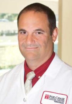 Philip Abbosh合作论文数Fox Chase Cancer Center, Temple Health3
 Lulla Amriti合作论文数Penn State Hershey Cancer Institute, Penn State College of Medicine3
 Arunasalam Navaraj合作论文数Department of Biochemistry, Indian Institute of Science3Saad Shaikh合作论文数3Francesca Di Cristofano合作论文数2
 Dinara Ryspayeva合作论文数National Cancer Institute2John Plastaras合作论文数Department of Radiation Oncology, Perelman School of Medicine, University of Pennsylvania2
 Steven Brem合作论文数Department of Neurosurgery, Perelman School of Medicine, University of Pennsylvania2Mintz Akiva合作论文数Dept Radiol, Wake Forest Sch Med2Melissa L. Dowling合作论文数2
 Andrew Tsourkas合作论文数Department of Bioengineering, School of Engineering and Applied Science, University of Pennsylvania2
 Ahmad Amirshaghaghi合作论文数Polymer Engineering Group, Tarbiat Modares University2Nolan Stubbs合作论文数1Yutong Xia合作论文数1Marina Hahn合作论文数1John C Santiago合作论文数1Alexander Raufi合作论文数1Rahmat K Sikder合作论文数1Ashley Sanchez Sevilla Uruchurtu合作论文数1Leiqing Zhang合作论文数1Roxanne Wood合作论文数1Bradley D DeNardo合作论文数1attila a seyhan合作论文数1Maximilian Pinho-Schwermann合作论文数1Shengling Zhang合作论文数1Jillian Strandberg合作论文数1Nataliia Verovkina合作论文数1
 Jiangbin Ye合作论文数Division of Radiation and Cancer Biology, Department of Radiation Oncology, School of Medicine, Stanford University1Nicholas R Liguori合作论文数1




