Artificial intelligence-generated content (AIGC) has shown remarkable performance in nuclear medicine imaging (NMI), offering cost-effective software solutions for tasks such as image enhancement, motion correction, and attenuation correction. However, these advancements come with the risk of hallucinations, generating realistic yet factually incorrect content. Hallucinations can misrepresent anatomic and functional information, compromising diagnostic accuracy and clinical trust. This paper presents a comprehensive perspective on hallucination-related challenges in AIGC for NMI, introducing the DREAM report, which covers recommendations for definition, representative examples, detection and evaluation metrics, and attributions and mitigation strategies. This position statement paper aims to initiate a common understanding for discussions and future research toward enhancing AIGC applications in NMI, thereby supporting their safe and effective deployment in clinical practice.
Longitudinal medical visual question answering (VQA) requires reasoning about anatomical differences between an image of a current time point and an image of a referred time point. We propose an attention-guided encoder-decoder for this task with chest X-rays. Instead of conventional direct contrast, we propose to include a lightweight affine registration module to reduce nuisance motion by co-registering the current image to the reference image with a small registration regularizer. The registered image pair is fed into the image encoder, followed by a frozen DINO-based mask generator and a trainable adaptive mask generator to produce masks applied to the original image pairs. The masked image pairs are again fed into the image encoder and concatenated with text features as the input to a multimodal transformer-based decoder to generate final answers. To facilitate learning stabilization and clarify the change signal, inspired by DINO-v3, we include additional auxiliary objectives, including a mask rebuilding loss, a pairwise Gram-style consistency loss, and a KoLeo uniformity loss, which enhances the geometry of the representation. On the Medical-Diff-VQA benchmark, the model delivers strong BLEU, ROUGE-L, CIDEr, and METEOR scores while offering intrinsic interpretability through the shared saliency mask. These results support saliency-conditioned generation with mild pre-alignment as a principled framework for longitudinal reasoning in medical VQA. Our training strategy also illustrates the potential of a paradigm in utilizing image foundation models in biomedicine: optimizing both supervised and unsupervised learning objectives simultaneously.
Artificial intelligence (AI)-driven histopathological image analysis has shown significant advantages for disease diagnosis, prognosis, and treatment planning, and it is receiving growing attention in modern healthcare. Due to the gigapixel size of whole slide images (WSIs), multiple instance learning (MIL) methods are widely employed in their analysis. Existing MIL approaches primarily rely on either instance-level or bag-level supervision, each facing challenges related to noisy pseudo-labels and suboptimal feature aggregation, respectively. In this paper, we present a novel MIL method for WSI analysis, termed CIB-MIL, which integrates collaborative instance-level and bag-level supervision. We introduce a label disambiguation module within the instance-level supervision channel that employs a noisy-label learning strategy to refine instance pseudo-labels and mitigate the impact of noisy labels. Additionally, we propose a collaborative supervision framework that promotes communication and interaction between the attention mechanism in the bag-level supervision channel and the pseudo-label mechanism in the instance-level supervision channel, enabling cooperative optimization of supervision in both channels. Extensive experiments conducted on five datasets, including three public datasets and two in-house datasets, demonstrate the state-of-the-art performance of CIB-MIL. The code is available at https://github.com/TencentAILabHealthcare/CIB-MIL.
Prompt-free adaptation of the Segment Anything Model (SAM) has emerged as a promising paradigm for automatic medical image segmentation. Existing methods mainly focus on prompt generation, while overlooking that prompt quality is fundamentally constrained by the expressiveness of anatomical representations. However, deterministic prototypes or semantic tokens are insufficient to jointly capture global anatomical priors, intra-structure diversity, and local structural reliability. To address this limitation, we propose the Hierarchical Probabilistic Representation (HPR) framework, which learns complementary anatomical representations through Distributional Anatomical Representation (DAR), Multi-component Anatomical Representation (MAR), and Local Reliability Representation (LRR), and integrates their predictions via Hierarchical Prediction Fusion (HPF) while remaining compatible with the original SAM decoder. Experiments on the Synapse, LA, and PROMISE12 datasets demonstrate that HPR-SAM achieves state-of-the-art performance on Synapse and the best performance under few-shot settings on LA and PROMISE12, validating the effectiveness of the proposed hierarchical probabilistic representation learning framework for prompt-free medical image segmentation. Code is available at https://anonymous.4open.science/r/HPR-SAM-E4AF.
Recent advances in semi-supervised medical image segmentation have achieved remarkable performance through prediction consistency, pseudo-label supervision, and hard-region supervision. However, these methods primarily improve supervision quality rather than explicitly enforcing semantic consistency in the learned representations of hard regions. Consequently, even under increasingly stronger prediction-level supervision, difficult regions exhibiting unstable semantic assignment often fail to establish semantically consistent representations during training, thereby limiting further segmentation improvement. To address this issue, we propose SHTA (Semantic Hard Token Correction and Center Alignment), a lightweight training-time semantic representation branch. Instead of introducing additional prediction supervision, SHTA refines intermediate semantic representations through Semantic Assignment, Hard Token Refinement, and Semantic Center Alignment, thereby improving semantic consistency in hard regions while preserving the original prediction pathway and introducing no additional inference cost. We integrate SHTA into representative semi-supervised segmentation frameworks, including GA-CPS, CPS, URPC, and MagicNet, and conduct evaluations on the Synapse and AMOS datasets. Experimental results demonstrate that SHTA delivers consistent paired improvements across frameworks, with especially clear gains in segmentation accuracy, weak-organ recovery, and semantic ambiguity reduction, while incurring only training-time overhead. The code is available at https://anonymous.4open.science/r/release_SHTA-42D5/.
General-purpose multimodal large language models (MLLMs) are increasingly applied to infrared images, where they are commonly scored by answer accuracy alone. However, a correct answer does not ensure that the model's explanation is grounded in infrared thermal evidence. We introduce an explanation-aware evaluation framework that separates answer correctness, output-level explanation groundedness, and thermal grounding for infrared visual questions. Using a Dual-LLM Consensus Judge with a preliminary human-anchor calibration check, we find that correct answers can still rely on weak or visible-light evidence; withholding the original infrared image and showing only a visible-like rendering erodes thermal grounding with little accuracy change; and this erosion is observed most strongly for more capable models but disappears when infrared remains available. We further propose Thermal-Grounded Feedback (TGF), a training-free feedback loop that diagnoses explanation-side failures and revises the explanation while preserving the selected answer. On local paired-input validation, TGF improves explanation-side grounding without changing answers. These findings suggest that future trustworthy MLLMs for infrared scene understanding should be evaluated and developed to produce thermally grounded explanations rather than merely accurate answers.
The synergistic interpretation of anatomical information from computed tomography (CT) and metabolic information from positron emission tomography (PET) is important to oncologic imaging. However, existing deep learning methods for PET/CT remain largely task-specific, are often trained on single-center cohorts, or adopt dual-branch fusion schemes that delay cross-modal interaction and underutilize early spatial correspondence between PET and CT. To address these limitations, we present an open-source, multi-center, whole-body FDG PET/CT foundation model utilizing 4,997 harmonized scans from four public datasets. Our framework employs hierarchical UNet-shaped backbones with early channel-wise concatenation, enabling anatomical and metabolic features to interact from the first embedding layer onward. We further introduce a masked autoencoding objective based on zero-mean imputation, combined with a weighted global reconstruction loss. This design avoids non-physical intensity discontinuities at masked-region boundaries that arise from learnable mask tokens. On downstream AutoPET lesion segmentation, the proposed models demonstrate strong label efficiency: with only 10% of the labeled training data, they achieve performance comparable to models trained from scratch on the full dataset. Under extreme 5-shot linear probing, joint PET/CT pretraining also achieves higher Dice scores than separated-modality pretraining. This multi-center foundation model demonstrates label efficiency and cross-modality representation learning for PET/CT tumor segmentation. It provides a robust, open-source basis for advancing automated oncologic imaging, significantly reducing the need for large-scale manual annotations in clinical practice.
Unmanned aerial vehicle (UAV) infrared image super-resolution aims to recover weak thermal structures for deployment on resource-constrained platforms; lightweight models are therefore preferred, but multi-loss training can be unstable. A common strategy combines pixel-domain and frequency-domain objectives; however, low contrast, limited high-frequency content, and sensor-specific noise often make their gradients weakly aligned or conflicting. To address this optimization ambiguity, we propose Orthogonal Gradient Gaming and Frequency Rectification (OGG-FR), a plug-and-play optimization framework that decomposes the frequency gradient into a redundant parallel component and an orthogonal innovation component relative to the pixel gradient. In the conflict regime, OGG-FR computes a safe base gradient using the Multiple Gradient Descent Algorithm (MGDA) and adds a variance-rectified orthogonal innovation; in the compatible regime, it discards redundant parallel information and injects the orthogonal innovation according to a confidence score estimated from the high-frequency residual. Experimental results on the UAV thermal benchmark show broad gains under BI and BD degradations at × 4 and × 8 scales, while gradient analyses support the effectiveness of the proposed conflict-aware update rule.
Understanding the relationship between vocal tract motion during speech and the resulting acoustic signal is crucial for aided clinical assessment and developing personalized treatment and rehabilitation strategies. Toward this goal, we introduce an audio-to-video generation framework for creating Real Time/cine-Magnetic Resonance Imaging (RT-/cine-MRI) visuals of the vocal tract from speech signals. Our framework first preprocesses RT-/cine-MRI sequences and speech samples to achieve temporal alignment, ensuring synchronization between visual and audio data. We then employ a modified stable diffusion model, integrating structural and temporal blocks, to effectively capture movement characteristics and temporal dynamics in the synchronized data. This process enables the generation of MRI sequences from new speech inputs, improving the conversion of audio into visual data. We evaluated our framework on healthy controls and tongue cancer patients by analyzing and comparing the vocal tract movements in synthesized videos. Our framework demonstrated adaptability to new speech inputs and effective generalization. In addition, positive human evaluations confirmed its effectiveness, with realistic and accurate visualizations, suggesting its potential for outpatient therapy and personalized simulation of vocal tract visualizations.
Semi-supervised 3D medical image segmentation reduces the need for dense voxel-level annotations by exploiting unlabeled volumes. Although existing methods such as consistency regularization, pseudo-labeling, and co-training improve prediction-level robustness, they often provide insufficient feature-space organization for anatomically complex structures, especially small organs and ambiguous boundary regions with large intra-class variations. To address this issue, we propose Variation-Conditioned Distributional Proxy Learning (VCDP), a plug-and-play training-only regularization module for semi-supervised 3D medical image segmentation. VCDP represents each class with a learnable Gaussian distribution for shared class semantics and multiple variation prototypes for fine-grained intra-class patterns. A unified variation-conditioned compatibility score is further formulated to fuse distributional similarity and soft variation aggregation, guiding voxel embeddings to align with both global organ identity and local anatomical variations. VCDP is attached to decoder features during training and removed during inference, introducing no additional inference cost. Experiments on multi-organ segmentation benchmarks show that VCDP improves most evaluated baselines, particularly for small, ambiguous, and highly variable organs. Our anonymous code is released at https://anonymous.4open.science/r/VCDP_code-41ED.
Objective. Deep learning has significantly advanced low-count positron emission tomography (PET) denoising. However, models trained on specific distributions often yield biased outputs when applied to scans with different activity distributions caused by anatomical and physiological variations (distribution shifts). Existing methods fail to generalize well across these scan-wise variations. Our goal is to formulate PET denoising as a scan-wise domain generalization (DG) problem to mitigate these variations and achieve robust, unbiased denoising for unseen scans. Approach. We propose a contrastive adversarial DG (CADG) framework to learn scan-invariant features efficiently. We leverage the property that multiple low-count noise realizations can be generated from a single raw list-mode PET scan to form a scan-wise domain distribution. Unlike conventional adversarial training with cross-entropy (CE) loss, we propose a contrastive adversarial framework that minimizes the mutual information between feature and scan-wise domains. Considering that each subject can have multiple scans in longitudinal studies, we further propose an ordered and memory-queued contrastive adversarial framework. This method efficiently includes realizations from the same scan as positive pairs, different scans of the same subject as pseudo-positive pairs, and different subjects as negative pairs in a memory batch. We explicitly exploit their ordered relationship as prior knowledge using a novel noisy-robust multi-positive ordinal contrastive loss. Main results. We systematically validated the effectiveness of our methods using 1920 noise realizations derived from 80 subjects with 192 longitudinal scans of MK-6240 tau PET data. The proposed CADG approach demonstrated superior denoising performance compared to CE-based adversarial methods and standard baselines. The ordered contrastive loss successfully improved the peak signal-to-noise ratio and structural similarity index consistently while reducing bias and standard deviation in Alzheimer-related regions and the whole brain. Significance. To our knowledge, this is the first attempt to alleviate the performance degradation in cross-scan denoising from the perspective of DG. Our study is also a pioneering work in utilizing longitudinal scans as pseudo-positives within an ordered contrastive learning scheme to exploit fine-grained relationships for robust clinical PET imaging applications.
Estimating individualized treatment effects (ITE) is critical for personalized medicine, yet it remains a challenge due to retrospective observational data, which suffer from selection bias in clinical practice and the complexity of multimodal data used for patient status depiction. In this work, the authors develop an end-to-end deep learning (DL) framework that incorporates multimodal patient data and multiple treatments for accurate ITE inference in a retrospective head and neck cancer (HNC) study. A possible solution is concatenating the factors and adapting adversarial training, which has shown great promise on tabular data, to disentangle patient characteristics from patient status features to mitigate treatment selection bias. However, this approach suffers from instability when applied to complex multimodal patient data and multiple treatment options. For flexible and efficient treatment-conditioned information fusion, they propose a bi-stage adaptive instance normalization (Bi-AdaIN) to inject relevant factors into corresponding layers, an approach that is also robust to missing values. Furthermore, they propose to disentangle status features from the multi-treatment variable using mutual information (MI) regularization, enabling more accurate predictions of patient-specific outcomes for both factual and counterfactual data. The authors evaluated their model on the RADCURE dataset, comprising 3,346 HNC cases with CT scans and multiple clinical variables who received radiotherapy or additional chemotherapy and EGFRI. The Bias-Adjusted Treatment Effect (BATE) is substantially reduced compared to the conventional direct ITE method (which does not consider treatment bias) and to adversarial training, indicating a more robust estimation of causal effects. This work is one of the first DL-based studies to address ITE estimation using multimodal medical imaging, offering a promising approach to counterfactual reasoning in clinical oncology for decision support.
In clinical practice, medical segmentation datasets are often limited and heterogeneous, with variations in modalities, protocols, and anatomical targets across institutions. Existing deep learning models struggle to jointly learn from such diverse data, often sacrificing either generalization or domain-specific knowledge. To overcome these challenges, we propose a joint training method called Universal Harmonization (U-Harmony), which can be integrated into deep learning-based architectures with a domain-gated head, enabling a single segmentation model to learn from heterogeneous datasets simultaneously. By integrating U-Harmony, our approach sequentially normalizes and then denormalizes feature distributions to mitigate domain-specific variations while preserving original dataset-specific knowledge. More appealingly, our framework also supports universal modality adaptation, allowing the seamless learning of new imaging modalities and anatomical classes. Extensive experiments on cross-institutional brain lesion datasets demonstrate the effectiveness of our approach, establishing a new benchmark for robust and adaptable 3D medical image segmentation models in real-world clinical settings.
Long-term modeling has proven effective for video-based depression recognition and has recently attracted growing attention. However, a key challenge is that long-term depression modeling often obscures crucial behavioral cues, making it difficult to learn global representations that faithfully reflect overall depressive states. To this end, the paper proposes a Long-short Behavioral Evolution for Depression (LS-BED) framework. It simultaneously captures long-term behavioral evolution and motion-enhanced short-term dynamics to form a discriminative representation for depression recognition. Specifically, the proposed LS-BED contains three components: 1) a Short-term Dynamic Booster (SDB) that focuses on diverse motion cues in shortterm dynamics, highlighting the capture of subtle motion movements and salient responses; 2) a Long-term Evolutionary Timeline (LET) that characterizes the temporal progression of depressive behaviors by detecting facial anchors defined as frames that capture pivotal transitions in facial dynamics, thereby effectively modeling long-term behavioral evolution while maintaining crucial local information; and 3) a Dual-stage Collaborative Fusion (DCF) that systematically integrates long-short bidirectional interaction and multiscale temporal aggregation, thereby enabling comprehensive modeling of temporal dependencies at varying scales. The experiments on the AVEC2013, AVEC2014 and DAIC-WOZ datasets demonstrate that the LS-BED achieves superior performance in depression recognition, yielding mean absolute error (MAE) and root mean square error (RMSE) of 5.01/6.97, 5.00/6.82, and 3.79/5.16, respectively.
Report-supervised (RSuper) learning seeks to alleviate the need for dense tumor voxel labels with constraints derived from radiology reports (e.g., volumes, counts, sizes, locations). In MRI studies of brain tumors, however, we often involve multi-parametric scans and substructures. Here, fine-grained modality/parameter-wise reports are usually provided along with global findings and are correlated with different substructures. Moreover, the reports often describe only the largest lesion and provide qualitative or uncertain cues (“mild,” “possible”). Classical RSuper losses (e.g., sum volume consistency) can over-constrain or hallucinate unreported findings under such incompleteness, and are unable to utilize these hierarchical findings or exploit the priors of varied lesion types in a merged dataset. We explicitly parse the global quantitative and modality-wise qualitative findings and introduce a unified, one-sided, uncertainty-aware formulation (MS-RSuper) that: (i) aligns modality-specific qualitative cues (e.g., T1c enhancement, FLAIR edema) with their corresponding substructures using existence and absence losses; (ii) enforces one-sided lower-bounds for partial quantitative cues (e.g., largest lesion size, minimal multiplicity); and (iii) adds extra- vs. intra-axial anatomical priors to respect cohort differences. Certainty tokens scale penalties; missing cues are down-weighted. On 1238 report-labeled BraTS-MET/MEN scans, our MS-RSuper largely outperforms both a sparsely-supervised baseline and a naive RSuper method.
Depression has become one of the most serious mental illnesses, leading to a substantial decline in quality of life, an elevated risk of suicide, and significant societal challenges. Despite significant progress in the application of deep learning for depression diagnosis, most prevalent methods rely on correlative rather than causal features, limiting their accuracy and interpretability. Here, we propose a causal graph learning (CGL) method for the hierarchical diagnosis of depression. Specifically, we first construct a novel depression facial graph (DFGraph) structure based on a prior knowledge, which collects information about subjects’ facial cues. Our CGL model leverages the DFGraph structure and incorporates a built-in masking mechanism, which is designed to effectively differentiate causal features from confounding ones. It employs backdoor adjustment techniques, which control for confounding variables by blocking noncausal paths, to identify and select pertinent causal features, thereby enhancing the accuracy of the hierarchical diagnosis of depression. We conducted extensive experiments on the collected depression dataset. Our results show that the proposed method provides better results and interpretability is further improved compared to the publicly available baseline.
Fully automated myocardial segmentation from cardiac magnetic resonance imaging (MRI) is vital for efficient diagnosis and treatment planning. Although numerous automated methods have been proposed, they typically focus on single MRI sequences and therefore have difficulties in generalizing across vendors and across cardiac MRI protocols. Simultaneous analysis of complementary cardiac MRI sequences, such as cine, T1 mapping, and late gadolinium enhancement (LGE) MRI, remains challenging due to their distinct image characteristics and scanner-specific variations. To address these issues, we propose an unsupervised domain adaptation approach that allows robust myocardial segmentation across multi-vendor cine, T1, and LGE MRI data. In particular, we introduce a class-imbalance self-training framework to transfer information learned from a source domain with labels to any unlabeled target domain, while maintaining consistent performance across different MRI sequences. Our framework iteratively refines segmentation accuracy by generating pseudo-labels for target data using a hardness-aware strategy, thus effectively addressing the problem of class imbalance in cardiac MRI segmentation. To mitigate data scarcity following pseudo-label selection, we employ a variance-guided vicinal feature extrapolation, which expands data points in the feature space into a probabilistic distribution. This, in turn, facilitates joint source-target training by generating a larger intersection in the feature space. Experimental results demonstrate that our framework outperforms existing methods when assessed using the Dice coefficient and Hausdorff distance. Our framework enables cardiac evaluation across MRI protocols without sequence-specific manual annotations.
Three-Dimensional Gaussian representation (3DGS) has shown substantial promise in the field of computer vision, but remains unexplored in the field of magnetic resonance imaging (MRI). This study explores its potential for the reconstruction of isotropic resolution 3D MRI from undersampled k-space data. We introduce a novel framework termed 3D Gaussian MRI (3DGSMR), which employs 3D Gaussian distributions as an explicit representation for MR volumes. Experimental evaluations indicate that this method can effectively reconstruct voxelized MR images, achieving a quality on par with that of well-established 3D MRI reconstruction techniques found in the literature. Notably, the 3DGSMR scheme operates under a self-supervised framework, obviating the need for extensive training datasets or prior model training. This approach introduces significant innovations to the domain, notably the adaptation of 3DGS to MRI reconstruction and the novel application of the existing 3DGS methodology to decompose MR signals, which are presented in a complex-valued format.
Low-light image super-resolution (LLSR) is a challenging task due to the coupled degradation of low resolution and poor illumination. To address this, we propose the Guided Texture and Feature Modulation Network (GTFMN), a novel framework that decouples the LLSR task into two sub-problems: illumination estimation and texture restoration. First, our network employs a dedicated Illumination Stream whose purpose is to predict a spatially varying illumination map that accurately captures lighting distribution. Further, this map is utilized as an explicit guide within our novel Illumination Guided Modulation Block (IGM Block) to dynamically modulate features in the Texture Stream. This mechanism achieves spatially adaptive restoration, enabling the network to intensify enhancement in poorly lit regions while preserving details in well-exposed areas. Extensive experiments demonstrate that GTFMN achieves the best performance among competing methods on the OmniNormal5 and OmniNormal15 datasets, outperforming them in both quantitative metrics and visual quality.
 Georges El Fakhri合作论文数Department of Biomedical Informatics & Data Science, Yale School of Medicine;Department of Radiology & Biomedical Imaging, Yale School of Medicine32
 Jonghye Woo合作论文数Department of Radiology, Harvard Medical School;Massachusetts General Hospital26
 Fangxu Xing合作论文数Mass General Research Institute, Massachusetts General Hospital;Harvard Medical School19
 Ouyang Jinsong合作论文数Department of Radiology, Harvard Medical School and Brigham and Women's Hospital, Boston, Massachusetts 02115, USA. ouyang@bwh.harvard.edu11
 Jia You合作论文数Department of Computing, Faculty of Engineering, The Hong Kong Polytechnic University9Maureen L. Stone合作论文数6
 Jerry L. Prince合作论文数Department of Electrical and Computer Engineering, Whiting School of Engineering, Johns Hopkins University;Sonavex, Inc.6
 C.-C. Jay Kuo合作论文数Thomas Lord Department of Computer Science, Viterbi School of Engineering, University of Southern California;Media Communications Lab, Viterbi School of Engineering, University of Southern California6Thibault Marin合作论文数Medical Imaging Research Center, lllinois Institute of Technology6
 Tomo Miyazaki合作论文数Grad. Sch. of Eng., Tohoku Univ.;c;Grad. Sch. of Eng., Tohoku Univ.5
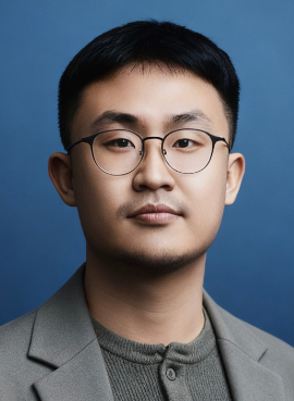 Xiaoqi (Jackie) Zhao (赵骁骐)合作论文数Yale University5
 Vijayakumar Bhagavatula合作论文数Carnegie Mellon University Africa;Carnegie Mellon University5
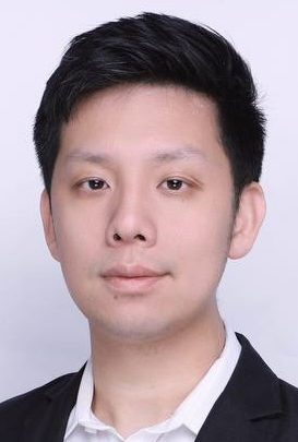 Harry Yang合作论文数The Hong Kong University of Science and Technology;Everlyn4
 Linghao Jin合作论文数Information Sciences Institute, University of Southern California4
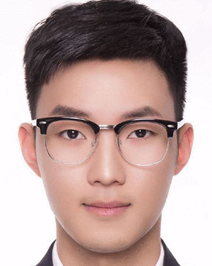 Yubin Ge合作论文数Amazon Web Services4
 Chi Liu合作论文数Department of Radiology and Biomedical Imaging, Yale School of Medicine, Yale University4Shinichiro Omachi合作论文数Graduate School of Engineering;Department of Management of Science and Technology4
 Jiachen Zhuo合作论文数Department of Diagnostic Radiology, University of Maryland Baltimore4Evans Xu Han合作论文数Yale University3Yuxuan Shi合作论文数3Li Wang合作论文数3HongMeng Yu (余洪猛)合作论文数3
 Yanzhong Wang合作论文数3
 Lihe Zhang (张立和)合作论文数School of Information and Communication Engineering, Dalian University of Technology3
 Huchuan Lu (卢湖川)合作论文数School of Information and Communication Engineering, Dalian University of Technology;School of Future Technology, Dalian University of Technology3Youwei Pang合作论文数School of Information and Communication Engineering, Dalian University of Technology3Qiuyuan Liu合作论文数2Samira Vafay Eslahi合作论文数2
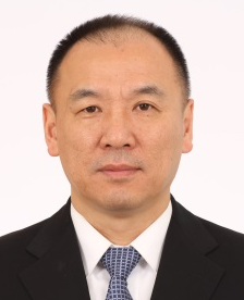 Ping Jia (贾平)合作论文数Changchun Institute of Optics, Fine Mechanics and Physics, Chinese Academy of Sciences;University of Chinese Academy of Sciences2
 Site Li (李斯特)合作论文数Cabot Corporation2Zhen Liang合作论文数2Mingmei Cheng合作论文数2Bingqing Bai合作论文数2J Wu合作论文数2Wanqing Xie合作论文数University of Science and Technology of China2
 Reimund Bayerlein合作论文数University of Siegen2
 Chae Hwa Yoo合作论文数Ewha Womans University2Qiao Mei (梅俏)合作论文数2
 Xu Han合作论文数2
 Chao Yang (杨超)合作论文数College of Computer Science and Electronic Engineering, Hunan University2Jihong Shen (沈继红)合作论文数College of Mathematical Sciences, Harbin Engineering University2
 Yao Lu (陆遥)合作论文数School of Computer Science and Engineering, Sun Yat-sen University;State Key Laboratory of Oncology in South China2
 Hui Luo (罗晖)合作论文数School of Computer Science, China University of Geosciences2
 Thomas Bortfeld合作论文数Division of Radiation Biophysics, Department of Radiation Oncology, Massachusetts General Hospital;University of Freiburg;Harvard Medical School;Faculty of Medicine, Harvard University;Mass General Research Institute2
 Keith Johnson合作论文数Division of Nuclear Medicine and Molecular Imaging, Massachusetts General Hospital;Harvard Medical School2Chao Ma (马超)合作论文数2Jing Hu (胡静)合作论文数2
 Shijian Lu合作论文数College of Engineering, Nanyang Technological University;School of Computer Science and Engineering, Nanyang Technological University2




