The landscapes of somatic mutation in normal cells inform us about the processes of mutation and selection operative throughout life, providing insight into normal ageing and the earliest stages of cancer development1. Here, by whole-genome sequencing of 238 microdissections2 from 30 individuals, including 18 with gastric cancer, we elucidate the developmental trajectories of normal and malignant gastric epithelium. We find that gastric glands are units of monoclonal cell populations that accrue roughly 28 somatic single-nucleotide variants per year, predominantly attributable to endogenous mutational processes. In individuals with gastric cancer, metaplastic glands often show elevated mutation burdens due to acceleration of mutational processes linked to proliferation and oxidative damage. Unusually for normal cells, gastric epithelial cells often carry recurrent trisomies of specific chromosomes, which are highly enriched in a subset of individuals. Surveying 829 polyclonal gastric microbiopsies by targeted sequencing, we find somatic 'driver' mutations in a distinctive repertoire of known cancer genes, including ARID1A, ARID1B, ARID2, CTNNB1 and KDM6A. The prevalence of mutant clones increases with age to occupy roughly 8% of the gastric epithelial lining by age 60 years and is significantly increased by the presence of severe chronic inflammation. Our findings provide insights into intrinsic and extrinsic influences on somatic evolution in the gastric epithelium in healthy, precancerous and malignant states.
Somatic variants accumulate in non-malignant tissues with age. Functional variants, leading to clonal advantage of hepatocytes, accumulate in the liver of patients with acquired chronic liver disease (CLD). Whether somatic variants are common to CLD from differing etiologies is unknown. We analyzed liver somatic variants in patients with genetic CLD from alpha-1 antitrypsin (A1AT) deficiency or hemochromatosis. We show that somatic variants in SERPINA1, the gene encoding A1AT, are strongly selected for in A1AT deficiency, with evidence of convergent evolution. Acquired SERPINA1 variants are clustered at the carboxyl terminus of A1AT, leading to truncation. In vitro and in vivo, C-terminal truncation variants reduce disease-associated Z-A1AT polymer accumulation and disruption of the endoplasmic reticulum, supporting the C-terminal domain swap mechanism. Therefore, somatic escape variants from a deleterious germline variant are selected for in A1AT deficiency, suggesting that functional somatic variants are disease-specific in CLD and point to disease-associated mechanisms.
Cancer predisposition syndromes mediated by recessive cancer genes generate tumors via somatic variants (second hits) in the unaffected allele. Second hits may or may not be sufficient for neoplastic transformation. Here we performed whole-genome and whole-exome sequencing on 479 tissue biopsies from a child with neurofibromatosis type 1, a multisystem cancer-predisposing syndrome mediated by constitutive monoallelic NF1 inactivation. We identified multiple independent NF1 driver variants in histologically normal tissues, but not in 610 biopsies from two nonpredisposed children. We corroborated this finding using targeted duplex sequencing, including a further nine adults with the same syndrome. Overall, truncating NF1 mutations were under positive selection in normal tissues from individuals with neurofibromatosis type 1. We demonstrate that normal tissues in neurofibromatosis type 1 commonly harbor second hits in NF1 , the extent and pattern of which may underpin the syndrome’s cancer phenotype.
Embryogenesis is a vulnerable time. Mutations in developmental cells can result in the wide dissemination of cells predisposed to disease within mature organs. We characterised the evolutionary history of four synchronous renal tumours from a 14-year-old girl using whole genome sequencing alongside single cell and bulk transcriptomic sequencing. Phylogenetic reconstruction timed the origin of all tumours to a multipotent embryonic cell committed to the right kidney, around 4 weeks post-conception. Biochemical and structural analysis of their shared MTOR mutation, absent from normal tissues, demonstrates enhanced protein flexibility, enabling a FAT domain hinge to dramatically increase activity of mTORC1 and mTORC2. Developmental mutations, not usually detected in traditional genetic screening, have vital clinical importance in guiding prognosis, targeted treatment, and family screening decisions for paediatric tumours.
Somatic variants accumulate in non-malignant tissues with age (1,2). Functional variants leading to clonal advantage of hepatocytes accumulate in the liver from patients with acquired chronic liver disease (CLD) (3-5). Whether these somatic variants are common to CLD from differing aetiologies is unknown. We analysed somatic variants in the liver from patients with genetic CLD from alpha-1 anti-trypsin (A1AT) deficiency or haemochromatosis. We show that somatic variants in SERPINA1, the gene encoding A1AT, are strongly selected for in A1AT deficiency, with evidence of convergent evolution. Acquired variants cluster at the 3-prime end of SERPINA1 leading to C-terminal missense or truncation variants of A1AT. In vitro, C-terminal truncation variants abrogate disease-associated A1AT polymerisation and retention in the endoplasmic reticulum, supporting the C-terminal domain swap mechanism. Therefore, somatic escape variants from a deleterious germline variant are selected for in A1AT deficiency, suggesting adaptive functional somatic variants are disease-specific in CLD and point to disease-associated mechanisms. ### Competing Interest Statement PJC is a co-founder, shareholder and consultant for Quotient Therapeutics Ltd. MH is a consultant for Quotient Therapeutics Ltd, AstraZeneca and Boston Scientific and has received unrestricted scientific grants from Pfizer.
Cancer predisposition syndromes mediated by recessive cancer genes generate tumours via somatic variants (second hits) in the unaffected allele. Second hits may or may not be sufficient for neoplastic transformation. Here, we performed whole genome and exome sequencing on 479 tissue biopsies from a child with neurofibromatosis type 1, a multi-system cancer-predisposing syndrome mediated by constitutive monoallelic NF1 inactivation. We identified multiple independent NF1 driver variants in histologically normal tissues, but not in 610 biopsies from two non-predisposed children. We corroborated this finding using targeted duplex sequencing, including a further nine adults with the same syndrome. Overall, truncating NF1 mutations were under positive selection in normal tissues from individuals with neurofibromatosis type 1. We demonstrate that normal tissues in neurofibromatosis type 1 commonly harbour second hits in NF1 , the extent and pattern of which may underpin the syndrome’s cancer phenotype. ### Competing Interest Statement IM is a co-founder and consultant of Quotient Therapeutics. DH provides consultancy to AstraZeneca/MedImmune, Alexion Pharmaceuticals, Bayer, Biodexa, Roche/Genentech, and Novartis, as well as expert testimony to AstraZeneca and Novartis, and his expenses are covered by Alexion Pharmaceuticals, Boehringer Ingelheim, Roche/Genentech, and Novartis.
The rates and patterns of somatic mutation in normal tissues are largely unknown outside of humans 1 – 7 . Comparative analyses can shed light on the diversity of mutagenesis across species, and on long-standing hypotheses about the evolution of somatic mutation rates and their role in cancer and ageing. Here we performed whole-genome sequencing of 208 intestinal crypts from 56 individuals to study the landscape of somatic mutation across 16 mammalian species. We found that somatic mutagenesis was dominated by seemingly endogenous mutational processes in all species, including 5-methylcytosine deamination and oxidative damage. With some differences, mutational signatures in other species resembled those described in humans 8 , although the relative contribution of each signature varied across species. Notably, the somatic mutation rate per year varied greatly across species and exhibited a strong inverse relationship with species lifespan, with no other life-history trait studied showing a comparable association. Despite widely different life histories among the species we examined—including variation of around 30-fold in lifespan and around 40,000-fold in body mass—the somatic mutation burden at the end of lifespan varied only by a factor of around 3. These data unveil common mutational processes across mammals, and suggest that somatic mutation rates are evolutionarily constrained and may be a contributing factor in ageing.
Tumour behaviour is intricately dependent on the oncogenic properties of cancer cells and their multi-cellular interactions. To better understand these dependencies within the wider micro-environment, we studied over 270,000 single cell transcriptomes and 100 micro-dissected whole exomes from 12 patients with kidney tumours, prior to validation through the use of spatial transcriptomics. Tissues were sampled from multiple regions of the tumour core, the tumour-normal interface, normal surrounding tissues, and peripheral blood. We found the tissue-type location of CD8+ T cell clonotypes largely defined their exhaustion state, with intra-tumoural spatial heterogeneity that is not well-explained by somatic heterogeneity. De novo mutation calling from single cell RNA sequencing data allows us to broadly infer the clonality of stromal cells and lineage-trace myeloid cell development. We discovered six conserved meta-programmes that distinguish tumour cell function. An epithelial-mesenchymal transition meta-programme, highly enriched at the tumour-normal interface appears co-localised with IL1B expressing macrophages, which could represent a plausible therapeutic target.Funding Information: This work was supported by Cancer Research UK/Royal College of Surgeons Clinician Scientist Fellowship (T.J.M: C63474/A27176), British Heart Foundation (R.L), the National Institute of Health Research (NIHR) Cambridge Biomedical Research Centre and the NIHR Blood and Transplant Research Unit (J.R.F. and M.R.C), Kidney Research UK Clinical PhD Fellowship (K.W.L.: TF_013_20171124), Wellcome Science Strategic Award for the Human Cell Atlas (G.S.B), Medical Research Council Human Cell Atlas Research Grant (M.R.C.: MR/S035842/1), Cancer Research UK Cambridge Centre (A.W.: C9685/A25177), Kidney Cancer UK and Facingup2Kidney cancer (M.G.B.T). The Wellcome Sanger Institute is supported by core funding from the Wellcome Trust (206194).Conflict of Interests:In the past 3 years, S.A.T has consulted for Roche and Genentech and is a Scientific Advisory Board member of Qiagen, Foresite labs, Biogen and GSK, as well as a consultant and equity holder as co-founder of Transition Bio. All other authors declare no competing interests.Ethical Approval: Human kidney and tumour tissues were collected through studies approved by UK NHS research ethics committees. All adult kidneys samples, except PD44967 were collected from patients enrolled in the DIAMOND study; Evaluation of biomarkers in urological disease (NHS National Research Ethics Service reference 03/018). Tumour PD44967 was collected from a patient enrolled in Characterisation of the immunological and biological markers of Renal cancer progression (NHS National Research Ethics Service reference 16/WS/0039).
Germ cell tumours (GCTs) are a collection of benign and malignant neoplasms derived from primordial germ cells. They are uniquely able to recapitulate embryonic and extraembryonic tissues, which carries prognostic and therapeutic significance. The developmental pathways underpinning GCT initiation and histogenesis are incompletely understood. Here, we study the relationship of histogenesis and clonal diversification in GCTs by analysing the genomes and transcriptomes of 547 microdissected histological units. We find no correlation between genomic and histological heterogeneity. However, we identify unifying features including the retention of fetal developmental transcripts across tissues, expression changes on chromosome 12p, and a conserved somatic evolutionary sequence of whole genome duplication followed by clonal diversification. While this pattern is preserved across all GCTs, the developmental timing of the duplication varies between prepubertal and postpubertal cases. In addition, tumours of younger children exhibit distinct substitution signatures which may lend themselves as potential biomarkers for risk stratification. Our findings portray the extensive diversification of GCT tissues and genetic subclones as randomly distributed, while identifying overarching transcriptional and genomic features.
Starting from the zygote, all cells in the human body continuously acquire mutations. Mutations shared between different cells imply a common progenitor and are thus naturally occurring markers for lineage tracing 1 , 2 . Here we reconstruct extensive phylogenies of normal tissues from three adult individuals using whole-genome sequencing of 511 laser capture microdissections. Reconstructed embryonic progenitors in the same generation of a phylogeny often contribute to different extents to the adult body. The degree of this asymmetry varies between individuals, with ratios between the two reconstructed daughter cells of the zygote ranging from 60:40 to 93:7. Asymmetries pervade subsequent generations and can differ between tissues in the same individual. The phylogenies resolve the spatial embryonic patterning of tissues, revealing contiguous patches of, on average, 301 crypts in the adult colonic epithelium derived from a most recent embryonic cell and also a spatial effect in brain development. Using data from ten additional men, we investigated the developmental split between soma and germline, with results suggesting an extraembryonic contribution to primordial germ cells. This research demonstrates that, despite reaching the same ultimate tissue patterns, early bottlenecks and lineage commitments lead to substantial variation in embryonic patterns both within and between individuals.
Mismatch repair (MMR) is a critical defence against mutation, but we lack quantification of its activity on different DNA lesions during human life. We performed whole-genome sequencing of normal and neoplastic tissues from individuals with constitutional MMR deficiency to establish the roles of MMR components, tissue type and disease state in somatic mutation rates. Mutational signatures varied extensively across genotypes, some coupled to leading-strand replication, some to lagging-strand replication and some independent of replication, implying that the various MMR components engage different forms of DNA damage. Loss of MSH2 or MSH6 (MutSα), but not MLH1 or PMS2 (MutLα), caused 5-methylcytosine-dependent hypermutation, indicating that MutSα is the pivotal complex for repairing spontaneous deamination of methylated cytosines in humans. Neoplastic change altered the distribution of mutational signatures, particularly accelerating replication-coupled indel signatures. Each component of MMR repairs 1-10 lesions/day per normal human cell, and many thousands of additional events during neoplastic transformation. Highlights MMR repairs 1-10 lesions/day in every normal cell and thousands more in tumor cells MMR patterns and rates are shaped by genotype, tissue type and malignant transformation MSH2 and MSH6 are pivotal for repairing spontaneous deamination of methylated cytosine Replication indels and substitutions vary by leading versus lagging strand and genotype
The ontogeny of the human haematopoietic system during fetal development has previously been characterized mainly through careful microscopic observations(1). Here we reconstruct a phylogenetic tree of blood development using whole-genome sequencing of 511 single-cell-derived haematopoietic colonies from healthy human fetuses at 8 and 18 weeks after conception, coupled with deep targeted sequencing of tissues of known embryonic origin. We found that, in healthy fetuses, individual haematopoietic progenitors acquire tens of somatic mutations by 18 weeks after conception. We used these mutations as barcodes and timed the divergence of embryonic and extra-embryonic tissues during development, and estimated the number of blood antecedents at different stages of embryonic development. Our data support a hypoblast origin of the extra-embryonic mesoderm and primitive blood in humans.
OBJECTIVES: Application of a new variable stiffness colonoscope (VSC) is expected to control loop formation and to lessen patient discomfort. The aim of this prospective study was to compare the efficacy of VSC with a conventional colonoscope (CC) in unsedated colonoscopy, based on the experience of examiners. METHODS: Four-hundred sixty-seven patients were randomly assigned to undergo colonoscopy with either VSC or CC by an endoscopist, including experienced and less-experienced examiners. The percentages of completed procedure and time to cecal intubation were recorded. Patients were asked to rate pain on a 5-point pain score. RESULTS: The percentages of completed procedure with VSC and CC were 98% and 95%, respectively, by less-experienced hands, and 99% and 98%, respectively, by experienced hands. Time for cecal intubation with VSC and CC was 15.7 and 18.5 min, respectively, by less-experienced hands, and 9.8 and 10.6 min, respectively, by experienced hands. A significantly lower mean pain score was noted in VSC patients compared with CC patients, irrespective of experience of the examiner. The percent of patients rating the procedure as moderately or severely painful was significantly lower with VSC than with CC, both in less-experienced (19% vs 40%; p < 0.01) and experienced hands (15% vs 26%; p < 0.05). CONCLUSIONS: Our results indicated that VSC allows favorable examination compared with CC regarding completeness, time to cecal intubation, and comfort of patients undergoing unsedated colonoscopy, irrespective of the examiner's experience. These features suggest VSC as the preferred colonoscope for patients undergoing unsedated colonoscopy.
The progression of chronic liver disease to hepatocellular carcinoma is caused by the acquisition of somatic mutations that affect 20-30 cancer genes(1-8). Burdens of somatic mutations are higher and clonal expansions larger in chronic liver disease(9-13) than in normal liver(13-16), which enables positive selection to shape the genomic landscape(9-13). Here we analysed somatic mutations from 1,590 genomes across 34 liver samples, including healthy controls, alcohol-related liver disease and non-alcoholic fatty liver disease. Seven of the 29 patients with liver disease had mutations in FOXO1, the major transcription factor in insulin signalling. These mutations affected a single hotspot within the gene, impairing the insulin-mediated nuclear export of FOXO1. Notably, six of the seven patients with FOXO1(S22W) hotspot mutations showed convergent evolution, with variants acquired independently by up to nine distinct hepatocyte clones per patient. CIDEB, which regulates lipid droplet metabolism in hepatocytes(17-19), and GPAM, which produces storage triacylglycerol from free fatty acids(20,21), also had a significant excess of mutations. We again observed frequent convergent evolution: up to fourteen independent clones per patient with CIDEB mutations and up to seven clones per patient with GPAM mutations. Mutations in metabolism genes were distributed across multiple anatomical segments of the liver, increased clone size and were seen in both alcohol-related liver disease and non-alcoholic fatty liver disease, but rarely in hepatocellular carcinoma. Master regulators of metabolic pathways are a frequent target of convergent somatic mutation in alcohol-related and non-alcoholic fatty liver disease.
Tumour behaviour is dependent on the oncogenic properties of cancer cells and their multi-cellular interactions. These dependencies were examined through 270,000 single cell transcriptomes and 100 micro-dissected whole exomes obtained from 12 patients with kidney tumours. Tissue was sampled from multiple regions of tumour core, tumour-normal interface, normal surrounding tissues, and peripheral blood. We found the principal spatial location of CD8+ T cell clonotypes largely defined exhaustion state, with clonotypic heterogeneity not explained by somatic intra-tumoural heterogeneity. De novo mutation calling from single cell RNA sequencing data allows us to lineage-trace and infer clonality of cells. We discovered six meta-programmes that distinguish tumour cell function. An epithelial-mesenchymal transition meta-programme, enriched at the tumour-normal interface appears modulated through macrophage expressed IL1B, potentially forming a therapeutic target.
Over the course of an individual's lifetime, normal human cells accumulate mutations(1). Here we compare the mutational landscape in 29 cell types from the soma and germline using multiple samples from the same individuals. Two ubiquitous mutational signatures, SBS1 and SBS5/40, accounted for the majority of acquired mutations in most cell types, but their absolute and relative contributions varied substantially. SBS18, which potentially reflects oxidative damage(2), and several additional signatures attributed to exogenous and endogenous exposures contributed mutations to subsets of cell types. The rate of mutation was lowest in spermatogonia, the stem cells from which sperm are generated and from which most genetic variation in the human population is thought to originate. This was due to low rates of ubiquitous mutational processes and may be partially attributable to a low rate of cell division in basal spermatogonia. These results highlight similarities and differences in the maintenance of the germline and soma.
ABSTRACTStarting from the zygote, all cells in the developing and adult human body continuously acquire mutations. A mutation shared between two different cells implies a shared progenitor cell and can thus be used as a naturally occurring marker for lineage tracing. Here, we reconstruct extensive phylogenies of normal tissues from three adult individuals using whole-genome sequencing of 511 laser capture microdissected samples from multiple organs. Early embryonic progenitor cells inferred from the phylogenies often contribute in different proportions to the adult body and the extent of this asymmetry is variable between individuals, with ratios between the first two reconstructed cells ranging from 56:44 to 92:8. Asymmetries also pervade subsequent cell generations and can differ between tissues in the same individual. The phylogenies also resolve the spatial embryonic origins and patterning of tissues, revealing a spatial effect in the development of the human brain. Supplemented by data on eleven men, we timed the split between soma and germline, with the earliest observed segregation occurring at the first cell divisions. This research demonstrates that, despite reaching the same ultimate tissue patterns, early bottlenecks and lineage commitments lead to substantial variation in embryonic patterns both within and between individuals.
To date, ontogeny of the human haematopoietic system during foetal development has been characterized mainly through careful microscopic observations. Here we used whole-genome sequencing (WGS) of 511 single-cell derived haematopoietic colonies from healthy human foetuses of 8 and 18 post-conception weeks (pcw) coupled with deep targeted sequencing of tissues of known embryonic origin to reconstruct a phylogenetic tree of blood development. We found that in healthy foetuses, individual haematopoietic progenitors acquire tens of somatic mutations by 18 pcw. Using these mutations as barcodes, we timed the divergence of embryonic and extra-embryonic tissues during development and estimated the number of blood antecedents at different stages of embryonic development. Our analysis has shown that ectoderm originates from a smaller set of blood antecedents compared to endoderm and mesoderm. Finally, our data support a hypoblast origin of the extra-embryonic mesoderm and primitive blood in humans.
 Peter Campbell合作论文数Quotient Therapeutics23
 Inigo Martincorena合作论文数Wellcome Sanger Institute19
 Tim H H Coorens合作论文数Wellcome Sanger Inst, Hinxton, England17
 Butler Timothy M合作论文数Department of Molecular and Medical Genetics, Oregon Health Sciences University16
 Mike Stratton合作论文数Wellcome Sanger Institute, Genome Research Limited16
 Luiza Moore合作论文数Wellcome Sanger Inst, Canc Ageing & Somat Mutat Programme, Hinxton CB10 1SA, Cambs, England14
 Thomas Mitchell合作论文数Department of Surgery, University of Cambridge11
 Mathijs A. Sanders合作论文数Department of Hematology, Erasmus University Medical Center10Philip S. Robinson合作论文数9
 Alex Cagan合作论文数Wellcome Sanger Inst8
 Sam Behjati合作论文数Department of Paediatrics, University of Cambridge8Matthew D. Young合作论文数7Andrew R. J. Lawson合作论文数7Henry Lee-Six合作论文数Wellcome Trust Sanger Inst, Canc Ageing & Somat Mutat Programme, Hinxton, England7
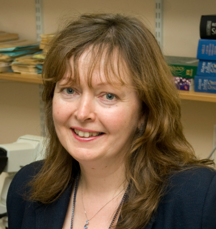 Anne Warren合作论文数Department of Pathology, University of Cambridge7Rashesh Sanghvi合作论文数6
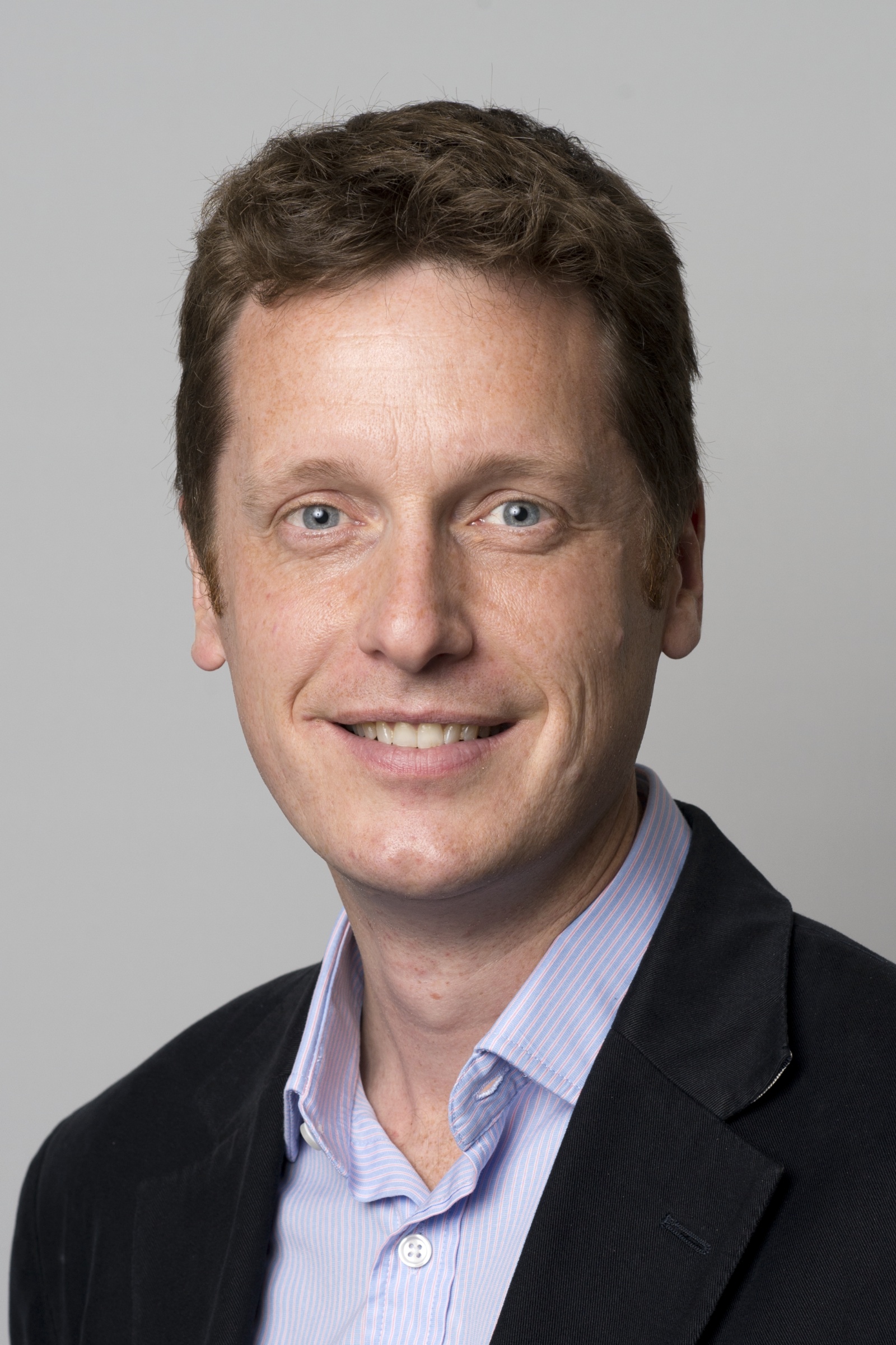 Matthew Hoare合作论文数Department of Medicine, University of Cambridge/Cancer Research UK Cambridge Institute, University of Cambridge6
 Raheleh Rahbari合作论文数Department of Genetics, University of Leicester6Federico Abascal合作论文数Department of Biochemistry, Genetics and Immunology, Universidad de Vigo6Natalia Brzozowska合作论文数5Nicholas Williams合作论文数Wellcome Trust Sanger Institute, Cambridge, UK.5Hyunchul Jung合作论文数5
 Thomas Oliver合作论文数School of Chemistry,University of Bristol5
 Grant Stewart合作论文数Selwyn College, University of Cambridge;Department of Surgery, University of Cambridge;Cancer Research UK Cambridge Institute;Cancer Research UK Cambridge Centre;NICE - National Institute for Health and Care Excellence;The Royal College of Surgeons of England;Kidney Cancer UK;Cambridge University Hospitals NHS Foundation Trust;The University of Edinburgh5
 Daniel Leongamornlert合作论文数Wellcome Sanger Institute5
 Mette Jørgensen合作论文数4Taryn D Treger合作论文数4
 Karp Natasha A合作论文数Department of Biochemistry, Cambridge University4
 Nangalia Jyoti合作论文数Wellcome - MRC Cambridge Stem Cell Institute, University of Cambridge4Brunner Simon F合作论文数4Burke Amos合作论文数4
 Isidro Cortes-Ciriano合作论文数Dept Biol Struct & Chim, Inst Pasteur4Jacqueline K. White合作论文数Addenbrooke's Hospital, University of Cambridge Clinical School4Kevin J. Dawson合作论文数4
 Kourosh Saeb-Parsy合作论文数Saeb-Parsy Lab, Department of Surgery, University of Cambridge;Revalia Bio;Excelect Ltd;Avatrial;Oxford Immune Algorithmics Ltd;Cambridge University Hospitals NHS Trust;Fitzwilliam College4
 Krishnaa Mahbubani合作论文数Lucy Cavendish College, University of Cambridge;Department of Haematology, University of Cambridge4Emma L. Cambridge合作论文数3Salsbury Grace合作论文数3Laura O'Neill合作论文数3
 Keiran Martin Raine合作论文数3Speak Anneliese O合作论文数3
 Mark Arends合作论文数Department of Pathology, University of Edinburgh;Department of Pathology, Fitzwilliam College, University of Cambridge3
 Vincent Gnanapragasam合作论文数Cambridge University Hospitals3
 Ramirez-Solis Ramiro合作论文数Wellcome Trust Sanger Institute3
 Gerstung Moritz合作论文数Dept Biosyst Sci & Engn, Swiss Fed Inst Technol3McIntyre Zoe合作论文数3Isherwood Christopher合作论文数3
 Loudon K W合作论文数Department of Renal Medicine, Addenbrooke's Hospital3




