Small cell lung cancer (SCLC) is an aggressive malignancy with limited effective therapeutic options. DLL3-targeted CAR-T therapy shows promising anti-tumor potential but is often restricted by T-cell exhaustion, which impairs its long-term efficacy. In the present study, we found that golidocitinib, a highly selective JAK1 inhibitor, induces apoptosis in SCLC cells in vitro via inhibiting STAT3 phosphorylation and regulating apoptosis‑associated genes. It also reduced the expression of exhaustion markers in anti-DLL3 CAR-T cells, promoted the formation of memory T cell phenotypes, and enhanced CAR-T cell persistence both in vitro and in vivo. When combined with anti-DLL3 CAR-T therapy, golidocitinib significantly augmented anti-tumor efficacy in both in vitro cytotoxicity assays and in vivo models, without obvious organ toxicity. These findings collectively demonstrate the dual anti-tumor effects of golidocitinib, thus providing a novel and promising strategy for SCLC treatment.
Receptor-interacting protein kinase 3 (RIPK3) has emerged as a central player in necroptosis and apoptosis activation in specific scenarios, concurrently modulating inflammatory responses. Here, we reveal that direct activation of RIPK3 concomitantly triggers mixed lineage kinase domain-like (MLKL) phosphorylation, caspase activation, and gasdermin cleavage within individual cells, inducing PANoptotic cell death. This process is orchestrated by the formation of RIPK3-MLKL-RIPK1-FADD-Caspase-8 complexes on progressively polymerized RIPK3 homo-aggregates, achieved through sequential recruitment dictated by the differential affinities of MLKL and Receptor-interacting protein kinase 1 (RIPK1) for distinct oligomeric states of RIPK3. In this process, MLKL- and GSDMD-mediated membrane rupture is respectively inhibited by Caspase-3-dependent cleavage of RIPK3 and GSDMD cleavage, while the pro-necrotic kinase activity of RIPK3 impedes RIPK1 recruitment and attenuates caspase activation. Cross-regulation between pathways results in unique cellular morphology, altered damage-associated molecular patterns (DAMPs) release profiles and distinct chemokine secretion paradigms that differ fundamentally from classical necroptosis, apoptosis and pyroptosis. This work highlights a common mechanism unveiling RIPK3 as a multimolecular platform to modulate and integrate different programmed cell death (PCD) pathways, thus providing a framework for targeting inflammatory cell death in disease.
Small cell lung cancer (SCLC) exhibits a high incidence of perineural invasion (PNI), a clinical feature associated with poor prognosis. Here, we establish PNI as an independent adverse prognostic factor in a surgical SCLC cohort. We further show that the neural microenvironment upregulates stathmin-2 (STMN2) in SCLC cells. STMN2, in a concentration-dependent manner, activates the β-alanine metabolic pathway, leading to intracellular β-alanine accumulation, which enhances tumor cell migration and invasion. In vivo, STMN2 knockdown suppresses neural invasion, an effect reversible upon β-alanine supplementation. This work defines a neural-STMN2-β-alanine-invasion axis that drives PNI in SCLC, providing mechanistic insights and highlighting a promising metabolic vulnerability for therapeutic intervention.
Profiling circulating extracellular vesicle-associated microRNAs (EVs-miRNA) is essential for improving lung cancer (LC) diagnosis and prognosis in clinical tests. However, the complexity of RNA extraction procedures and the lack of an LC-specific EVs-miRNA signature limit the clinical applicability of the current liquid biopsy tools. Herein, we develop a fusogenic liposome nanoreactor for the direct profiling of plasma EVs-miRNAs. This platform integrates reagent-encapsulating liposomes, isothermal rolling circle amplification, and CRISPR-Cas12a endonuclease cleavage in a one-pot manner, allowing for ultrasensitive measurements down to fM levels. A LC-specific EVs-miRNA signature was identified through a designed four-phase screening procedure, which was validated by using both public databases and clinical samples. In a proof-of-concept cross-sectional and longitudinal study involving a clinical cohort (n = 141), the profiled signature enabled early diagnosis, therapy monitoring, and prognosis evaluation with an accuracy of up to 93.1%. The platform streamlines the diagnostic workflow into a single fusogenic step, providing an RNA extraction-free and proof-of-concept liquid biopsy framework for potential LC management.
8106 Background: SCLC and LCNEC are characterized by aggressive clinical courses and limited therapeutic options following progression on platinum-based chemotherapy. DLL3(Delta-like ligand 3) is highly expressed and selectively on the surface of these high-grade neuroendocrine tumors, making it an ideal therapeutic target. SNC115 is a novel CAR-T cell armored with CD70 to target DLL3-expressing cells. Methods: This phase 1, open-label study utilized an accelerated titration design followed by a standard 3+3 escalation to evaluate the safety, tolerability, and pharmacokinetics of SNC115 in patients (pts) with R/R SCLC or LCNEC who progressed after ≥1 line of standard therapy. Following leukapheresis and a 3-day lymphodepletion regimen (fludarabine/cyclophosphamide), pts received a single infusion of SNC115 at one of five planned dose levels (DL): 1.0×10^5 (DL1), 3.0×10^5 (DL2), 1.0×10^6 (DL3), 3.0×10^6 (DL4), and 6.0×10^6 (DL5) CAR + T cells/kg. The primary endpoints were the determination of the maximum tolerated dose (MTD) and the recommended dose (RD). Results: As of 23 January 2026, 8 pts with R/R SCLC were treated across four DLs: DL1 (n = 1), DL2 (n = 3), DL3 (n = 2) and DL4 (n = 2). Median age was 53.5 (range 40-69) years, with a median of three prior therapy lines (range 2-6). 6 pts received bridging therapy. SNC115 demonstrated a manageable safety profile. CRS occurred in 3 pts (DL1: 1pt, DL4: 2pts), all Grade 1 or 2 and resolved with tocilizumab/corticosteroids. Grade ≥3 hematologic TEAEs included lymphocyte count decreased (8/8), white blood cell count decreased (2/8) and anaemia (1/8), all attributed to LD. Only one patient (DL4) experienced Grade ≥3 non-hematologic TEAEs related to SNC115, including alanine aminotransferase increased (G3), Gamma-glutamyltransferase increased (G3) and diarrhea (G3), which resolved with symptomatic care. No DLTs, SAEs or ICANS were reported. Among evaluable pts (n = 8), the ORR and DCR were 37.5% and 75%, respectively. All patients (n = 2) in the higher-dose level (DL4) achieved PR. CAR-T cells expansion peaked at a median of 7 days post-infusion, with Cmax ranging from 68.77 to 4359.89 copies/µg DNA. Conclusions: SNC115 demonstrated a manageable safety profile and encouraging preliminary antitumor activity in heavily pretreated pts with SCLC. The absence of DLTs and ICANS, combined with low-grade transient CRS, supports continued dose escalation. These early efficacy signals suggest that SNC115 may provide a novel therapeutic avenue for R/R SCLC. Clinical trial information: NCT06384482 .
11530 Background: Advanced bone and soft-tissue sarcomas have limited treatment options. Tumor-selective methionine dependency is a known metabolic vulnerability. SGN1 is designed to deplete methionine locally within tumors via intratumoral colonization of an attenuated Salmonella vector expressing L-methioninase. Preclinical studies have demonstrated safety and targeted antitumor activity. Here, we report the results from the escalation and expansion phases of the Phase I study in patients (pts) with advanced bone and soft-tissue sarcomas. Methods: The Phase I study includes a dose-escalation phase where pts received intravenous (IV) (2.0×10⁸ to 4.0×10⁸ CFU) or intra-arterial (IA) infusion (2.0×10⁸ to 6.0×10⁸ CFU) once a week (QW) in 28-day cycles, followed by an expansion phase with the putative recommended Phase 2 dose (RP2D) (2.0×10⁸ CFU for IV or 6.0×10⁸ CFU for IA QW) in 6 cohorts, including pts with recurrent either bone or soft tissue sarcoma after at least 2 lines of chemo therapies. SGN1 was administered in combination with physician's choice systemic therapy. Administration of SGN1 was via one of three routes: IV, IA, or a combined IV/IA regimen. Efficacy was assessed by RECIST version 1.1. Results: At the cut-off of Aug 19, 2024, 25 pts with advanced bone and soft-tissue sarcomas were enrolled in the escalation and expansion phases (median follow-up of 5.6 months; range, 0.5-18.2), including pts with bone sarcoma (n=10) and soft tissue sarcoma (n=15). Treatment was administered via IV (n=13), IA (n=8), or a combined IV/IA (n=4) route. The disease control rate (DCR) was 76.0% (95% CI: 56.5, 93.0) in all, 70.0% (95% CI: 34.8, 93.3) in bone sarcoma, 80.0% (95% CI: 52.0, 95.6) in soft tissue sarcoma pts, and 69.2% (95% CI: 38.5, 90.9) in IV, 75.0% (95% CI: 34.8, 93.3) in IA, 100% in combined IV/IA route. Quantitative PCR analysis confirmed significant intra-tumoral colonization by SGN1 post-treatment. The median progression-free survival (mPFS) was 11.5 months (95% CI: 11.1, NE) in all, 11.1 months (95% CI: 1.1, NE) in bone sarcoma, not reached (NR) (95% CI: 3.2, NE) in soft tissue sarcoma pts, and NR (95% CI: 2.7, NE) in IV, 11.5 months (95% CI: 1.3, NE) in IA, NR (95% CI: 11.1, NE) in combined IV/IA route. Complete tumor necrosis was observed in at least two soft tissue sarcoma patients. In all pts, the most frequent treatment-related adverse events (TRAEs) were pyrexia (32.0%) and elevation of blood LDH increased (20.0%). No TRAEs leading to temporary drug discontinuation, no serious TRAEs resulting in withdrawal from the study, and no treatment-related deaths occurred. Conclusions: SGN1 exhibits a manageable safety profile, and showed encouraging clinical benefit in advanced bone and soft-tissue sarcoma, as evidenced by mPFS and DCR. The promising data from this phase I study supports further testing of SGN1 in Phase II studies. Clinical trial information: ChiCTR2400085361.
11523 Background: Previous studies have demonstrated that preoperative intra-arterial cisplatin infusion elicits a favorable histologic response—a key prognostic factor for survival in osteosarcoma. Recombinant human endostatin (rh-endostatin; Endostar), an anti-angiogenic agent, has been shown to significantly improve survival when administered intravenously in combination with chemotherapy for osteosarcoma. Herein, we report preliminary results on the efficacy and safety of continuous intra-arterial cisplatin and rh-endostatin combined with systemic chemotherapy in osteosarcoma (NCT06562673). Methods: This open-label, single-arm, single-center, phase II clinical trial enrolled patients with histologically confirmed primary localized extremity conventional osteosarcoma. Patients received 2 cycles of high-dose methotrexate and anthracyclines intravenously. Intra-arterial infusion was performed concurrently with anthracyclines. Rh-endostatin was administered intra-arterially (150 mg over 6 hours); then, cisplatin was administered intra-arterially (100-120 mg/m² over 6 hours). Efficacy and safety were evaluated after surgery. Results: Ten patients were enrolled from December 2024, to April 2025, including 7 males and 3 females with a mean age of 17.8 years (range, 12-42 years). Tumor locations included the femur (n=6), tibia (n=3), and humerus (n=1). Preoperative arterial infusions were administered 3 times (n=1), 2 times (n=8), and 1 time (n=1). All patients underwent limb-sparing surgery. The tumor necrosis rate was >90% in 5 cases (50%) and ≤90% in 5 cases (50%). Adverse events included grade 3 vomiting and nausea (n=1), grade 1 renal impairment (n=2), fever (n=1), and skin induration (n=1). Due to grade 3 gastrointestinal toxicity observed in the first enrolled female patient, the cisplatin dose was uniformly reduced to 100 mg/m² for all subsequent patients in the trial. Conclusions: Intra-arterial administration of high-dose rh-endostatin and cisplatin combined with systemic chemotherapy in osteosarcoma appears safe and achieves a good histologic response rate of 50%, compared with a historical rate of 30% with intravenous chemotherapy alone. Clinical trial information: NCT06562673 .
BACKGROUND:Multiple high-risk human papillomavirus (hrHPV) infections are frequently detected in cervical screening, but their biological interactions and clinical significance remain unclear. Because HPV DNA detection indicates viral presence but not transcriptional activity, this exploratory study evaluated viral load, genotype-specific E6/E7 mRNA expression, localization, and follow-up. METHODS:Among 12,000 cervical specimens tested for hrHPV, 598 were positive and 72 showed multiple genotypes. Viral load was assessed using the BioPerfectus Multiplex Real-Time assay. A tissue-available subset of 36 dual HPV16+52 or HPV16+58 cases was analyzed by duplex RNAscope for genotype-specific E6/E7 mRNA expression and localization. Diagnoses were categorized as benign, LSIL, or HSIL. Viral load and RNAscope analyses used incompletely matched subsets and were interpreted as complementary, not paired. RESULTS:Elevated viral load (≥4.00 log10 copies/10,000 cells) was associated with HSIL. In cohort-level analyses, diffuse (+++) E6/E7 mRNA expression was more frequent in HSIL than elevated viral load (95.2% vs 55.5%, P = 0.01). RNAscope-negative cases were mostly benign or LSIL and showed no progression. Genotype-specific signals were spatially distinct, with rare overlap. Dual-positive cells showed fewer RNA signals than adjacent single-positive cells (P < 0.001). Higher E6/E7 mRNA expression showed possible association with LSIL progression, but events were limited. CONCLUSIONS:In multiplex hrHPV infection, transcriptional activity may better reflect cervical lesion biology than viral DNA burden alone. Genotype-specific E6/E7 mRNA expression may complement HPV DNA load. Spatially distinct signals and reduced signal intensity in rare dual-positive cells suggest possible interactions between co-infecting hrHPV genotypes, but require prospective validation in fully matched cohorts.
Reduced lymphoid enhancer-binding factor 1 (LEF1) expression in patients with adenomyosis during the mid-secretory phase leads to impaired endometrial receptivity, affecting embryo implantation. This study investigated the molecular mechanisms underlying reduced endometrial receptivity in 25 adenomyosis patients and 25 controls. Functional experiments were conducted using human endometrial stromal cells (HESCs) and TERT-immortalized HESCs(T-HESCs), with final validation performed using a mouse model. Western blot and quantitative real-time polymerase chain reaction (RT-qPCR) analyses revealed that patients with adenomyosis showed a marked decrease in LEF1 expression in the stromal cells of the endometrium during the mid-secretory phase. In vitro experiments demonstrated that LEF1 knockdown in stromal cells led to impaired decidualization. Transcriptome sequencing, dual-luciferase reporter assays, and chromatin immunoprecipitation (ChIP) experiments showed that LEF1 could bind to the promoter region of interleukin (IL)-11 and promote its transcription, and IL-11 expression was also found to be downregulated in adenomyosis patients. Overexpression of IL-11 rescued the impaired decidualization caused by decreased LEF1 expression. In the in vitro co-culture model, LEF1/IL-11 knockdown led to a reduction in embryo implantation area, which was partially restored upon IL-11 overexpression. In the adenomyosis mouse model, we observed a decrease in LEF1 expression and a reduction in implantation sites compared to control mice, accompanied by impaired decidualization and receptivity. Notably, supplementation with IL-11 restored the number of implantation sites. The decrease in fertility due to reduced endometrial receptivity in adenomyosis patients is a significant clinical issue in assisted reproductive technology. This research provides insights into one potential molecular mechanism underlying this decreased receptivity, with a specific focus on the reduced expression of LEF1 in the endometrial stromal cells during the mid-secretory phase in adenomyosis patients. Our findings offer new perspectives for clinical strategies to improve endometrial receptivity in patients with adenomyosis, potentially enhancing their chances of successful pregnancy.
OBJECTIVE:Aloin, the main active component in Aloe vera (L.) Burm. f., has shown promising anti-tumor effects. This study investigated the impact of aloin in lung squamous cell carcinoma (LUSC) and explored its functional mechanism. METHODS:We analyzed the viability, migration, invasion, proliferation, and apoptosis of two LUSC cell lines after treatment with aloin. Target molecules of aloin and downstream target transcripts of nuclear receptor subfamily 3 group C member 2 (NR3C2) were predicted by bioinformatics. The biological functions of NR3C2 and metallothionein 1 M (MT1M) in the malignant properties of LUSC cells were determined. A co-culture system of LUSC cells with monocyte-derived macrophages was constructed. Mouse xenograft tumor models were generated to analyze the functions of aloin and NR3C2 in the tumorigenic activity of LUSC cells and macrophage polarization in vivo. RESULTS:Aloin suppressed malignant properties of LUSC cells in vitro. However, these effects were negated by the silencing of NR3C2. NR3C2 was found to activate MT1M transcription by binding to its promoter. Additional upregulation of MT1M suppressed the malignant behavior of LUSC cells augmented by NR3C2 silencing. Analysis of the M1 and M2 markers/cytokines in the macrophages or the culture supernatant revealed that aloin treatment or MT1M overexpression in LUSC cells enhanced M1 polarization while suppressing M2 polarization of macrophages, whereas NR3C2 silencing led to reverse trends. Consistent findings were reproduced in vivo. CONCLUSION:This study demonstrated that aloin activates the NR3C2/MT1M axis to suppress the malignant behavior of LUSC cells and M2 macrophage polarization. Please cite this article as: Chen YN, Lu JY, Gao CF, Fang ZR, Zhou Y. Aloin blocks the malignant behavior of lung squamous cell carcinoma cells and M2 macrophage polarization by modulating the NR3C2/MT1M axis. J Integr Med. 2025; 23(2): 195-208.
Schwann cells (SCs) are the primary glial cells of the Peripheral Nervous System (PNS), which insulate and provide protection and nutrients to the axons. Technological and experimental advances in neuroscience, focusing on the biology of SCs, their interactions with other cells, and their role in the pathogenesis of various diseases, have paved the way for exploring new treatment strategies that aim to harness the direct protective or causative properties of SCs in neurological disorders. SCs express cytokines, chemokines, neurotrophic growth factors, matrix metalloproteinases, extracellular matrix proteins, and extracellular vesicles, which promote the inherent potential of the injured neurons to survive and accelerate axonal elongation. The ability of SCs to support the development and functioning of neurons is lost in certain hereditary, autoimmune, metabolic, traumatic, and toxic conditions, suggesting their role in specific neurological diseases. Thus, targeting, modifying, and replacing SC strategies, as well as utilizing SC-derived factors and exosomes, have been considered novel therapeutic opportunities for neuropathological conditions. Preclinical and clinical data have demonstrated that SCs and SC-derived factors can serve as viable cell therapy for reconstructing the local tissue microenvironment and promoting nerve anatomical and functional recovery in both peripheral and central nerve injury repair, as well as in peripheral neuropathies. However, despite the promising successes of genetic engineering of SCs, which are now in preclinical and clinical trials, improving tactics to obtain ‘repair’ SCs and their products from different sources is the key goal for future clinical success. Finally, further development of innovative therapeutic approaches to target and modify SC survival and function in vivo is also urgently needed.
PANoptosis, an immunogenic programmed cell death (PCD) modality, integrates features of pyroptosis, apoptosis, and necroptosis through assembly of the PANoptosome complex. Despite its conceptualization as a distinct PCD form, PANoptosis remains controversial due to insufficient characterization of its morphological hallmarks and molecular regulation. This study aimed to investigate the molecular mechanisms underlying the assembly of PANoptosomes in the PANoptotic pathway. We identified a novel receptor-interacting protein kinase 3 (RIPK3)-initiated PANoptotic pathway that functions without pattern recognition receptors (PRRs) and the ASC (apoptosis-associated speck-like protein containing a CARD) inflammasome. Using multimodal imaging and biochemical approaches, we identify unique morphological signatures distinguishing PANoptotic cells from canonical pyroptotic, apoptotic, or necroptotic counterparts. Mechanistically, RIPK3 forms round homopolymeric scaffolds, distinct from necroptotic amyloid-like fibers, to sequentially recruit MLKL and RIPK1, forming a dynamic RIPK3-MLKL-RIPK1-FADD-caspase-8 complex (RIPK3-PANoptosome). This platform coordinates concurrent activation of pyroptotic, apoptotic, and necroptotic effectors. Cross-regulatory interactions between these pathways establish a homeostatic system where perturbations bias death modality into a certain cell death type, altering the death process and outcomes. Functionally, PANoptotic cells orchestrate chemokine secretion through parallel kinase-dependent (RIPK3-MLKL) and kinase-independent (RIPK1-IKK-NF-κB) mechanisms, driving macrophage recruitment. Our findings resolve the molecular logic of PANoptosome assembly, redefine PANoptosis as a tunable PCD paradigm, and establish its role in immunomodulation, providing a framework for targeting inflammatory cell death in disease. ### Competing Interest Statement The authors have declared no competing interest.
This letter provides a review of the report by Peng et al on a unique case of non-small cell lung cancer (NSCLC), specifically lung adenocarcinoma, featuring reactive oxygen species proto-oncogene 1-receptor (ROS1) co-mutation. The case involves a 64-year-old patient who exhibited both epidermal growth factor receptor (EGFR) L858R mutation and ROS1 rearrangement, achieving significant disease stabilization following treatment with crizotinib. This rare EGFR/ROS1 co-mutation poses distinct challenges for clinical management and highlights the necessity of personalized treatment strategies. While third-generation EGFR tyrosine kinase inhibitors (TKIs), such as osimertinib, are commonly regarded as first-line therapies, recent studies indicate that crizotinib may offer superior disease control in certain EGFR-mutant patients, particularly those who exhibit poor responses to EGFR TKIs. The case also examines the influence of tumor cell genetic heterogeneity on treatment response, underscoring the importance of evaluating tumor characteristics. In patients with EGFR/ROS1 co-mutation, gefitinib is generally effective as a first-line treatment; however, its efficacy can be limited, whereas crizotinib has demonstrated improved disease control. Future research should focus on identifying optimal treatment strategies for patients with EGFR/ROS1 co-mutation to enhance patient outcomes. In conclusion, this case report not only illustrates the effectiveness of crizotinib in managing patients with EGFR/ROS1 co-mutation but also underscores the importance of personalized treatment approaches, offering valuable insights for improving clinical outcomes in NSCLC patients with complex genetic profiles.
 Haiyan Hu (胡海燕)合作论文数Shanghai Sixth People's Hospital38
 Hua Zhong (钟华)合作论文数Shanghai Chest Hospital26
 Zan Shen (沈赞)合作论文数Shanghai Sixth People's Hospital Affiliated to Shanghai Jiao Tong University School of Medicine24
 Yang Yao (姚阳)合作论文数Department of Oncology, Shanghai Sixth People's Hospital Affiliated to Shanghai Jiao Tong University;Shanghai GoBroad Cancer Hospital, China Pharmaceutical University20
 Aina He (何爱娜)合作论文数18Yujing Huang合作论文数16
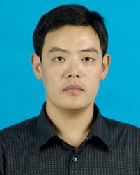 Yonggang Wang (王永刚)合作论文数Renji Hospital, Shanghai Jiao Tong University School of Medicine16Li-Na Tang合作论文数15
 Wenxi Yu (余文熙)合作论文数15Guowei Qian合作论文数14Xiaomin Ding合作论文数<institution>Oncology Department of Shanghai Jiao Tong University Affiliated Sixth People’s Hospital</institution>, Shanghai; No 600 Yishan Road Xuhui District of Shanghai City, <country>China</country>, 20023313Yaling Wang合作论文数Shanghai Jiao Tong Univ, Affiliated Peoples Hosp 6, Dept Oncol, Shanghai, Peoples R China13Hongtao Li (李洪涛)合作论文数13Shui-Er Zheng合作论文数13Shuhui Cao合作论文数12Junyi Yin合作论文数Oncology Department of Shanghai Jiao Tong University Affiliated Sixth People's Hospital of Shanghai, Shanghai 200233, China.12
 Qingcheng Yang (杨庆诚)合作论文数Shanghai Sixth People's Hospital Affiliated to Shanghai Jiao Tong University12Yue Wang合作论文数11Jingwen Li合作论文数11
 Yao Zhang合作论文数10
 Yang Dong (董扬)合作论文数Department of Orthopaedics, Shanghai Jiao Tong University Affiliated Sixth People's Hospital, Shanghai Jiao Tong University10Han Kun合作论文数9
 Daliu Min (闵大六)合作论文数Shanghai Sixth People's Hospital9Xuxinyi Ling合作论文数8Cheng Yang合作论文数8Yang Su合作论文数Shanghai Jiao Tong Univ, Sch Med, Rui Jin Hosp, Dept Surg,Shanghai Inst Digest Surg, Shanghai 200030, Peoples R China8Chenliang Zhou合作论文数Shanghai Jiao Tong Univ Affiliated Peoples Hosp 6, Dept Oncol, 600 Yishan Rd, Shanghai 200233, Peoples R China8
 Xiao-Bin Lv合作论文数8Yawen Zhang合作论文数7
 Qiyuan Bao (鲍其远)合作论文数Dept Physiol, Northwestern Univ7
 Baohui Han (韩宝惠)合作论文数Shanghai Chest Hospital;Shanghai GoBroad Cancer Hospital China Pharmaceutical University7
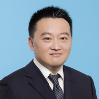 Runbo Zhong (钟润波)合作论文数Chest Hospital, Shanghai Jiao Tong University School of Medicine7
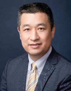 Tong Ji (季彤)合作论文数Department of Oral Maxillofacial-Head Neck Oncology, Shanghai Ninth People’s Hospital, School of Medicine, Shanghai Jiao Tong University;College of Stomatology, Shanghai Jiao Tong University7
 Yingqi Hua (华莹奇)合作论文数School of Health Science and Engineering, University of Shanghai for Science and Technology;Shanghai General Hospital;Shanghai Jiao Tong University School of Medicine7
 Yuanjue Sun (孙元珏)合作论文数Tongji University7Jinrong Liang合作论文数6Yuanliang Zhang合作论文数6Yang Yu合作论文数6DAI Xiangrong (戴向荣)合作论文数6Gan Zhi-Hua合作论文数6
 Zhichang Zhang合作论文数Shanghai Jiao Tong Univ, Peoples Hosp 6, Dept Orthoped, Shanghai 200233, Peoples R China5Yuqing Lou合作论文数5GV Shurin合作论文数4Shuchen Lin合作论文数4Wei Meng合作论文数Southern Med Univ, Inst Genet Engn, Guangzhou, Guangdong, Peoples R China4LiWen Xiong (熊丽纹)合作论文数4
 Michael R. Shurin合作论文数Department of Pathology, University of Pittsburgh;Department of Immunology, University of Pittsburgh4
 Jianjun Zhang (张建军)合作论文数College of Stomatology, Shanghai Jiao Tong University4




