Atomic force microscopy (AFM) provides quantitative capabilities for mapping the mechanical properties of living cells with nanometer resolution, yet its potential for monitoring a range of cell mechanical states remains largely underexplored. Here, we employed AFM to map the mechanical state progression of endothelial cells (ECs) across physiological and stress conditions. Distinct mechanical signatures emerged for each physiological state: cells stiffen during division, soften in response to cell stress such as inflammation, and exhibit mechanical collapse when cytoskeletal integrity is compromised. High-resolution AFM imaging of the cytoskeletal architecture revealed changes in its organization and coherency. By simultaneously tracking the mechanical and topographical changes, AFM enables the prediction of cellular viability and functional state in real-time. AFM measurements provide a real-time assessment of endothelial cell state in vitro, with broader implications for understanding endothelial dysfunction in vascular disease and enabling new tools in tissue engineering.
Dysfunction of specialized adipose endothelial cells has been implicated in the metabolic derangements observed in individuals with obesity and type 2 diabetes. In this issue of Nature Metabolism, AlZaim et al. provide a single-cell molecular atlas of the human subcutaneous adipose tissue and highlight the possible contributions of adipose endothelial cells to adipose tissue pathology.
Microfluidic platforms offer a powerful approach for ultimately replicating vascularization in vitro, enabling precise microscale control and manipulation of physical parameters. Despite these advances, the real-time ability to monitor and quantify mechanical forces—particularly pressure—within microfluidic environments remains constrained by limitations in cost and compatibility across diverse device architectures. Our work presents an advanced experimental module for quantifying pressure within a vascularizing microfluidic platform. Equipped with an integrated Arduino microcontroller and image monitoring, the system facilitates real-time remote monitoring to access temporal pressure and flow dynamics within the device. This setup provides actionable insights into the hemodynamic parameters driving vascularization in vitro. In-line pressure sensors, interfaced through I2C communication, are employed to precisely record inlet and outlet pressures during critical stages of microvasculature tubulogenesis. Flow measurements are obtained by analyzing changes in reservoir volume over time (dV/dt), correlated with the change in pressure over time (dP/dt). This quantitative assessment of various pressure conditions in a microfluidic platform offers insights into their impact on microvasculature perfusion kinetics. Data acquisition can help inform and finetune functional vessel network formation and potentially enhance the durability, stability, and reproducibility of engineered in vitro platforms for organoid vascularization in regenerative medicine.
Tissue-specific endothelial cells (ECs) are critical for the homeostasis of pancreatic islets and most other tissues. In vitro recapitulation of islet biology and therapeutic islet transplantation both require adequate vascularization, which remains a challenge. Using human reprogrammed vascular ECs (R-VECs), human islets were functionally vascularized in vitro, demonstrating responsive, dynamic glucose-stimulated insulin secretion and Ca2+ influx. Subcutaneous transplantation of islets with R-VECs reversed hyperglycemia in diabetic mice, with high levels of human insulin detected within recipient serum and relapses of hyperglycemia following graft removal. Examination of retrieved grafts demonstrated that engrafted human islets were mainly vascularized by the cotransplanted R-VECs, which had anastomosed with the host microcirculation. Notably, single-cell RNA-sequencing revealed that R-VECs, when cocultured with islets, acquired islet EC-specific characteristics. Together, R-VECs establish an adaptable vascular niche that supports islet homeostasis both in vitro and in vivo.
Tissue-specific endothelial cells (ECs) regulate metabolism, inflammation, coagulation, organ development and regeneration. However, therapeutic application of EC transplantation requires scalable expansion of engraftable ECs that sustain their angiogenic and angiocrine functions. Here we identify a non-canonical aryl hydrocarbon receptor (AHR) pathway switched on by canonical AHR inhibitors that reactivates quiescent EC proliferation. Incubation of tissue-specific human ECs with AHR inhibitors, such as StemRegenin1 (SR1), increased EC proliferation by three-fold within an 8-day period. AHR inhibitors induced 100-fold greater expansion of 200,000 primary human adipose ECs to 2.4 × 10¹² ECs, retaining in vivo vessel-forming and homeostatic functions in the recipient mice. AHR inhibitors induce a non-canonical AHR pathway by ornithine decarboxylase 1 (ODC1)-dependent synthesis of polyamines that drives EC cell cycle progression, detoxification of reactive oxygen species and oxidative phosphorylation metabolism, thereby recruiting hibernating ECs to accompany expanding EC populations without imposing replicative senescence. Therefore, AHR inhibitors, through transcriptional-independent protein–protein interactions, shepherd unrestricted human-scalable functional EC expansion, enabling cell therapies. Lin, Geng and colleagues identify a non-canonical AHR pathway that is activated by canonical AHR inhibitors, promoting the proliferation of quiescent endothelial cells with potential applications in cell therapy.
The human adenovirus serotype 5 E4ORF1 (Ad5E4ORF1) protein promotes primary endothelial cell survival and angiocrine functions by hijacking the cellular phosphatidylinositol 3-kinase (PI3K)/AKT signaling pathway. However, the mechanism by which E4ORF1 activates PI3K in vascular cells remains largely unknown. Here, we show that Ad5E4ORF1 recruits multiple host scaffold proteins, including DLG1, which facilitates AKT activation in response to both Ad5E4ORF1 and endogenous receptor agonists in human endothelial cells. Furthermore, Ad5E4ORF1 specifically engages the human PI3K isoform p110α-p85β through multidomain interactions exclusively with p110α. Notably, E4ORF1 proteins from different adenoviral serotypes differentially interact with p110α, resulting in varying levels of AKT activation in endothelial cells. We propose that E4ORF1 specifically recognizes and allosterically activates p110α-p85β via direct multisite contacts with p110α.
Transition between activation and quiescence states in hematopoietic stem and progenitor cells (HSPCs) is tightly governed by cell-intrinsic means and microenvironmental co-adaptation. Although this balance is fundamental for lifelong hematopoiesis and immunity, the underlying molecular mechanisms remain poorly defined. Multimodal analysis divulging differential transcriptional activity between distinct HSPC states indicates the presence of Fli-1 transcription factor binding motif in activated hematopoietic stem cells. We reveal that Fli-1 activity is essential during regenerative hematopoiesis in mice. Fli-1 directs activation programs while priming cellular sensory and output machineries, enabling HSPCs co-adoptability with a stimulated vascular niche through propagation of niche-derived angiocrine Notch1 signaling. Constitutively induced Notch1 signaling is sufficient to recuperate functional hematopoietic stem cells impairments in the absence of Fli-1, without leukemic transformation. Applying FLI-1 transient modified-mRNA transduction into latent adult human mobilized HSPCs, enables their niche-mediated expansion and superior engraftment capacities. Thus, decryption of stem cell activation programs offers valuable insights for immunological regenerative medicine.
BACKGROUND:Most organs are maintained lifelong by resident stem/progenitor cells. During development and regeneration, lineage-specific stem/progenitor cells can contribute to the growth or maintenance of different organs, whereas fully differentiated mature cells have less regenerative potential. However, it is unclear whether vascular endothelial cells (ECs) are also replenished by stem/progenitor cells with EC-repopulating potential residing in blood vessels. It has been reported recently that some EC populations possess higher clonal proliferative potential and vessel-forming capacity compared with mature ECs. Nevertheless, a marker to identify vascular clonal repopulating ECs (CRECs) in murine and human individuals is lacking, and, hence, the mechanism for the proliferative, self-renewal, and vessel-forming potential of CRECs is elusive.METHODS:We analyzed colony-forming, self-renewal, and vessel-forming potential of ABCG2 (ATP binding cassette subfamily G member 2)-expressing ECs in human umbilical vessels. To study the contribution of Abcg2-expressing ECs to vessel development and regeneration, we developed Abcg2CreErt2;ROSA TdTomato mice and performed lineage tracing during mouse development and during tissue regeneration after myocardial infarction injury. RNA sequencing and chromatin methylation chromatin immunoprecipitation followed by sequencing were conducted to study the gene regulation in Abcg2-expressing ECs.RESULTS:In human and mouse vessels, ECs with higher ABCG2 expression (ABCECs) possess higher clonal proliferative potential and in vivo vessel-forming potential compared with mature ECs. These cells could clonally contribute to vessel formation in primary and secondary recipients after transplantation. These features of ABCECs meet the criteria of CRECs. Results from lineage tracing experiments confirm that Abcg2-expressing CRECs (AbcCRECs) contribute to arteries, veins, and capillaries in cardiac tissue development and vascular tissue regeneration after myocardial infarction. Transcriptome and epigenetic analyses reveal that a gene expression signature involved in angiogenesis and vessel development is enriched in AbcCRECs. In addition, various angiogenic genes, such as Notch2 and Hey2, are bivalently modified by trimethylation at the 4th and 27th lysine residue of histone H3 (H3K4me3 and H3K27me3) in AbcCRECs.CONCLUSIONS:These results are the first to establish that a single prospective marker identifies CRECs in mice and human individuals, which holds promise to provide new cell therapies for repair of damaged vessels in patients with endothelial dysfunction.
Most circulating endothelial cells are apoptotic, but rare circulating endothelial colony-forming cells (C-ECFCs), also known as blood outgrowth endothelial cells, with proliferative and vasculogenic activity can be cultured; however, the origin and naive function of these C-ECFCs remains obscure. Herein, detailed lineage tracing revealed murine C-ECFCs emerged in the early postnatal period, displayed high vasculogenic potential with enriched frequency of clonal proliferative cells compared with tissue-resident ECFCs, and were not committed to or derived from the BM hematopoietic system but from tissue-resident ECFCs. In humans, C-ECFCs were present in the CD34bright cord blood mononuclear subset, possessed proliferative potential and in vivo vasculogenic function in a naive or cultured state, and displayed a single cell transcriptome sharing some umbilical venous endothelial cell features, such as a higher protein C receptor and extracellular matrix gene expression. This study provides an advance for the field by identifying the origin, naive function, and antigens to prospectively isolate C-ECFCs for translational studies.
Human-induced pluripotent stem cells (hiPSCs) cells have the proliferative potential and ability to differentiate into numerous cell types [...]
Transition between activation and quiescence programs in hematopoietic stem and progenitor cells (HSC/HSPCs) is perceived to be governed intrinsically and by microenvironmental co-adaptation. However, HSC programs dictating both transition and adaptability, remain poorly defined. Single cell multiome analysis divulging differential transcriptional activity between distinct HSPC states, indicated for the exclusive absence of Fli-1 motif from quiescent HSCs. We reveal that Fli-1 activity is essential for HSCs during regenerative hematopoiesis. Fli-1 directs activation programs while manipulating cellular sensory and output machineries, enabling HSPCs co-adoptability with a stimulated vascular niche. During regenerative conditions, Fli-1 presets and enables propagation of niche-derived Notch1 signaling. Constitutively induced Notch1 signaling is sufficient to recuperate functional HSC impairments in the absence of Fli-1. Applying FLI-1 modified-mRNA transduction into lethargic adult human mobilized HSPCs, enables their vigorous niche-mediated expansion along with superior engraftment capacities. Thus, decryption of stem cell activation programs offers valuable insights for immune regenerative medicine.
Current dogma dictates that, during adulthood, endothelial cells (ECs) are locked in an immutable stable homeostatic state. By contrast, herein we show that maintenance of EC fate and function are linked and active processes, which depend on the constitutive cooperativity of only two ETS transcription factors (TFs), ERG and Fli1. Although deletion of either ERG or Fli1 manifests subtle vascular dysfunction, their combined genetic deletion in adult ECs results in acute vasculopathy and multi-organ failure, due to loss of EC fate and integrity, hyperinflammation and spontaneous thrombosis, leading to death. ERG and Fli1 co-deficiency causes rapid transcriptional silencing of pan and organotypic vascular core genes, with dysregulation of inflammation and coagulation pathways. Vascular hyperinflammation leads to impaired hematopoiesis with myeloid skewing. Accordingly, enforced ERG and FLI1 expression in adult human mesenchymal stromal cells activates vascular programs and functionality, enabling in vivo engraftment of a perfusable vascular network. Genome-wide association study analysis identified vascular diseases that are associated with FLI1/ERG mutations. Constitutive expression of ERG and Fli1 upholds EC fate, physiological function and resilience in adult vasculature, whereas their functional loss can contribute to systemic human diseases. Gomez-Salinero, Itkin et al. demonstrate the cooperative role of two ETS transcriptor factors, ERG and Fli1, in the active maintenance of endothelial cell homeostatic function. Loss of these two genes in adult mice leads to multi-organ failure, hyperinflammation, systemic thrombosis and death. In vitro, expression of both ERG and FLI1 induces human adult non-vascular mesenchymal stromal cells to acquire endothelial-like properties. In humans, several cardiovascular disorders and inflammatory-related diseases are linked to mutations in both genes.
Human induced pluripotent stem cells (hiPSCs) were differentiated into a specific mesoderm subset characterized by KDR + CD56 + APLNR + (KNA + ) expression. KNA + cells had high clonal proliferative potential and specification into endothelial colony-forming cell (ECFCs) phenotype. KNA + cells differentiated into perfused blood vessels when implanted subcutaneously into the flank of nonobese diabetic/severe combined immunodeficient mice and when injected into the vitreous of type 2 diabetic mice ( db/db mice). Transcriptomic analysis showed that differentiation of hiPSCs derived from diabetics into KNA + cells was sufficient to change baseline differences in gene expression caused by the diabetic status and reprogram diabetic cells to a pattern similar to KNA + cells derived from nondiabetic hiPSCs. Proteomic array studies performed on retinas of db/db mice injected with either control or diabetic donor–derived KNA + cells showed correction of aberrant signaling in db/db retinas toward normal healthy retina. These data provide “proof of principle” that KNA + cells restore perfusion and correct vascular dysfunction in db/db mice.
The liver vascular network is patterned by sinusoidal and hepatocyte co-zonation. How intra-liver vessels acquire their hierarchical specialized functions is unknown. We study heterogeneity of hepatic vascular cells during mouse development through functional and single-cell RNA-sequencing. The acquisition of sinusoidal endothelial cell identity is initiated during early development and completed postnatally, originating from a pool of undifferentiated vascular progenitors at E12. The peri-natal induction of the transcription factor c-Maf is a critical switch for the sinusoidal identity determination. Endothelium-restricted deletion of c-Maf disrupts liver sinusoidal development, aberrantly expands postnatal liver hematopoiesis, promotes excessive postnatal sinusoidal proliferation, and aggravates liver pro-fibrotic sensitivity to chemical insult. Enforced c-Maf overexpression in generic human endothelial cells switches on a liver sinusoidal transcriptional program that maintains hepatocyte function. c-Maf represents an inducible intra-organotypic and niche-responsive molecular determinant of hepatic sinusoidal cell identity and lays the foundation for the strategies for vasculature-driven liver repair.
The self-renewal ability is a unique property of fetal-derived innate-like B-1a lymphocytes, which survive and function without being replenished by bone marrow (BM) progenitors. However, the mechanism by which IgM-secreting mature B-1a lymphocytes self-renew is poorly understood. In this study, we showed that Bmi1 was critically involved in this process. Although Bmi1 is considered essential for lymphopoiesis, the number of mature conventional B cells was not altered when Bmi1 was deleted in the B cell lineage. In contrast, the number of peritoneal B-1a cells was significantly reduced. Peritoneal cell transfer assays revealed diminished self-renewal ability of Bmi1-deleted B-1a cells, which was restored by additional deletion of Ink4-Arf, the well-known target of Bmi1. Fetal liver cells with B cell-specific Bmi1 deletion failed to repopulate peritoneal B-1a cells, but not other B-2 lymphocytes after transplantation assays, suggesting that Bmi1 may be involved in the developmental process of B-1 progenitors to mature B-1a cells. Although Bmi1 deletion has also been shown to alter the microenvironment for hematopoietic stem cells, fat- associated lymphoid clusters, the reported niche for B-1a cells, were not impaired in Bmi1(-/- )mice. RNA expression profiling suggested lysine demethylase 5B (Kdm5b) as another possible target of Bmi1, which was elevated in Bmi1(-/-) B-1a cells in a stress setting and might repress B-1a cell proliferation. Our work has indicated that Bmil plays pivotal roles in self-renewal and maintenance of fetal-derived B-1a cells.
Endothelial cells adopt tissue-specific characteristics to instruct organ development and regeneration1,2. This adaptability is lost in cultured adult endothelial cells, which do not vascularize tissues in an organotypic manner. Here, we show that transient reactivation of the embryonic-restricted ETS variant transcription factor 2 (ETV2)3 in mature human endothelial cells cultured in a serum-free three-dimensional matrix composed of a mixture of laminin, entactin and type-IV collagen (LEC matrix) ‘resets’ these endothelial cells to adaptable, vasculogenic cells, which form perfusable and plastic vascular plexi. Through chromatin remodelling, ETV2 induces tubulogenic pathways, including the activation of RAP1, which promotes the formation of durable lumens4,5. In three-dimensional matrices—which do not have the constraints of bioprinted scaffolds—the ‘reset’ vascular endothelial cells (R-VECs) self-assemble into stable, multilayered and branching vascular networks within scalable microfluidic chambers, which are capable of transporting human blood. In vivo, R-VECs implanted subcutaneously in mice self-organize into durable pericyte-coated vessels that functionally anastomose to the host circulation and exhibit long-lasting patterning, with no evidence of malformations or angiomas. R-VECs directly interact with cells within three-dimensional co-cultured organoids, removing the need for the restrictive synthetic semipermeable membranes that are required for organ-on-chip systems, therefore providing a physiological platform for vascularization, which we call ‘Organ-On-VascularNet’. R-VECs enable perfusion of glucose-responsive insulin-secreting human pancreatic islets, vascularize decellularized rat intestines and arborize healthy or cancerous human colon organoids. Using single-cell RNA sequencing and epigenetic profiling, we demonstrate that R-VECs establish an adaptive vascular niche that differentially adjusts and conforms to organoids and tumoroids in a tissue-specific manner. Our Organ-On-VascularNet model will permit metabolic, immunological and physiochemical studies and screens to decipher the crosstalk between organotypic endothelial cells and parenchymal cells for identification of determinants of endothelial cell heterogeneity, and could lead to advances in therapeutic organ repair and tumour targeting. The transient reactivation of ETV2 in adult human endothelial cells reprograms these cells to become adaptable vasculogenic endothelia that in three-dimensional matrices self-assemble into vascular networks that can transport blood and physiologically arborize organoids and decellularized tissues.
Human induced pluripotent stem cells (hiPSC) differentiated into a specific mesoderm subset expressing vascular endothelial growth factor receptor 2, neural cell adhesion molecule 1, and apelin G protein-coupled receptor APJ (called KNA+), possessed all of the phenotypic and functional endothelial colony forming cell potential that resides in differentiating hiPSC. Thus, we postulated KNA+ mesoderm treatment would correct diabetic retinal capillary vasodegeneration. KNA+ cells derived from diabetic (D) and non-diabetic (N) patient-derived hiPSC displayed comparable phenotypic and functional properties. N-KNA+ or D-KNA+ intravitreal injections into type II diabetic ( db/db ) murine eyes led to retinal vascular engraftment, showed no toxicity, visual acuity was significantly improved, and diabetes-induced scotopic and photopic ERG defects were corrected. Proteomic arrays revealed that hiPSC-derived KNA+ cell administration restored several aberrant cell signaling pathways in the neural retina. These results support the efficacy of both hiPSC-derived D-KNA+ and N-KNA+ cells in correcting vasodegeneration in the diabetic murine retina.
Human umbilical cord blood contains highly proliferative circulating endothelial colony-forming cells (ECFC). These cells have promising therapeutic potential for various cardiovascular diseases by possessing robust in vitro clonal expansion potential and the ability to form functional blood vessels in vivo upon transplantation into recipient immunodeficient mice. However whether similar cells also exist in murine blood remains unresolved, which impedes the study of circulating ECFC biology using murine models. Here we describe a method to identify and culture murine embryonic peripheral blood-derived circulating ECFC through co-culture with OP9 stromal cells. Using this method, embryonic circulating ECFC can be identified by the formation of sheet-like or network-like endothelial colonies upon OP9 stromal cell monolayers.
Cell migration initiates by traction generation through reciprocal actomyosin tension and focal adhesion reinforcement, but continued motility requires adaptive cytoskeletal remodeling and adhesion release. Here, we asked whether de novo gene expression contributes to this cytoskeletal feedback. We found that global inhibition of transcription or translation does not impair initial cell polarization or migration initiation, but causes eventual migratory arrest through excessive cytoskeletal tension and over-maturation of focal adhesions, tethering cells to their matrix. The transcriptional coactivators YAP and TAZ mediate this feedback response, modulating cell mechanics by limiting cytoskeletal and focal adhesion maturation to enable persistent cell motility and 3D vasculogenesis. Motile arrest after YAP/TAZ ablation was partially rescued by depletion of the YAP/TAZ-dependent myosin phosphatase regulator, NUAK2, or by inhibition of Rho-ROCK-myosin II. Together, these data establish a transcriptional feedback axis necessary to maintain a responsive cytoskeletal equilibrium and persistent migration.
It is generally considered that mouse embryonic stem cell (ESC) differentiation into blood cells in vitro recapitulates yolk sac (YS) hematopoiesis. As such, similar to YS-derived B-progenitors, we demonstrate here that ESC-derived B-progenitors differentiate into B-1 and marginal zone B cells, but not B-2 cells in immunodeficient mice after transplantation. ESC-derived B-1 cells were maintained in the recipients for more than 6 months, secreting natural IgM antibodies in vivo. Gene expression profiling displayed a close relationship between ESC- and YS-derived B-1 progenitors. Because there are no hematopoietic stem cells (HSCs) detectable in our ESC differentiation culture, successful long-term engraftment of ESC-derived functional B-1 cells supports the presence of HSC-independent B-1 cell development.
 Mervin Yoder合作论文数Department of Surgery, School of Medicine, University of Pittsburgh15
 Shahin Rafii合作论文数Cell & Developmental Biology, Weill Cornell Graduate School of Medical Sciences, Cornell University12
 Chang-Hyun Gil合作论文数School of Medicine, Konkuk University9
 Ryan Schreiner合作论文数Margaret Dyson Vision Research Institute, Cornell University9
 David Redmond合作论文数Joan and Sanford I. Weill Department of Medicine, Weill Cornell Medicine, Cornell University9Jesus Maria Gomez Salinero合作论文数Joan and Sanford I. Weill Department of Medicine, Weill Cornell Medicine, Cornell University8
 Momoko Yoshimoto合作论文数Wells Center for Pediatric Research, Indiana University School of Medicine7
 Kimihiko Banno合作论文数Dept Physiology 2, Nara Med Univ6
 Ying Liu合作论文数Weill Cornell Med Coll, Ansary Stem Cell Inst, Dept Med Genet, New York, NY 10065 USA6Fuqiang Geng合作论文数6Prasain Nutan合作论文数6
 Sean Houghton合作论文数Weill Cornell Med, Ansary Stem Cell Inst5Tomer Itkin合作论文数Joan and Sanford I. Weill Department of Medicine, Weill Cornell Medicine, Cornell University5Maria B. Grant合作论文数5
 Sina Y. Rabbany合作论文数Fred DeMatteis School of Engineering and Applied Science, Hofstra University;Department of Engineering, Hofstra University5
 Michael Murphy合作论文数Massachusetts Institute of Technology4Chaitanya R. Badwe合作论文数Weill Cornell Med, Ansary Stem Cell Inst4
 Sergio Li Calzi合作论文数Dept Pharmacol & Therapeut, Univ Florida4Darrell N. Kotton合作论文数3Micheli S Sielski合作论文数3Jason L Floyd合作论文数3Ping Hu合作论文数3Ge Li合作论文数3Mariana Dupont合作论文数3
 Chao Huang合作论文数Shanghai Key Lab Artificial Intelligence Med Imag, Shanghai 200051, Peoples R China3
 Matthew Wingo合作论文数Weill Cornell Medicine, Ansary Stem Cell Institute3
 Vieira Cristiano Pedrozo合作论文数Institute of Biology, State University of Campinas-UNICAMP3
 Ana Leda F Longhini合作论文数3
 Jenny Zhaoying Xiang合作论文数Department of Microbiology and Immunology, Weill Cornell Medical College, Cornell University3
 Masataka Yokoyama (横山真隆)合作论文数Graduate School of Medicine, Chiba University3
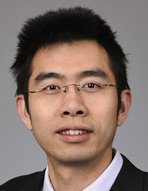 Xinxin Huang (黄鑫欣)合作论文数Institutes of Biomedical Sciences, Fudan University3
 Yunlong Liu合作论文数Department of Medical and Molecular Genetics, Indiana University School of Medicine3
 Hyung-Min Chung合作论文数CHA University;CHA Stemcell Institute3Michael E. Boulton合作论文数3
 Trevor J. Mcgill合作论文数Department of Otolaryngology, Children's Hospital3
 Mohamed Jamal合作论文数University of Texas MD Anderson Cancer Center, Houston, United States3
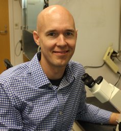 Brandon K. Hadland合作论文数Clinical Research Division, Fred Hutchinson Cancer Research Center3Dibyendu Chakraborty合作论文数University College of Science, University of Calcutta3
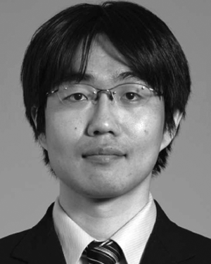 Michihiro Kobayashi合作论文数3Jae-Hung Shieh合作论文数3
 Sherry L. Harbin合作论文数Weldon School of Biomedical Engineering, College of Engineering, Purdue University;Department of Basic Medical Sciences, College of Veterinary Medicine, Purdue University2James H Dawahare合作论文数2W Christopher Shelley合作论文数2
 Negar Seyedhassantehrani合作论文数NSF Crest Center for Biomolecular Machines, University of California Merced2Hongyu Gao合作论文数2
 Stefan P Tarnawsky合作论文数Indiana Univ, Sch Med, Dept Pediat, Indianapolis, IN 46202 USA2
 Alex Murison合作论文数Princess Margaret Canc Ctr, Univ Hlth Network2
 Gregory T Booth合作论文数Cornell Univ, Dept Mol Biol & Genet, Ithaca, NY 14853 USA2Ellen Go合作论文数2




