Somatic stem cell pools comprise diverse, highly specialized subsets whose individual contribution is critical for the overall regenerative function. In the bone marrow, myeloid-biased hematopoietic stem cells (myHSCs) are indispensable for replenishment of myeloid cells and platelets during inflammatory response but, at the same time, become irreversibly damaged during inflammation and aging. Here we identify an extrinsic factor, semaphorin 4A (Sema4A), which non-cell-autonomously confers myHSC resilience to inflammatory stress. We show that, in the absence of Sema4A, myHSC inflammatory hyper-responsiveness in young mice drives excessive myHSC expansion, myeloid bias and profound loss of regenerative function with age. Mechanistically, Sema4A is mainly produced by neutrophils, signals via a cell surface receptor, plexin D1, and safeguards the myHSC epigenetic state. Our study shows that, by selectively protecting a distinct stem cell subset, an extrinsic factor preserves functional diversity of somatic stem cell pool throughout organismal lifespan. Toghani, Gupte et al. identify semaphorin 4A as a cell-extrinsic factor that protects myeloid-biased hematopoietic stem cells from inflammaging, mainly produced by their progeny—neutrophils—and acting via a negative feedback loop.
Transition between activation and quiescence states in hematopoietic stem and progenitor cells (HSPCs) is tightly governed by cell-intrinsic means and microenvironmental co-adaptation. Although this balance is fundamental for lifelong hematopoiesis and immunity, the underlying molecular mechanisms remain poorly defined. Multimodal analysis divulging differential transcriptional activity between distinct HSPC states indicates the presence of Fli-1 transcription factor binding motif in activated hematopoietic stem cells. We reveal that Fli-1 activity is essential during regenerative hematopoiesis in mice. Fli-1 directs activation programs while priming cellular sensory and output machineries, enabling HSPCs co-adoptability with a stimulated vascular niche through propagation of niche-derived angiocrine Notch1 signaling. Constitutively induced Notch1 signaling is sufficient to recuperate functional hematopoietic stem cells impairments in the absence of Fli-1, without leukemic transformation. Applying FLI-1 transient modified-mRNA transduction into latent adult human mobilized HSPCs, enables their niche-mediated expansion and superior engraftment capacities. Thus, decryption of stem cell activation programs offers valuable insights for immunological regenerative medicine.
Preparative regimens before Hematopoietic Cell Transplantation (HCT) damage the bone marrow (BM) microenvironment, potentially leading to secondary morbidity and even mortality. The precise effects of cytotoxic preconditioning on bone and BM remodeling, regeneration, and subsequent hematopoietic recovery over time remain unclear. Moreover, the influence of recipient age and cytotoxic dose have not been fully described. In this study, we longitudinally investigated bone and BM remodeling after busulfan treatment with low intensity (LI) and high intensity (HI) regimens as a function of animal age. As expected, higher donor chimerism was observed in young mice in both LI and HI regimens compared to adult mice. Noticeably in adult mice, significant engraftment was only observed in the HI group. The integrity of the blood-bone marrow barrier in calvarial BM blood vessels was lost after busulfan treatment in the young mice and remained altered even 6 weeks after HCT. In adult mice, the severity of vascular leakage appeared to be dose-dependent, being more pronounced in HI compared to LI recipients. Interestingly, no noticeable change in blood flow velocity was observed following busulfan treatment. Ex vivo imaging of the long bones revealed a reduction in the frequency and an increase in the diameter and density of the blood vessels shortly after treatment, a phenomenon that largely recovered in young mice but persisted in older mice after 6 weeks. Furthermore, analysis of bone remodeling indicated a significant alteration in bone turnover at 6 weeks compared to earlier timepoints in both young and adult mice. Overall, our results reveal new aspects of bone and BM remodeling, as well as hematopoietic recovery, which is dependent on the cytotoxic dose and recipient age.
Somatic stem cell pools are comprised of diverse, highly specialized subsets whose individual contribution is critical for the overall regenerative function. In the bone marrow, myeloid-biased HSC (myHSC) are indispensable for replenishment of myeloid cells and platelets during inflammatory response but at the same time, become irreversibly damaged during inflammation and aging. Here, we identify an extrinsic factor, Semaphorin 4A (Sema4A), which non cell-autonomously confers myHSC resilience to inflammatory stress. We show that the absence of Sema4A, myHSC inflammatory hyper-responsiveness in young mice drives excessive myHSC expansion, myeloid bias and profound loss of regenerative function with age. Mechanistically, Sema4A is mainly produced by neutrophils, signals via a cell surface receptor Plexin D1 and safeguards myHSC epigenetic state. Our study shows that by selectively protecting a distinct stem cell subset, an extrinsic factor preserves functional diversity of somatic stem cell pool throughout organismal lifespan. ### Competing Interest Statement The authors have declared no competing interest.
The thymus plays a critical role in the adaptive immune system as the site of T cells maturation. Unfortunately, the thymus is susceptible to damage by acute insults which can negatively impact patient health outcomes. Despite significant medical interest, research into the thymus has been limited due to a lack of intravital imaging methods to study the thymus in situ. Our lab previously developed a method of intravital adhesive-stabilized two-photon microscopy of the thymus in mice to overcome the limitations of previous imaging methods. However, this method was limited by the permanent attachment of a support structure to the thymus, which prevented repeat imaging of the same thymus across multiple timepoints. To enable longitudinal study of the thymus, we developed a vacuum-stabilized video-rate two-photon endoscope capable of imaging the native thymus in mice. The optical performance and ability of the endoscope to visualize the thymus was described. The stability of the endoscope to visualize the thymus was also described. Future longitudinal examination of the thymus is likely to expand the scientific communities' understanding of thymus biology and its relationship to overall human health.
The thymus, a key organ in the adaptive immune system, is sensitive to a variety of insults including cytotoxic preconditioning, which leads to atrophy, compression of the blood vascular system, and alterations in hemodynamics. Although the thymus has innate regenerative capabilities, the production of T cells relies on the trafficking of lymphoid progenitors from the bone marrow through the altered thymic blood vascular system. Our understanding of thymic blood vascular hemodynamics is limited due to technical challenges associated with accessing the native thymus in live mice. To overcome this challenge, we developed an intravital two-photon imaging method to visualize the native thymus in vivo and investigated functional changes to the vascular system following sublethal irradiation. We quantified blood flow velocity and shear rate in cortical blood vessels and identified a subtle but significant increase in vessel leakage and diameter ~24 hrs post-sublethal irradiation. Ex vivo whole organ imaging of optically cleared thymus lobes confirmed a disruption of the thymus vascular structure, resulting in an increase in blood vessel diameter and vessel area, and concurrent thymic atrophy. This novel two-photon intravital imaging method enables a new paradigm for directly investigating the thymic microenvironment in vivo.
Transition between activation and quiescence programs in hematopoietic stem and progenitor cells (HSC/HSPCs) is perceived to be governed intrinsically and by microenvironmental co-adaptation. However, HSC programs dictating both transition and adaptability, remain poorly defined. Single cell multiome analysis divulging differential transcriptional activity between distinct HSPC states, indicated for the exclusive absence of Fli-1 motif from quiescent HSCs. We reveal that Fli-1 activity is essential for HSCs during regenerative hematopoiesis. Fli-1 directs activation programs while manipulating cellular sensory and output machineries, enabling HSPCs co-adoptability with a stimulated vascular niche. During regenerative conditions, Fli-1 presets and enables propagation of niche-derived Notch1 signaling. Constitutively induced Notch1 signaling is sufficient to recuperate functional HSC impairments in the absence of Fli-1. Applying FLI-1 modified-mRNA transduction into lethargic adult human mobilized HSPCs, enables their vigorous niche-mediated expansion along with superior engraftment capacities. Thus, decryption of stem cell activation programs offers valuable insights for immune regenerative medicine.
Hematopoietic Cell Transplantation (HCT) is a common treatment for patients suffering from a variety of malignant or benign diseases, reconstituting the hematopoietic system after preconditioning treatment. In patients undergoing HCT, the capacity for the thymus to produce functional T cells is inhibited due to damage from the cytotoxic preconditioning. Endogenous thymus regeneration depends on the complex relationship between thymus stromal cells (including vascular endothelial cells (EC)) and the recruitment of de novo "seeding" thymic progenitor cells (TPCs) from the regenerated bone marrow (BM). It has been demonstrated that ECs are radio-resistant, however in the context of HCT, it is not clear which type of EC (arterial, venous, or capillary) can survive following injury. Moreover, ECs are critical cells in endogenous thymic regeneration and are involved in recruiting TPCs from the BM, but functional damage to the vascular network may alter the hemodynamics and negatively impact TPC homing and thymus regeneration. Flow cytometry, immunohistochemistry, and ex vivo imaging techniques have been applied to study the thymus in preclinical mouse models since direct visualization of the native thymus in live mice was deemed impossible. Teleost fish are an alternative model organism for direct thymus observation in vivo but redundant cytokine pathways are present which do not exist in humans and mice. Unlike fish models, in mammals T cell development critically depends on the IL-7 cytokine signaling pathway. Therefore, tools to directly visualize the mouse thymus are needed in order to study functional changes to the vascular system after cytotoxic preconditioning in an immunologically relevant preclinical animal model. We hypothesize that cytotoxic preconditioning causes functional and anatomical changes in blood vessel architecture, especially cortical vasculature, that negatively impacts TPC homing and leads to long-term changes in the thymus microenvironment. In this project, we developed intravital imaging techniques to visualize the native thymus in live mice. Using our methodology, we were able to quantify the changes to the blood vessel network after sublethal irradiation (4.5 Gy). Our results showed an increase in blood vessel diameter in mice one day after receiving irradiation. We validated this result using tissue clearing and ex vivo imaging. In addition, most cortical blood flow velocity is <500 µm/s in both control and one day after sublethal irradiation, although no significant changes were observed in blood flow velocity and shear rate between the groups at this timepoint. Taken together, our study suggests that native intravital thymus imaging is a powerful technique enabling functional and anatomical characterization of the thymus vascular network. We believe, further work will help clarify the changes to the vascular system at later timepoints and in the context of higher irradiation doses. This method enables a whole new paradigm for studying thymus biology not previously possible.
Tissue stem cells are hierarchically organized. Those that are most primitive serve as key drivers of regenerative response but the signals that selectively preserve their functional integrity are largely unknown. Here, we identify a secreted factor, Semaphorin 4A (Sema4A), as a specific regulator of myeloid-biased hematopoietic stem cells (myHSC), which are positioned at the top of the HSC hierarchy. Lack of Sema4A leads to exaggerated myHSC (but not downstream “balanced” HSC) proliferation after acute inflammatory stress, indicating that Sema4A enforces myHSC quiescence. Strikingly, aged Sema4A knock-out myHSC expand but almost completely lose reconstitution capacity. The effect of Sema4A is non cell-autonomous, since upon transplantation into Sema4A-deficient environment, wild-type myHSC excessively proliferate but fail to engraft long-term. Sema4A constrains inflammatory signaling in myHSC and acts via a surface receptor Plexin-D1. Our data support a model whereby the most primitive tissue stem cells critically rely on a dedicated signal from the niche for self-renewal and life-long persistence.
The biology of haematopoietic stem cells (HSCs) has predominantly been studied under transplantation conditions1,2. It has been particularly challenging to study dynamic HSC behaviour, given that the visualization of HSCs in the native niche in live animals has not, to our knowledge, been achieved. Here we describe a dual genetic strategy in mice that restricts reporter labelling to a subset of the most quiescent long-term HSCs (LT-HSCs) and that is compatible with current intravital imaging approaches in the calvarial bone marrow3-5. We show that this subset of LT-HSCs resides close to both sinusoidal blood vessels and the endosteal surface. By contrast, multipotent progenitor cells (MPPs) show greater variation in distance from the endosteum and are more likely to be associated with transition zone vessels. LT-HSCs are not found in bone marrow niches with the deepest hypoxia and instead are found in hypoxic environments similar to those of MPPs. In vivo time-lapse imaging revealed that LT-HSCs at steady-state show limited motility. Activated LT-HSCs show heterogeneous responses, with some cells becoming highly motile and a fraction of HSCs expanding clonally within spatially restricted domains. These domains have defined characteristics, as HSC expansion is found almost exclusively in a subset of bone marrow cavities with bone-remodelling activity. By contrast, cavities with low bone-resorbing activity do not harbour expanding HSCs. These findings point to previously unknown heterogeneity within the bone marrow microenvironment, imposed by the stages of bone turnover. Our approach enables the direct visualization of HSC behaviours and dissection of heterogeneity in HSC niches.
PURPOSE OF REVIEW:Recent advances in genomics and gene editing have expanded the range of model organisms to include those with interesting biological capabilities such as regeneration. Among these are the classic models of regeneration biology, the salamander. Although stimulating endogenous regeneration in humans likely is many years away, with advances in stem cell biology and biomedical engineering (e.g. bio-inspired materials), it is evident that there is great potential to enhance regenerative outcomes by approaching the problem from an engineering perspective. The question at this point is what do we need to engineer?RECENT FINDINGS:The value of regeneration models is that they show us how regeneration works, which then can guide efforts to mimic these developmental processes therapeutically. Among these models, the Accessory Limb Model (ALM) was developed in the axolotl as a gain-of-function assay for the sequential steps that are required for successful regeneration. To date, this model has identified a number of proregenerative signals, including growth factor signaling associated with nerves, and signals associated with the extracellular matrix (ECM) that induce pattern formation.SUMMARY:Identification of these signals through the use of models in highly regenerative vertebrates (e.g. the axolotl) offers a wide range of possible modifications for engineering bio-inspired, biomimetic materials to create a dynamic stem cell niche for regeneration and scar-free repair.
Astrocytes play a critical role in supporting the normal physiological function of neurons. Recent studies have revealed that astrocyte transplantation can promote axonal regeneration and functional recovery after spinal cord injury. Biomaterial can be designed as a growth-permissive substrate and serve as a carrier for astrocyte transplantation into injured spinal cord. In this study, we developed a method to generate collagen microspheres encapsulating astrocytes by injecting a mixture of collagen and astrocytes into a cell culture medium with a syringe controlled by a syringe pump. The collagen microspheres were crosslinked with poly(ethylene glycol) ether tetrasuccinimidyl glutarate (4S-StarPEG) to reduce the degradation rate. The viability of cells in the crosslinked microspheres was higher than 90%. Astrocytes were transfected with plasmids encoding nerve growth factor (NGF)-ires-enhanced green fluorescent protein (EGFP) genes by electroporation and encapsulated in crosslinked microspheres. The level of NGF released into the cell culture medium was higher than that remaining in the microspheres or astrocytes. When microspheres encapsulating astrocytes transfected with plasmids encoding NGF-ires-EGFP genes were added into the cultured rat dorsal root ganglion, the axonal growth was significantly enhanced. This study shows that the microspheres can be potentially used as a carrier of astrocytes to promote nerve regeneration in injured neural tissue.
Thesis (M.S.)--Wichita State University, Fairmount College of Liberal Arts and Sciences, Dept. of Biological Sciences
Astrocytes play a critical role in supporting the normal physiological function of neurons in the central nervous system (CNS).
Problems commonly associated with using nanoemulsions are their cytotoxic effects and low stability profiles. Here, for the first time, concentrations of ingredients of a nanoemulsion system were investigated to obtain the most stable nanoemulsion system with the least cytotoxic effect on MCF7 cell line. Artificial neural networks (ANNs) were used to model the experimentally obtained data. Surfactant concentration was found to be the dominant factor in determining the stability - surfactant concentration above a critical point made the preparation unstable, while it appeared not to be influencing the cytotoxicity. Concentration of oil showed a direct relationship to the cytotoxicity with a minimum value required to provide an acceptable safety profile for the preparation. Co-surfactant appeared not to be considerably effective on neither stability nor cytotoxicity. To obtain the optimum preparation with maximum stability and minimum cytotoxicity, surfactant and oil values need to be kept at their maximum and minimum possible, respectively.
 Joel A. Spencer合作论文数Departments of Medicine and Biological Chemistry, University of California, Irvine10
 Christian Burns合作论文数7
 Steven Josefowicz合作论文数Immunology & Microbial Pathoge, Weill Cornell Graduate School of Medical Sciences, Cornell University4Dorsa Toghani合作论文数3Ruth Verrinder合作论文数3Sharon Zeng合作论文数3Elmir Mahammadov合作论文数Inst Epigenet & Stem Cells, Helmholtz Zentrum Munchen3Amogh Pradeep合作论文数3Nicola K Wilson合作论文数3Kinston Sarah J合作论文数3
 David M Gravano合作论文数3Li Yao合作论文数3
 Stefan Radtke合作论文数Institute for Transfusion Medicine, University Hospital Essen3
 Lev Silberstein合作论文数3Rodriguez Sonia合作论文数Indiana University Simon Cancer Center, Indiana University School of Medicine3
 Antonio Scialdone合作论文数Dipartimento di Scienze Fisiche, Università di Napoli "Federico II"and INFN;Dipartimento di Scienze Fisiche, Università di Napoli "Federico II" and INFN3
 David Scadden合作论文数Department of Stem Cell and Regenerative Biology, Harvard University;Center for Regenerative Medicine, Massachusetts General Hospital3
 Hans-Peter Kiem合作论文数Clinical Research Division, Fred Hutchinson Cancer Center;Vaccine and Infectious Disease Division, Fred Hutchinson Cancer Center3
 Bertie Göttgens合作论文数Wellcome - MRC Cambridge Stem Cell Institute, Jeffrey Cheah Biomedical Centre, University of Cambridge;Department of Haematology, University of Cambridge3
 Nadia Carlesso合作论文数Dana-Farber Cancer Institute and Department of, Medicine, Harvard Medical School3
 Eric M. Pietras合作论文数David Geffen School of Medicine, UCLA3Marion Mesnieres合作论文数2Le Zong合作论文数2Jin-Gyu Cheong合作论文数2
 Nastaran Abbasizadeh合作论文数Dept Bioengn, Univ Calif Merced2
 Edie I Crosse合作论文数2
 Sean Houghton合作论文数Weill Cornell Med, Ansary Stem Cell Inst2
 Marcus N Berndt合作论文数Dept Biol Sci, Wichita State Univ2Chaitanya R. Badwe合作论文数Weill Cornell Med, Ansary Stem Cell Inst2
 Alex Murison合作论文数Princess Margaret Canc Ctr, Univ Hlth Network2
 Gregory T Booth合作论文数Cornell Univ, Dept Mol Biol & Genet, Ithaca, NY 14853 USA2
 Ying Liu合作论文数Weill Cornell Med Coll, Ansary Stem Cell Inst, Dept Med Genet, New York, NY 10065 USA2Tomer Itkin合作论文数Joan and Sanford I. Weill Department of Medicine, Weill Cornell Medicine, Cornell University2Jesus Maria Gomez Salinero合作论文数Joan and Sanford I. Weill Department of Medicine, Weill Cornell Medicine, Cornell University2Fuqiang Geng合作论文数2
 Peter Kharchenko合作论文数Altos Labs;Department of Biomedical Informatics, Harvard Medical School2Yang Lin合作论文数Department of Pediatrics, Wells Center for Pediatric Research and;Department of Biochemistry and Molecular Biology, Indiana University School of Medicine;Wells Center for Pediatric Research and;Department of Biochemistry and Molecular Biology, Indiana University School of Medicine2
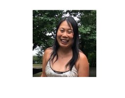 Xie Stephanie Z合作论文数2Li Yongchao合作论文数2
 Laura Garcia-Prat合作论文数CIBER Neurodegenerat Dis CIBERNED, Pompeu Fabra Univ UPF2Veronique Voisin合作论文数2
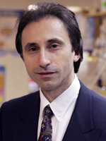 Shahin Rafii合作论文数Cell & Developmental Biology, Weill Cornell Graduate School of Medical Sciences, Cornell University2
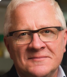 John Dick合作论文数Department of Molecular Genetics, Temerty Faculty of Medicine, University of Toronto2
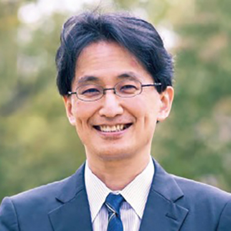 Atsushi Kumanogoh合作论文数Departmet of Respiratory Medicine and Clinical Immunology, Osaka University;Immunology Frontier Research Center, Osaka University2Thomas Worzfeld合作论文数2
 Ryan Schreiner合作论文数Margaret Dyson Vision Research Institute, Cornell University2David Redmond合作论文数Joan and Sanford I. Weill Department of Medicine, Weill Cornell Medicine, Cornell University2
 Cole Trapnell合作论文数Department of Genome Sciences, University of Washington2
 Kerstin Kaufmann合作论文数Plant Research International;Business unit Bioscience;Business unit Bioscience, Plant Research International2




