Astragalus membranaceus, as an important medicinal herb, is widely used all over the world. But, the presence of toxic heavy metals in herbal chains due to the rapid industrialization possesses a serious threat on human health. For rapid screening the positive samples in medicinal herbs, an environment friendly strategy is reported for the determination of heavy metals by using the square wave anodic stripping voltammetry method. The method showed good selectivity, linearity, recovery and precision. It is demonstrated that the intensity of the anodic peak at -0.87 V, -0.61V and -0.24V is proportional (R-2 = 0.9978, 0.9805, 0.9870) to the concentration of Cd2+ in the electrolyte over the range of 0.010.10 mu g.mL(-1), and Pb2+, Cu2+ in the range of 0.10-1.00 mu g.mL(-1). The proposed method has been successfully applied to the determination of the three heavy metal ions in Astragalus membranaceus samples with satisfactory recoveries of 88.00-110.00 %. Meanwhile, the detection limits achieved 0.0010, 0.0007 and 0.0028 mu g.mL(-1) for Cd2+, Pb2+ and Cu2+, respectively, which are much lower than the guideline values in herbs given by the Chinese pharmacopoeia. Real sample analysis of Cd2+, Pb2+ and Cu2+ in Astragalus membranaceus by the electrochemical method of anodic stripping voltammetry has demonstrated that the proposed method can be applied in rapid screening the positive samples of Astragalus membranaceus.
Integrating multiple discrete function-related theranostic modalities into one platform for effective cancer treatments has been considered to be a challenge for current nanomedicine design. In this work, a "four-in-one" theranostic system was simply prepared and developed for photoacoustic (PA) imaging-guided synergistic targeting chemo-gene-thermo trimodal therapy of breast cancer. In this system, polyethylene glycol (PEG)-bridged polyethylenimine (PEI) and a memHsp70 receptor-targeting peptide (TKD), PPT, was uniformly capped on doxorubicin (DOX)-loaded oxidized mesoporous carbon nanospheres (OMCN) to encapsulate therapeutic genes into cancer cells via active targeting accumulation. Taking both the advantages of OMCN (high photothermal conversion, strong PA contrast, and controllable drug loading) and the hydrophilic polymer (gene vector, switchable pores' cap, and targeting ability), the "four-in-one" theranostic system exhibited distinct PA imaging visualization, NIR/pH sensitive drug/gene release, and synergistic targeting therapeutic outcome, which were much superior than the single therapy or the combination of two treatments.
Combining controllable photothermal therapy and efficacious gene therapy in a single platform holds great promise in cancer therapy due to the enhanced combined therapeutic effects. Herein, polyethyleneimine-grafted oxidized mesoporous carbon nanospheres (OP) were developed for combined photothermal combined gene therapy in vitro and in vivo. The synthesized OP was characterized to have three dimensional spherical structure with uniformed diameter, ordered mesopores with graphitic domains, high water dispersion with zeta potential of +22 mV, and good biocompatibility. Consequently, OP was exploited as the photothermal convertor with strong NIR absorption and the gene vector via electrostatic interaction, which therefore cannot only deliver the therapeutic gene (pING4) to tumors for gene therapy, but also can eliminate the tumors by photothermal ablation. Moreover, the improved gene therapy accompanied by the NIR photothermally enhanced gene release was also well achieved based on OP. The excellent combined therapeutic effects demonstrated in vitro and in vivo suggested the OP's potential for cancer therapy.
A simple method was developed to synthesize Ag@graphene nanocomposites with rough Ag nanoparticles (AgNPs) conjugated with graphene nanosheets, and the nanocomposites could be used as substrates for effective surface-enhanced Raman spectroscopy (SERS) of fluorescent anticancer drug (Dox) since they could not only enhance the Raman signals but also suppress the fluorescent signals. (C) 2016 Elsevier B.V. All rights reserved.
C31H27ZnFN3O9.5S, triclinic, P1̅ (no. 2), a = 10.6434(4) Å, b = 12.6191(4) Å, c = 12.8532(5) Å, α = 93.998(3)°, β = 109.121(3)°, γ = 112.773(3)°, V = 1465.16(9) Å3, Z = 2, Rgt(F) = 0.0354, wRref(F2) = 0.1012, T = 293 K.
Glioma has been considered to be the most frequent primary tumor within the central nervous system (CNS). The complexity of glioma, especially the existence of the blood-brain barrier (BBB), makes the survival and prognosis of glioma remain poor even after a standard treatment based on surgery, radiotherapy, and chemotherapy. This provides a rationale for the development of some novel therapeutic strategies. Among them, receptor-mediated drug delivery is a specific pattern taking advantage of differential expression of receptors between tumors and normal tissues. The strategy can actively transport drugs, such as small molecular drugs, gene medicines, and therapeutic proteins to glioma while minimizing adverse reactions. This review will summarize recent progress on receptor-mediated drug delivery systems targeting to glioma, and conclude the challenges and prospects of receptor-mediated glioma-targeted therapy for future applications.
Multiple diagnosis of cancer by a facile fluorescent sensor is extremely attractive. Herein, a Cy3-labeled ssDNA probe (P0-Cy3) was π-π stacked on the surface of oxidized mesoporous carbon nanospheres (OMCN) to construct the fluorescent "turn-on" aptasensor. Attributing to the intrinsic properties of OMCN, the OMCN-based aptasensor not only can be used to detect mucin1 protein in liquid with a wide range of 0.1-10.6 μmol/L, a low detection limit of 6.52 nmol/L, and good selectivity, but also can quantify the cancer cells in solution with the linear range of 10(4)-2 × 10(6) cells/mL and a detection limit of 8500 cells/mL. Fascinatingly, this OMCN-based aptasensor was exploited to image cancer via solid tissues such as cells, tissue sections, and ex vivo and in vivo tumors, in which the obvious distinguishability between cancer and normal tissues was clearly demonstrated. This is a robust and simple detection technique, which can well achieve the multiple diagnosis of cancer in vitro and in vivo.
Cancer imaging requires biocompatible and bright contrast-agents with selective and high accumulation in the tumor region but low uptake in normal tissues. Herein, 1-methyl-2-pyrrolidinone (NMP)-derived polymer-coated nitrogen-doped carbon nanodots (pN-CNDs) with a particle size in the range of 5-15 nm are prepared by a facile direct solvothermal reaction. The as-prepared pN-CNDs exhibit stable and adjustable fluorescence and excellent water solubility. Results of a cell viability test (CCK-8) and histology analysis both demonstrate that the pN-CNDs have no obvious cytotoxicity. Most importantly, the pN-CNDs can expediently enter glioma cells in vitro and also mediate glioma fluorescence imaging in vivo with good contrast via elevated passive targeting.
Breast cancer has been considered as a serious threat to females' life. Active targeting drug delivery is a potential strategy in cancer therapy, which however is hindered by the targeting efficiency. Herein, a 14-mer peptide (TKD) derived from the oligomerization domain of membrane heat-shock protein 70 (memHsp70), for the first time, was exploited as a tumor-targeting ligand to modify polymeric micelles. NMR results demonstrated the successful synthesis of TKD-PEG-PLGA polymer. No difference was observed in the drug release between TKD-modified doxorubicin (DOX)-loaded micelles (TKD-D-M) and unmodified counterparts. The modification of TKD mediated apparently higher cellular uptake within memHsp70-positive MCF-7 cells, compared to normal MCF-10A cells. Excessive TKD pretreatment significantly inhibited the cellular uptake of TKD-D-M, indicating the receptor-mediated mechanism. Enhanced accumulation of TKD-D-M within the tumor of MCF-7 bearing mice further demonstrated the targeting ability of TKD in vivo. CCK-8 assay showed that the modification of TKD significantly increase the anti-proliferation effect against MCF-7 cells. The findings demonstrated that TKD peptide is a potential ligand which can target drug delivery systems to memHsp70-positive breast cancer.
Tumor-specific therapeutic platforms designed for combined tumor therapy has recently received wide attention. In this work, a new HB5 aptamer-functionalized mesoporous silica–carbon based doxorubicin (DOX)-loaded system (MSCN-PEG-HB5/DOX) was successfully constructed and characterized for chemo-photothermal combined therapy of human epithelial growth factor receptor 2 (HER2)-positive breast cancer cells. The in vitro release result showed that MSCN-PEG-HB5/DOX exhibited pH-sensitive and NIR-triggered release manner. HB5-modified nanoparticles showed significant higher cellular uptake in HER2-positive breast cancer cells (SK-BR-3) but not in normal breast epithelial cells (MCF-10A), compared to unmodified counterparts. The intracellular uptake of functional nanoparticles was mainly based on the receptor-mediated mechanism which was energy-dependent. Cytotoxicity experiments demonstrated that combined therapy induced highest cell killing effect compared to chemotherapy and photothermal therapy alone. The combination index (CI) was 0.253 indicating the synergistic effect of chemotherapy and photothermal therapy. These findings suggested that MSCN-PEG-HB5/DOX was a potential chemo-photothermal therapeutic platform targeting to HER2-positive breast cancers.
 Rongqin Huang (黄容琴)合作论文数School of Pharmacy, Fudan University8
 Chengyi Li合作论文数7
 Min Qian (钱忞)合作论文数The Second Norman Bethune Hospital of Jilin University6
 Shanshan Wang (王珊珊)合作论文数Department of Environmental Science and Engineering, Fudan University6
 Xueying Yan合作论文数5
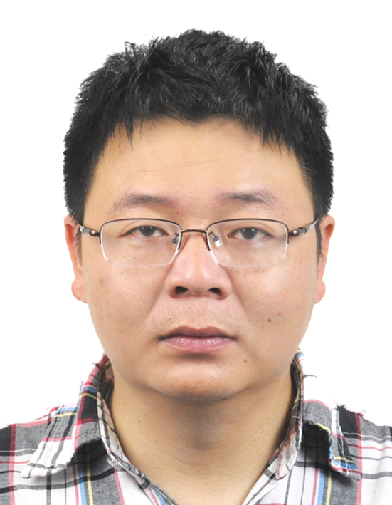 Yi Wang (王义)合作论文数College of Chemistry, Chemical Engineering and Biotechnology, Donghua University4Peng Xiyue合作论文数2ZhuoWen Fan (范卓文)合作论文数2
 Jianxin Wang (王建新)合作论文数School of Pharmaceutical Sciences, Fudan University2Hui Yao合作论文数1Kuo Cui合作论文数1Dandan Kong合作论文数1
 Yilin Du合作论文数1
 Yufan Zheng合作论文数State Key Lab Med Neurobiol, Fudan Univ1Yu-Feng Huang合作论文数1Huiling Jiang合作论文数1Liang-Liang Wang合作论文数1Kaiyuan Wang合作论文数1Ji-Zhi Liu合作论文数1
 Meihua Yang (杨美华)合作论文数The Institute of Medicinal Plant Development, Chinese Academy of Medical Sciences1Jian Chen合作论文数1
 Wei Shi (施炜)合作论文数Affiliated Hospital, Nantong University1
 Weiyue Lu (陆伟跃)合作论文数School of Pharmacy, Fudan University;Key Laboratory of Smart Drug Delivery, Ministry of Education, China, Fudan University1
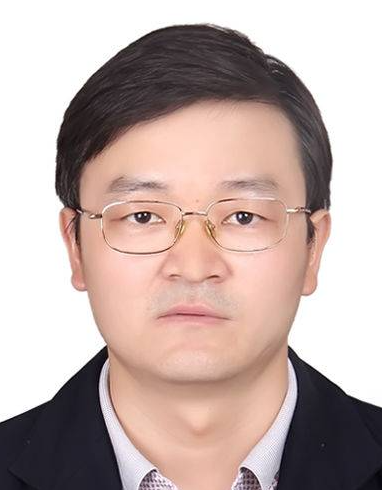 Weijun Kong (孔维军)合作论文数Institute of Medicinal Plant Development, Chinese Academy of Medical Sciences1




