Salivary secretion is essential for maintaining oral and systemic health. In parotid acinar cells, activation of the cAMP-protein kinase A (PKA) pathway following β-adrenergic receptor stimulation triggers the exocytosis of amylase. However, the downstream signaling pathways from PKA activation to exocytotic membrane fusion remain to be fully elucidated. In this review, we focused on Myristoylated alanine-rich C kinase substrate (MARCKS), a major protein kinase C (PKC) substrate, from the perspective of cell membrane homeostasis. Western blot analysis confirmed high MARCKS expression across various exocrine tissues, including the parotid, submandibular, sublingual, and lacrimal glands, as well as the exocrine pancreas. Our results revealed that β-adrenergic receptor stimulation activates PKCδ via PKA, leading to MARCKS phosphorylation in the parotid gland. Phosphorylated MARCKS dissociates from lipid rafts (detergent-resistant membranes) and translocates to the cytosol. Notably, stimulus-induced translocation from lipid rafts was observed in both cAMP-dependent (parotid) and Ca2+-dependent (pancreas) secretion. The inhibitory effect of a MARCKS-related peptide (MANS) on amylase secretion suggests that MARCKS functions as a physical or spatial regulator of secretion-related molecules, such as SNARE proteins, on lipid rafts. These findings indicate that MARCKS-mediated membrane domain regulation is a fundamental system underlying exocrine function. This review proposes a novel model of secretion regulated by MARCKS and discusses its pathophysiological significance as a potential therapeutic target for exocrine disorders.
Epithelial acinar cells of the salivary gland establish apical-basal polarity and secretory competence during development, processes that require tightly coordinated membrane trafficking. The Rho-family GTPase Cdc42 is a key regulator of epithelial polarity, but it has rarely been linked to the vesicle-fusion machinery in vivo. Using mice with acinar cell-specific deletion of Cdc42, we found that Cdc42 loss caused the accumulation of small vesicles near the apical surface, where the water channel AQP5 was retained in clusters and lumen formation was defective. Cdc42-deficient glands showed a selective reduction of two vesicle-associated SNAREs, VAMP2 and VAMP4, whereas VAMP8, the t-SNAREs, and an endocytic marker were unchanged. The corresponding mRNAs were not decreased, indicating that Cdc42 maintains these v-SNAREs through post-transcriptional mechanisms. Cdc42-deficient acinar cells also accumulated condensing vacuoles, a form of immature secretory granule, indicating impaired granule maturation. We propose that Cdc42 coordinates two distinct membrane-remodeling processes by maintaining these v-SNAREs: VAMP2 supports the fusion of AQP5-containing vesicles with the apical membrane, whereas VAMP4 supports secretory granule maturation. These findings identify the vesicle-fusion machinery as a target of Cdc42-dependent polarity control, with potential implications for secretory gland disorders.
Epithelial polarity is essential for proper tissue organization and function, yet the molecular mechanisms governing apical membrane formation during secretory epithelial development remain incompletely understood. Here, we investigate the role of the small GTPase Cdc42 in salivary gland acinar cell development using a mouse model designed to knock out Cdc42 specifically at the onset of acinar cell formation. Loss of Cdc42 resulted in defective apical membrane formation accompanied by accumulation of vesicles around the apical lumen. These vesicles contained the apical water channel AQP5 and the apical recycling endosome (ARE) marker Rab11a, while the basolateral transporter NKCC1 retained normal localization, indicating an apical-selective trafficking defect. Importantly, Cdc42 deficiency caused a selective 40% reduction in the expression of the SNARE protein VAMP2, while other vesicle trafficking proteins including VAMP8, SNAP23, and EEA1 remained unchanged. Our findings reveal that Cdc42 controls apical membrane formation by maintaining VAMP2 expression, which is essential for the fusion of Rab11a-positive recycling endosomes. The accumulation of fusion-incompetent AREs near the apical surface demonstrates the critical role of the Cdc42-VAMP2 pathway in epithelial development. These results provide new insights into how polarity regulators integrate vesicle trafficking and fusion machinery, and may have implications for understanding glandular diseases involving epithelial polarity defects.
This in vivo mouse model study was conducted to investigate the temporal alteration of the function of CD36 in salivary secretion. CD36 was highly expressed in the parotid gland of BALB/c mice. No significant variations were shown in the CD36 levels in the 8-, 48-, and 72-week-old animals. However, pilocarpine-induced salivary secretion was reduced in an age-dependent manner, showing a significantly low level at the age of 72 weeks. Pilocarpine-induced salivary secretion was significantly reduced by pretreatment with a CD36 inhibitor at 8 and 48 weeks, but not at 72 weeks. In senescence-accelerated mice (SAM), the pilocarpine-induced salivary secretion was significantly reduced at the age of 56 weeks, and a significantly lower amount of CD36 was demonstrated in the parotid gland, compared with the control. These results suggest that the involvement of parotid CD36 in mouse salivary secretion is altered with age.
BACKGROUND:Sjögren's syndrome (SS) is known to cause dry eyes and mouth due to inflammation of the lacrimal and salivary glands. However, some reports imply that other factors trigger dry eyes and mouth. We previously investigated various factors using RNA-sequencing analysis of lacrimal glands from male non-obese diabetic (NOD) mice, an SS model. In this review, we described (1) the exocrine features of male and female NOD mice, (2) the up- and down-regulated genes in the lacrimal glands of male NOD mice as revealed by our RNA-sequencing data, and (3) comparisons between these genes and data in the Salivary Gland Gene Expression Atlas. HIGHLIGHTS:Male NOD mice exhibit a steady worsening of lacrimal hyposecretion and dacryoadenitis, whereas females exhibit a complex pathophysiological condition that includes diabetic disease, salivary hyposecretion, and sialadenitis. Ctss, an up-regulated gene, is a potential inducer of lacrimal hyposecretion and is also expressed in salivary glands. Two other up-regulated genes, Ccl5 and Cxcl13, may worsen the inflammation of SS in both the lacrimal and salivary glands. The genes Esp23, Obp1a, and Spc25 were detected as down-regulated, but judging the relationship between these genes and hyposecretion is difficult as only limited information is available. Another down-regulated gene, Arg1, is involved in lacrimal hyposecretion, and it also has the potential to cause salivary hyposecretion in NOD mice. CONCLUSION:In NOD mice, males may be better than females at evaluating the pathophysiology of SS. Some regulated genes revealed by our RNA-sequencing data might be potential therapeutic targets for SS.
Xerostomia is common in middle-aged and elderly patients. In rodents, adipose tissue increases around the aging parotid gland (PG). Triglycerides are stored in adipose tissue and metabolized into fatty acids and glycerol as needed in the cells. It has been reported that CD36, a fatty acid transporter, is expressed on the tongue and plays a role in some taste sensations through the uptake of dietary fatty acids such as palmitic acid. However, there are few reports on CD36 in salivary glands. In this study, we investigated the role of CD36 in salivary secretion using male mice. In salivary glands of BALB/c mice, CD36 mRNA was highly expressed in the PG compared with submandibular and sublingual glands. The mice pretreated with a CD36 inhibitor showed decreased muscarinic agonist-induced salivary secretion at 8 and 48 weeks but not 72 weeks of age. In vitro [3H]-palmitic acid uptake assay, the amount of [3H] from isolated PG of mice at 8 weeks of age was significantly reduced in the CD36 inhibitor-pretreated group. In highly aged mice, senescence-accelerated mice of 56 weeks of age, there was no change in salivary secretion after pretreatment with CD36 inhibitor due to low protein expression of PG CD36. These results suggest that the importance of PG CD36 in mouse salivary secretion changes with age.
Abstract Background/Aim: This study evaluated the effect of blueberry leaf hot water extract (BLEx) on Sjögren’s syndrome (SS)-like lacrimal hyposecretion in male non-obese diabetic (NOD) mice. Materials and Methods: NOD or BALB/c mice were fed 1% BLEx or control (AIN-93G) for 2 weeks from the age of 4 to 6 weeks. Pilocarpine-induced tear volume was measured using a phenol red-impregnated thread. The lacrimal glands were evaluated histologically by H&E staining. The IL-1β and TNF-α levels in the lacrimal gland tissue were measured by ELISA. The mRNA expression levels of secretion-related proteins were measured by real-time PCR. LC3 I/II and arginase 1 expression levels were measured by western blot. Results: After feeding with BLEx, pilocarpine-induced tear secretion in NOD mice was increased. In contrast, the mRNA expression levels of the cholinergic muscarinic M3 receptor, aquaporin 5, and ion channels related to lacrimal secretion were not changed by BLEx administration. In addition, the protein expression of arginase 1, which was recently reported to be involved in tear hyposecretion in NOD mice, was also not improved by BLEx administration. Although infiltration in the lacrimal gland of NOD mice was not decreased, the levels of TNF-α and the autophagy-related protein LC3 were significantly suppressed by BLEx treatment. Conclusion: BLEx treatment may ameliorate lacrimal hyposecretion in NOD mice by delaying the progression of autoimmune disease by suppressing autophagy in lacrimal glands.
BACKGROUND/AIM:Tears secreted from the lacrimal gland are essential for preserving the ocular surface. Thus, dysfunction of the lacrimal gland in Sjögren's syndrome (SS) can lead to dry eye, resulting in a reduced quality of life. We previously reported that blueberry 'leaf' water extract prevents lacrimal hyposecretion in male non-obese diabetic (NOD) mice in a SS-like model. In this study, we investigated the effect of blueberry 'stem' water extract (BStEx) on lacrimal hyposecretion in NOD mice.MATERIALS AND METHODS:Male NOD mice were fed 1% BStEx or control (AIN-93G) for 2, 4, or 6 weeks from 4 weeks of age. Pilocarpine-induced tear secretion was measured using a phenol red-impregnated thread. The lacrimal glands were histologically evaluated by HE staining. Inflammatory cytokine levels in the lacrimal glands were measured using ELISA. Immunostaining was performed to examine aquaporin 5 (AQP5) localization. The expression levels of autophagy-related proteins, AQP5, and phosphorylated AMPK were measured using western blotting.RESULTS:After feeding BStEx to mice for 4 or 6 weeks, tear volume was observed to have increased in the BStEx group compared with that in the control group. There were no significant differences in inflammatory cell infiltration, autophagy-related protein expression, or the localization and expression of AQP5 in the lacrimal glands between the two groups. In contrast, AMPK phosphorylation increased in the BStEx group.CONCLUSION:BStEx prevented lacrimal hyposecretion in the SS-like model of male NOD mice, probably by opening tight junctions via the activation of AMPK in lacrimal acinar cells.
Cdc42 is a small GTPase essential for the cell cycle, morphogenesis, and cell adhesion, and it is involved in the polarity of epithelial cells. However, the functional roles of Cdc42 in exocrine glands, such as the maintenance of acini and water secretion, are not yet well understood. In this study, we generated acinar-cell-specific Cdc42 conditional knockout (Cdc42cKO) mice to assess their maintenance of acinar cells and physiological functions in the salivary glands (SGs) and lacrimal glands (LGs). Our data revealed that the loss of Cdc42 altered the luminal structures to bulging structures and induced acinar cell apoptosis in both the parotid glands (PGs) and LGs of Cdc42cKO mice. Interestingly, saliva secretion in response to pilocarpine stimulation was decreased in the Cdc42cKO group, whereas tear secretion was increased. Consistent with the water secretion results, protein expression of the water channel AQP5 in acinar cells was also decreased in the PGs but conversely increased in the LGs. Moreover, the changes that increased AQP5 expression in LGs occurred in the acinar cells rather than the duct cells. The present study demonstrates that Cdc42 is involved in the structural and survival maintenance of acinar cells in SGs and LGs. On the other hand, depletion of Cdc42 caused the opposite physiological phenomena between PGs and LGs.
Dry mouth is observed commonly in middle-aged and elderly patients. Although the deterioration of parotid gland (PG) function in those patients is considered, the relationship between aging and hyposecretion has been unclear. We hypothesized that the reduction in expression of fatty acid translocase FAT/CD36, which facilitates the transport of fatty acids, induced dry mouth through the hyposecretion in PG. In this study, the level of CD36 expression was detected by real-time RT-PCR in male 8- and 48-weeks BALB/c mice. Also, effect of CD36 inhibitor sulfosuccinimidyl oleate (SSO) on salivary secretion of male 48-weeks BALB/c mice was assessed. Moreover, the involvement of PG CD36 in the salivary secretion of male 48-weeks senescence-accelerated mouse (SAM) was investigated. The RNA expression level of CD36 in PG was superior to other salivary glands in BALB/c mice. SSO reduced pilocarpine-induced salivation in BALB/c mice. Compared with SAM resistant 1 (SAMR1), the pilocarpine-induced salivation in age-matched SAM prone 1 (SAMP1) was significantly decreased. In addition, the CD36 protein expression in PG was investigated by western blotting, and the expression of CD36 of SAMP1 was significantly lower than that of SAMR1. These results suggest that the CD36 plays an important role in the aging-induced hyposecretion of PG.
Xerostomia is commonly observed in middle-aged and elderly patients. Since the adipose tissue infiltration is frequently observed in the parotid gland (PG) of alder animals. We hypothesized that the alteration in expression of fatty acid translocase (FAT/CD36), which facilitates fatty acids transportation, induces xerostomia through the hyposecretion in the PG. We firstly examined the CD36 expression in the three major salivary glands of male BALB/c mice. The PG expressed significantly substantial CD36 among them. In addition, the immunohistochemical analysis revealed that the CD36 protein localized in duct cells, but not acinar cells, in the PG of BALB/c mice. Then, the effect of CD36 inhibitor, sulfosuccinimidyl oleate, treatment on the salivary secretion in 48-weeks BALB/c mice was assessed. The inhibitor reduced pilocarpine (Pilo)-induced salivation. Moreover, the involvement of the PG CD36 in the salivary secretion of male 48-weeks senescence-accelerated mouse (SAM) was investigated. Compared with SAM resistant 1 (SAMR1), the Pilo-induced salivation in age-matched SAM prone 1 (SAMP1) was decreased. In addition, the protein expression of PG CD36 in SAMP1 was significantly lower than that of SAMR1. These results suggest that the CD36 in ducts of PG plays an important role in the aging-induced hyposecretion.
Epithelial cells of exocrine glands responsible for saliva and tear secretion bear cell polarity. Cdc42, essential for the polarity of epithelial cells, is required for the formation and maintenance of luminal structures which is important site for the secretion. However, it is still unclear whether Cdc42 plays the identical role in different epithelial tissues in vivo. In this study, we generated exocrine epithelial cell-specific Cdc42 conditional knockout (KO) mice and analyzed the difference of Cdc42 roles between salivary glands and lacrimal glands.
Sjogren's syndrome induces salivary and lacrimal hyposecretion, which leads to reduced quality of life. Although many studies have been conducted from the perspective of inflammation, the development of a causal treatment has not been achieved yet. We previously investigated the cause from the perspective of non-inflammation, and identified arginase 1 as a novel non-inflammatory regulator of exocrine function. However, the mechanism of arginase 1 regulating the function remains unknown, so we aimed to elucidate the mechanism.
Early detection of such retinal diseases as glaucoma and age-related macular degeneration (AMD) is important to prevent blindness. There have been reports of changes in some components in the tears of glaucoma and AMD patients, suggesting tears' potential usefulness in screening for retinal diseases. We hypothesized that retinal damage might alter gene expression in the lacrimal gland, leading to those changes in tear components. We caused retinal damage in mice by intravitreal injection of N-methyl-d-aspartate (NMDA) or excessive light exposure. Hematoxylin and eosin staining showed no histological changes in the lacrimal glands of animals whose retinas had been damaged. However, RNA sequencing of lacrimal glands on the 3rd day after NMDA injection or light exposure revealed changes in the expression of 491 genes (268 up-regulated; 223 down-regulated) in the NMDA group and 531 genes (311 up-regulated; 220 down-regulated) in the light group. Further gene-set enrichment analysis indicated that both types of retinal damage activated the immune system in the lacrimal glands. This is the first demonstration that retinal damage can alter gene expression in the lacrimal glands, and it might lead to a novel non-invasive screening method for early detection of retinal diseases.
Senescence induces exocrine insufficiency which leads to decreased quality of life because of depletion of saliva and/or tears. These secretions have been considered to share the similar mechanism, although the detailed differences in functions have not been clarified. In this study, we generated acinar cells-specific Cdc42-knockout mice by Cre-loxP system to elucidate the secretion mechanism among these organs. The results showed tissue weights of the salivary and lacrimal glands were reduced by a deficiency of Cdc42. Moreover, actin staining revealed that the luminal structure was destroyed in both glands. The volume of pilocarpine-stimulated secretion of saliva decreased after knockout of Cdc42, correlating with tissue weight results. On the other hand, the tear volume was unexpectedly maintained even though the tissue weight was decreased. These results suggest that saliva and tear may have different secretory mechanisms. While the identification of factors which maintain exocrine secretion must await further investigation, the factors might be a therapeutic target of xerostomia.
Lacrimal fluid (tears) is important for preservation of the ocular surface, and thus dry eye induced by lacrimal hyposecretion in Sjögren's syndrome (SS) leads to reduced quality of life. However, the cause of lacrimal hyposecretion remains unknown, even though many studies have been conducted from the perspective of inflammation. Here, we hypothesized that a non-inflammatory factor induces lacrimal hyposecretion in SS pathophysiology. To elucidate such a factor, we conducted transcriptome analysis of the lacrimal glands in male non-obese diabetic (NOD) mice as a SS model. The result revealed that only four genes, including arginase 1, were downregulated in the lacrimal glands of male NOD mice after onset of lacrimal hyposecretion and dacryoadenitis. Furthermore, non-dacryoadenitis-type NOD mice were used to investigate the relationships among arginase 1 expression, lacrimal hyposecretion and dacryoadenitis. Non-dacryoadenitis-type NOD mice showed reduced tear secretion and low expression level of arginase 1. In addition, in BALB/c mice, an arginase 1 inhibitor reduced tear secretion. In conclusion, a non-inflammatory factor, arginase 1, is involved in lacrimal hyposecretion in male NOD mice, regardless of dacryoadenitis status. These results shed light on the pathophysiological role of arginase 1 in SS (dry eye).
Key points Few reports have explored the possibility of involvement of non‐inflammatory factors in lacrimal hyposecretion in Sjögren's syndrome (SS). RNA‐sequencing analysis revealed that only four genes, including arginase 1, were downregulated in the lacrimal gland of SS model male mice (NOD mice) after onset of lacrimal hyposecretion and dacryoadenitis. Even in non‐dacryoadenitis‐type NOD mice, tear secretion and arginase 1 expression remained low. An arginase 1 inhibitor reduced tear secretion and partially reduced saliva secretion in BALB/c mice. The results indicate that a non‐inflammatory factor, arginase 1, is involved in lacrimal hyposecretion in male NOD mice, regardless of dacryoadenitis status. AbstractLacrimal fluid (tears) is important for preservation of the ocular surface, and thus lacrimal hyposecretion in Sjögren's syndrome (SS) leads to reduced quality of life. However, the cause(s) of lacrimal hyposecretion remains unknown, even though many studies have been conducted from the perspective of inflammation. Here, we hypothesized that a non‐inflammatory factor induces lacrimal hyposecretion in SS pathology, and to elucidate such a factor, we conducted transcriptome analysis of the lacrimal glands in male non‐obese diabetic (NOD) mice as an SS model. The NOD mice showed inflammatory cell infiltration and decreased pilocarpine‐induced tear secretion at and after 6 weeks of age compared to age‐matched BALB/c mice. RNA‐sequencing analysis revealed that only four genes, including arginase 1, were downregulated, whereas many genes relating to inflammation were upregulated, in the lacrimal glands of male NOD mice after onset of lacrimal hyposecretion and dacryoadenitis (lacrimal gland inflammation). Changes in the level of arginase 1 expression were confirmed by real‐time RT‐PCR and western blot analysis. Furthermore, non‐dacryoadenitis‐type NOD mice were used to investigate the relationships among arginase 1 expression, lacrimal hyposecretion and dacryoadenitis. Interestingly, these NOD mice retained the phenotype of dacryoadenitis with regard to tear secretion and arginase 1 expression level. An arginase 1 inhibitor reduced tear secretion and partially reduced saliva secretion in BALB/c mice. In conclusion, a non‐inflammatory factor, arginase 1, is involved in lacrimal hyposecretion in male NOD mice, regardless of dacryoadenitis status. These results shed light on the pathophysiological role of arginase 1 in SS (dry eye).
JPH203 is a novel anti-cancer drug targeting L-type amino acid transporter 1 (LAT1), which plays a primary role in the uptake of essential amino acids in tumor cells. Although a co-incubation inhibitory effect of JPH203 has been shown in a conventional uptake assay, its preincubation inhibitory effects have remained undetermined. Therefore, we aimed to characterize the preincubation inhibitory effects of JPH203 on LAT1 function using leucine uptake assays in LAT1-positive human colon cancer HT-29 cells. Preincubation of the cells with JPH203 (0.3 μM for 120 min) decreased the activity level to 30% of that in dimethylsulfoxide-treated cells. Similarly, in time-dependency analysis, preincubation of HT-29 cells with 10 μM JPH203 for 30, 60, and 120 min decreased the leucine uptake activity (42%, 32%, and 28% of that in control cells, respectively). Furthermore, the IC50 value of the combination of preincubation and co-incubation effects was lower than that of co-incubation inhibition alone (34.2 ± 3.6 nM vs. 99.2 ± 11.0 nM). In conclusion, we revealed that JPH203 has the capability to inhibit LAT1 function through preincubation effects. Moreover, preincubation synergistically enhances the co-incubation inhibitory effects. These findings provide a novel insight into the anti-cancer effects of JPH203 in cancer therapy.
Sjogren's syndrome (SS) is a disease that presents dry eye and mouth with chronic inflammation of exocrine glands, such as lacrimal gland (LG). The causal treatment of SS has not been established since the mechanism of exocrine secretion remains unclear. Non-obese diabetes (NOD) mice, which present a leukocytic infiltrate of exocrine glands, are used as SS model. Here, we investigated lacrimal hyposecretion in male NOD mice and characterized the LG. Male mice were used at ages 4, 6, and 10 weeks. In NOD mice, tear flow rate after pilocarpine treatment was decreased at ages 6 and 10 weeks, compared with age-matched control (BALB/c) mice. In addition, LG weight/body weight in NOD mice was increased as compared with that in control mice. On hematoxylin-eosin-stained LG sections in NOD mice, the inflammatory cells were observed from 6 weeks old, and the infiltration area was increased at 10 weeks old. By transcriptome (RNA-seq) analysis, expression changes of 827 genes in LG among samples were revealed. These results suggested that the onset of dacryoadenitis and lacrimal hyposecretion occur simultaneously. Furthermore, while identification of the responsible molecule must await further investigation, a large-scale change of gene expressions seems to underlie tear hyposecretion.
Background/Aim: Cryopreservation of cell lines has been widely used in the laboratory; however, cryopreservation of organs is still considered to be difficult. The submandibular gland (SMG) of fetal mice is one of the best-characterized organs. We investigated the conditions for cryopreserving SMG rudiments. Materials and Methods: Embryonic day 13 SMG rudiments were cryopreserved with or without a cryoprotectant. They were thawed and incubated in DMEM/F12 medium. Moreover, the influence of EGF stimulation on the signaling cascade after frozen-thawing the rudiments was analyzed by Western blotting. Results: When SMG rudiments were cryopreserved without a cryoprotectant, all cells in the rudiments died. However, the SMG rudiments that had been preserved in a cryoprotectant showed branching morphogenesis. Additionally, the responsiveness of signaling cascades to EGF did not differ between frozen with a cryoprotectant and non-frozen rudiments. Conclusion: Cryopreservation might be a useful technology for preserving tissues from small organs, such as fetal SMG rudiments.
 Hideaki Hara (原英彰)合作论文数Gifu Pharmaceutical University20
 Shimazawa Masamitsu合作论文数17Keitaro Satoh合作论文数17Masanori Kashimata合作论文数16Kazuhiro Tsuruma合作论文数16Haruna Nagase合作论文数11
 Akiko Shitara合作论文数9Otsuka Tomohiro合作论文数8Inoue Yuki合作论文数7
 Naohiko Anzai (安西尚彦)合作论文数Graduate School of Medicine, School of Medicine, Chiba University7Yusuke Otsuka合作论文数6Ouchi Motoshi合作论文数6Yoshinori Itoh合作论文数5Tomohiro Nakanishi合作论文数5
 Naoyuki Otani合作论文数4Tadashi Sugiyama合作论文数4Imamura Tomoyo合作论文数4Suzuki Akio合作论文数4Shuichi Tsuruoka合作论文数4
 Toru Iwama (岩間亨)合作论文数Department of Neurosurgery, Graduate School of Medicine, Gifu University;Department of Neurosurgery, Gifu University Hospital4
 Kazunori Adachi合作论文数4Tomomi Furihata合作论文数4Asuka Morita合作论文数3takashi deguchi合作论文数3
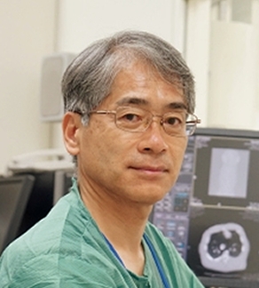 Hideki Hayashi (林秀樹)合作论文数Center for Frontier Medical Engineering, Chiba University3Sugitani Sou合作论文数3Yamada Yuto合作论文数3Kuse Yoshiki合作论文数3Ojino Kazuki合作论文数3Promsuk Jutabha合作论文数Department of Pharmacology and Toxicology, Kyorin University School of Medicine3Hidefumi Wakashin合作论文数2
 Kenjirou Ogawa合作论文数2Hiroshi Izawa合作论文数2Yoshie Reien合作论文数2Karin Urata合作论文数2Shiho Ogami合作论文数2Yo Goto合作论文数2Mika Yamauchi合作论文数2Takashi Niwa合作论文数2Yu Kato合作论文数2Kiyoshi Nakagawa合作论文数2Mizutani Kosuke合作论文数2Kobayashi Ryo合作论文数2Yamane Shinsaku合作论文数2
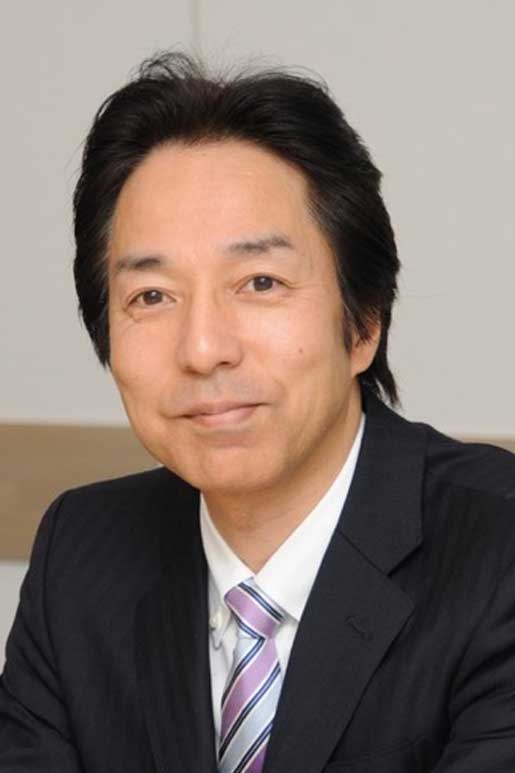 Shin-Ichi Yoshimura合作论文数Department of Neurosurgery, Hyogo College of Medicine2
 Hisato Kunitake合作论文数2
 Hirotoshi Iihara合作论文数2Masao Yamasaki合作论文数Department of Physiology, Fukushima Medical College2Masanao Saio合作论文数2




