BackgroundImmunocompromised patients are at particular risk of Severe Acute Respiratory Syndrome Corona Virus 2 (SARS-CoV-2) infection and previous findings suggest that the infection or vaccination induced immune response decreases over time. Our main goal was to investigate the SARS-CoV-2-specific immune response in rheumatoid arthritis patients and healthy controls over prolonged time.MethodsThe SARS-CoV-2-specific humoral immune response was measured by Elecsys Anti-SARS-CoV-2 Spike (S) immunoassay, and antibodies against SARS-CoV-2 nucleocapsid protein (NCP) were also evaluated by Euroimmun enzyme-linked immunosorbent assay (ELISA) test. The SARS-CoV-2-specific T-cell response was detected by an IFN- γ release assay.ResultsWe prospectively enrolled 84 patients diagnosed with rheumatoid arthritis (RA) and 43 healthy controls in our longitudinal study. Our findings demonstrate that RA patients had significantly lower anti-S antibody response and reduced SARS-CoV-2-specific T-cell response compared to healthy controls (p<0.01 for healthy controls, p<0.001 for RA patients). Furthermore, our results present evidence of a notable increase in the SARS-CoV-2-specific humoral immune response during the follow-up period in both study groups (p<0.05 for healthy volunteers, p<0.0001 for RA patients, rank-sum test). Participants who were vaccinated against Coronavirus disease-19 (COVID-19) during the interim period had 2.72 (CI 95%: 1.25–5.95, p<0.05) times higher anti-S levels compared to those who were not vaccinated during this period. Additionally, individuals with a confirmed SARS-CoV-2 infection exhibited 2.1 times higher (CI 95%: 1.31–3.37, p<0.01) anti-S levels compared to those who were not infected during the interim period. It is worth noting that patients treated with targeted therapy had 52% (CI 95%: 0.25–0.94, p<0.05) lower anti-S levels compared to matched patients who did not receive targeted therapy. Concerning the SARS-CoV-2-specific T-cell response, our findings revealed that its level had not changed substantially in the study groups.ConclusionOur present data revealed that the level of SARS-CoV-2-specific humoral immune response is actually higher, and the SARS-CoV-2-specific T-cell response remained at the same level over time in both study groups. This heightened humoral response, the nearly permanent SARS-CoV-2-specific T-cell response and the coexistence of different SARS-CoV-2 variants within the population, might be contributing to the decline in severe COVID-19 cases.
Introduction There is a critical gap in understanding which SARS-CoV-2 patients would benefit most from venovenous extracorporeal membrane oxygenation (VV-ECMO) support. The potential role of a dysregulated immune response is still unclear in this patient population. Objectives To assess the potential predictive value of SARS-CoV-2 specific cellular and humoral immune responses for survival in critically ill COVID-19 patients requiring VV-ECMO. Methods We conducted a prospective single-center observational study of unvaccinated patients requiring VV-ECMO support treated at the intensive care unit of Semmelweis University Heart and Vascular Center between March and December 2021. Peripheral blood samples were collected to measure the humoral and cellular immune statuses of the patients at the VV-ECMO cannulation. Patients were followed until hospital discharge. Results Overall, 35 COVID-19 patients (63% men, median age 37 years) on VV-ECMO support were included in our study. The time from COVID-19 verification to ECMO support was a median (IQR) of 10 (7-14) days. Of the patients, 9 (26%) were discharged alive and 26 (74%) died during their hospital stay. Immune tests confirmed ongoing SARS-CoV-2 infection in all the patients, showing an increased humoral immune response. SARS-CoV-2-specific cellular immune response was significantly higher among survivors compared to the deceased patients. A higher probability of survival was observed in patients with markers indicating a higher T cell response detected by both QuantiFeron (QF) and flow cytometry (Flow) assays. (Flow S1 CD8+ ≥ 0.15%, Flow S1 CD4+ ≥ 0.02%, QF CD4 ≥ 0.07, QF whole genome ≥ 0.59). In univariate Cox proportional hazard regression analysis BMI, right ventricular (RV) failure, QF whole genome T cell level, and Flow S1 CD8+ T cell level were associated with mortality, and we found that an increased T cell response showed a significant negative association with mortality, independent of BMI and RV failure. Conclusion Evaluation of SARS-CoV-2 specific T cell response before the cannulation can aid the risk stratification and evaluation of seriously ill COVID-19 patients undergoing VV-ECMO support by predicting survival, potentially changing our clinical practice in the future.
Rapidly evolving clinical data suggest that the novel coronavirus (SARS-CoV-2) and vaccination against COVID-19 might be associated with thyroid disturbances. However, studies remain limited among the pediatric population . Our aim was to assess the prevalence and permanence of thyroid autoimmunity (TA) and dysfunction in children after an acute infection and its potential association with vaccination. A prospective, multicenter registry analysis was performed among 458 children (mean age: 12.4 ± 3,8 years, 45.4% male) with preceding COVID-19. Patient inclusion lasted from 24 th March, 2021 to 23 rd March, 2022 at three pediatric outpatient facilities at Semmelweis University, Budapest. Primary outcomes were the rate of thyroid disturbances assessed by laboratory parameters (thyroid function tests, antithyroglobulin [ATG] and anti-thyroid peroxidase [ATPO] antibodies) and thyroid ultrasound. TA rate among vaccinated and unvaccinated children was determined. Children with newly diagnosed thyroid alterations were followed up for 12.7 ± 4.3 months. Six children had previous thyroid disease. Out of 452 children, 30 cases (6.6%) of newly diagnosed TA (six of them had abnormal thyroid-stimulating hormone [TSH] levels) and eight cases (1.8%) of isolated TSH elevation were observed. Ultrasound-proven autoimmune thyroiditis (AIT) was 4.0%. No association was found between COVID-19 vaccination and thyroid autoimmunity (χ 2 (1,N = 452) = 0.138, p = 0.815). Among children with TA, 73.3% had long-lasting alterations. Conclusion : Vaccination had no effect on the prevalence of TA. Until further controlled studies state otherwise, children with preceding COVID-19 might benefit from thyroid screening. What is Known: • Numerous case reports implicate that coronavirus disease-2019 (COVID-19) and vaccination against SARS-CoV-2 can be responsible for thyroid disturbances. • Thyroid alterations discovered during acute COVID-19 tend to cease by time and only incidental thyroid autoimmunity (TA) is diagnosed after COVID-19. In adults, no increase in vaccine-related hyper- or hypothyroidism was found. What is New: • TA rate after COVID-19 vaccination among children was not increased. TA had no role in long COVID syndrome. • We discovered a considerable rate of TA (6.6%) and ultrasound-proven autoimmune thyroiditis (AIT) (4.0%) after SARS-CoV-2 infection, and the majority of these alterations remained positive after 6 months.
Introduction Although myocarditis after anti-SARS-CoV-2 vaccination is increasingly recognized, we have little data regarding the course of the disease and, consequently, the imaging findings, including the tissue-specific features. The purpose of this study is to describe the clinical, immunological, and cardiac magnetic resonance (CMR) features of myocarditis after COVID-19 immunization in the acute phase and during follow-up. We aimed to compare the trajectory of the disease to myocarditis cases unrelated to COVID-19. Methods We assembled a CMR-based registry of potentially COVID-19 vaccination-related myocarditis cases. All patients who experienced new-onset chest pain and troponin elevation after COVID-19 vaccination and imaging confirming the clinical suspicion of acute myocarditis were enrolled in our study. Participants underwent routine laboratory testing and testing of their humoral and cellular immune response to COVID-19 vaccination. Clinical and CMR follow-up was performed after 3–6 months. We included two separate, sex- and age-matched control groups: (1) individuals with myocarditis unrelated to COVID-19 infection or vaccination confirmed by CMR and (2) volunteers with similar immunological exposure to SARS-CoV-2 compared to our group of interest (no difference in the number of doses, types and the time since anti-SARS-CoV-2 vaccination and no difference in anti-nucleocapsid levels). Results We report 16 CMR-confirmed cases of myocarditis presenting (mean ± SD) 4 ± 2 days after administration of the anti-SARS-CoV-2 vaccine (male patients, 22 ± 7 years), frequently with predisposing factors such as immune-mediated disease and previous myocarditis. We found that 75% received mRNA vaccines, and 25% received vector vaccines. During follow-up, CMR metrics depicting myocardial injury, including oedema and necrosis, decreased or completely disappeared. There was no difference regarding the CMR metrics between myocarditis after immunization and myocarditis unrelated to COVID-19. We found an increased T-cell response among myocarditis patients compared to matched controls (p < 0.01), while there was no difference in the humoral immune response. Conclusion In our cohort, myocarditis occurred after both mRNA and vector anti-SARS-CoV-2 vaccination, frequently in individuals with predisposing factors. Upon follow-up, the myocardial injury had healed. Notably, an amplified cellular immune response was found in acute myocarditis cases occurring 4 days after COVID-19 vaccination.
Background:To investigate the factors that have significant impact on the Severe Acute Respiratory Syndrome Corona Virus 2 (SARS-CoV-2) infection and vaccination induced immune response in rheumatoid arthritis (RA).Methods:Serological response was measured by quantifying anti-SARS-CoV-2 specific antibodies, while the cell-mediated response was measured by a whole-blood test quantifying the interferon (IFN)-γ response to different SARS-CoV-2-specific domains.Results:We prospectively enrolled 109 RA patients and 43 healthy controls. The median time (IQR) between the confirmed infection or the last vaccination dose and the day when samples were taken ("sampling interval") was 3.67 (2.03, 5.50) months in the RA group. Anti-Spike (anti-S) specific antibodies were detected in 94% of RA patients. Among the investigated patient related variables, age (p<0.004), sampling interval (p<0.001), the brand of the vaccine (p<0.001) and targeted RA therapy (TNF-inhibitor, IL-6 inhibitor, anti-CD20 therapy) had significant effect on the anti-S levels. After covariate adjustment TNF-inhibitor therapy decreased the anti-S antibody concentrations by 80% (p<0.001). The same figures for IL-6 inhibitor and anti-CD20 therapy were 74% (p=0.049) and 97% (p=0.002), respectively. Compared to subjects who were infected but were not vaccinated, the RNA COVID-19 vaccines increased the anti-S antibody levels to 71.1 (mRNA-1273) and 36.0 (BNT162b2) fold (p<0.001). The corresponding figure for the ChAdOx1s vaccine is 18.1(p=0.037). Anti-CCP (anti-cyclic citrullinated peptides) positive patients had 6.28 times (p= 0.00165) higher anti-S levels, than the anti-CCP negative patients. Positive T-cell response was observed in 87% of the healthy volunteer group and in 52% of the RA patient group. Following vaccination or infection it declined significantly (p= 0.044) but more slowly than that of anti-S titer (6%/month versus 25%). Specific T-cell responses were decreased by 65% in patients treated with anti-CD20 therapy (p=0.055).Conclusion:Our study showed that the SARS-CoV-2-specific antibody levels were substantially reduced in RA patients treated with TNF-α-inhibitors (N=51) and IL-6-inhibitor (N=15). In addition, anti-CD20 therapy (N=4) inhibited both SARS-CoV-2-induced humoral and cellular immune responses. Furthermore, the magnitude of humoral and cellular immune response was dependent on the age and decreased over time. The RNA vaccines and ChAdOx1s vaccine effectively increased the level of anti-S antibodies.
Összefoglaló. A SARS-CoV-2-fertőzés ritka gyermekkori szövődménye a sokszervi gyulladás, angol terminológiával paediatric inflammatory multisystem syndrome (PIMS). Két vagy több szerv érintettségével járó, súlyos tünetekkel induló betegségről van szó, amelynek tünetei átfedést mutatnak a Kawasaki-betegséggel, a toxikus sokk szindrómával és a makrofágaktivációs szindrómával. A PIMS-betegek intenzív terápiás osztályon vagy intenzív terápiás háttérrel rendelkező intézményben kezelendők, ahol biztosítottak a kardiológiai ellátás feltételei is. A szükséges immunterápia a klinikai prezentációtól függ. A jelen közleményben a szerzők a releváns nemzetközi irodalom áttekintését követően ajánlást tesznek a PIMS diagnosztikai és terápiás algoritmusára. Orv Hetil. 2021; 162(17): 652-667. Summary. Pediatric inflammatory multisystem syndrome (PIMS) is a rare complication of SARS-CoV-2 infection in children. PIMS is a severe condition, involving two or more organ systems. The symptoms overlap with Kawasaki disease, toxic shock syndrome and macrophage activation syndrome. PIMS patients should be treated in an intensive care unit or in an institution with an intensive care background, where cardiological care is also provided. The required specific immunotherapy depends on the clinical presentation. In this paper, after reviewing the relevant international literature, the authors make a recommendation for the diagnostic and therapeutic algorithm for PIMS. Orv Hetil. 2021; 162(17): 652-667.
Serological testing is a tool to predict protection against later infection. This potential heavily relies on antibody levels showing acceptable agreement with gold standard virus neutralization tests. The aim of our study was to investigate diagnostic value of the available serological tests in terms of predicting virus neutralizing activity of serum samples drawn 5-7 weeks after onset of symptoms from 101 donors with a history of COVID-19. Immune responses against Receptor Binding Domain (RBD), Spike1 and 2 proteins and Nucleocapsid antigens were measured by various ELISA tests. Neutralizing antibody activity in serum samples was assessed by a cell-based virus neutralization test. Spearman correlation coefficients between serological and neutralization results ranged from 0.41 to 0.91 indicating moderate to strong correlation between ELISA test results and virus neutralization. The sensitivity and specificity of ELISA tests in the prediction of neutralization were 35-100% and 35-90% respectively. No clear cut off levels can be established that would reliably indicate neutralization activity. For some tests, however, a value below which the sample is not expected to neutralize can be established. Our data suggests that several of the ELISA kits tested may be suitable for epidemiological surveys 1-2 months after the infection, estimating whether a person may have recently exposed to the virus. Sensitivities considerably superseding specificity at the cut-off values proposed by the manufacturers suggest greater potential in the identification of insufficient antibody responses than in confirming protection. Nevertheless, the former might be important in assessing response to vaccination and characterizing therapeutic plasma preparations.
Pilates exercises have several demonstrated physical and psychological benefits. To date, most research in this context was conducted with symptomatic or elderly people with few dependent measures. The current study examined the chronic or longitudinal effects of very low frequency, once a week, Pilates training on several physical and psychological measures, over a 10-week intervention, in young, healthy, and sedentary women. Further, the study gauged the acute effects of Pilates exercises on positive- and negative affect in 10 exercise sessions. Compared to a control group, the Pilates group exhibited significant improvements in skeletal muscle mass, flexibility, balance, core- and abdominal muscle strength, body awareness, and negative affect. This group also showed favorable changes in positive (22.5% increase) and negative affect (12.2% decrease) in nine out of ten exercise sessions. This work clearly demonstrates the acute and chronic benefits of Pilates training on both physical and psychological measures. It also reveals that even only once a week Pilates training is enough to trigger detectable benefits in young sedentary women. While this frequency is below the required levels of exercise for health, it may overcome the 'lack of time' excuse for not exercising and subsequently its tangible benefits may positively influence one's engagement in more physical activity.
 Hajnalka Vago合作论文数Semmelweis Univesity5
 Zsuzsanna Ulakcsai合作论文数4
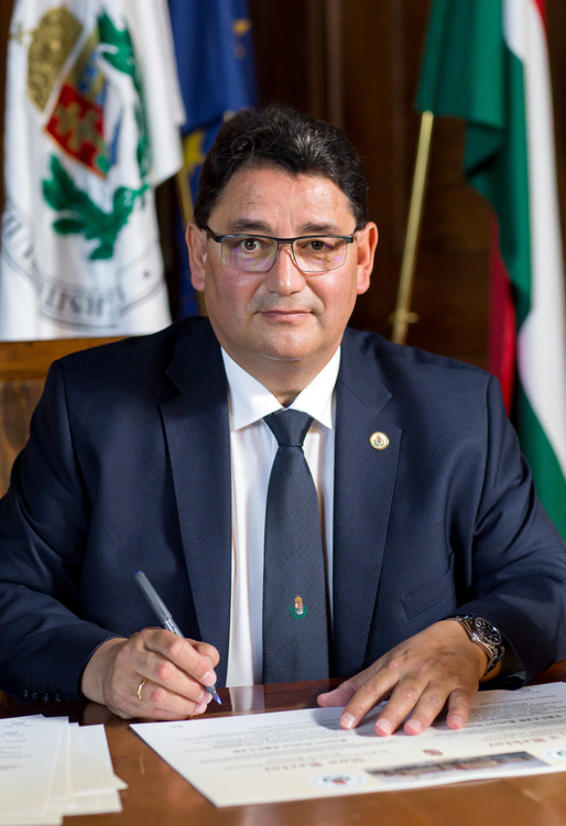 Bela Merkely合作论文数Department of Aviation and Space Medicine, Faculty of Medicine, Semmelweis University4
 Gyorgy Nagy合作论文数Department of Rheumatology and Genetics, Cell and Immunobiology, Semmelweis University;Faculty of Medicine, Semmelweis University4Dóra Németh合作论文数3
 David Becker合作论文数3Bernadett Rojkovich合作论文数2Liliana Szabo合作论文数2
 Attila Szabo合作论文数Department of Paediatrics, Semmelweis University2
 Attila Tóth合作论文数Centre for Neuroscience, University of Pécs2Laszlo Tothfalusi合作论文数Semmelweis Medical University;Department of Pharmacodynamics;Department of Pharmacodynamics, Semmelweis Medical University2
 Tamas Szabo合作论文数Center for Cancer Research, National Cancer Institute2Lilla Gunkl-Toth合作论文数1
 Nikolett Beniczky合作论文数1
 Vivien Herczeg合作论文数1Péter Krivácsy合作论文数1Nóra Tolnai合作论文数1Agnes Nemeth合作论文数1Fanni Kovacs合作论文数1Noémi Andrási合作论文数1Ádám Goschler合作论文数1Judit Kincs合作论文数1Krisztina Kalocsai合作论文数1Rita Káposzta合作论文数1Kinga Kardics合作论文数1Viktória Kemény合作论文数1Tamás Pék合作论文数1Andrea Tölgyesi合作论文数1Beáta Onozó合作论文数1Csaba Vilmanyi合作论文数1
 Peter Weiser合作论文数1Kálmán Tory合作论文数1Alexandra Vereb合作论文数1Réka Garai合作论文数1Nóra Kovács合作论文数1
 E. Hosszu合作论文数High Speed Networks Lab, MTA BME Future Internet Res Grp1
 Vencel Juhasz合作论文数Heart and Vascular Center, Semmelweis University1
 Kornelia Bodo合作论文数1
 Bálint Egyed合作论文数Semmelweis Egyet, Altalanos Orvostud Kar1Zsofia Dohy合作论文数Heart & Vasc Ctr, Semmelweis Univ1A Ponyi合作论文数1
 Gabor Geza Kovacs合作论文数Department of Laboratory Medicine & Pathobiology, Temerty Faculty of Medicine, University of Toronto1
 Ferenc Koteles合作论文数Institute for Health Promotion and Sport Sciences, Eötvös Loránd University1
 Földes Fanni合作论文数János Szentágothai Research Center, University of Pécs;Institute of Biology, University of Pécs;University of Pécs, University of Pécs1
 Johanna Takacs合作论文数Magatartastudomanyi Intezet, Semmelweis Egyetem1
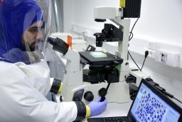 Gabor Kemenesi合作论文数Szentagothai Research Center, University of Pecs1Barabás Eszter合作论文数Department of Laboratory Medicine, Semmelweis University1Eniko Kovacs合作论文数Coronary Intensive Care Unit, Semmelweis University Heart Centre1Mosdósi Bernadett合作论文数1




