目的:探讨三维动态增强磁共振血管成像(3D CE-MRA)在双下肢动脉病变成像中对比剂最佳用量.方法:45例我院进行双下肢动脉3D CE-MRA血管成像的患者,根据就诊顺序随机分为A、B和C三组,三组钆对比剂用量分别为0.1、0.2和0.3mmol/kg,分别与盐水进行1:1配比,分别对不同对比剂用量的后处理图像进行主观及客观评分,并对小腿及足部血管进行整体静脉重叠评分,最后采用Wilcoxon检验比较3种扫描方案差异.客观评分是对3组的原始增强图像进行信号强度(SI)、信噪比(SNR)、对比噪声比(CNR)的测量及计算,比较股动脉、胭动脉及小腿动脉水平信号强度差异,进行t检验比较3种扫描方案差异.结果:对比剂用量为0.1mmol/kg时股动脉、胭动脉及小腿动脉的图像质量评分分别为(3.35±0.25)、(2.97±0.25)、(2.35±1.15);对比剂用量为0.2mmol/kg时,上述部位的评分分别为(3.75±0.35)、(3.55±0.32)、(2.97±0.70);对比剂用量为0.3mmol/kg 时,上述部位的评分分别为(3.90±0.41)、(3.83±0.52)、(3.10±0.75).A 组有33.33%图像不满足诊断需要,B组和C组图像全部满足诊断需要,A组与B组差异有统计学意义,B组与C组差异无统计学意义.B和C组的股动脉、胭动脉的SI、SNR、CNR明显高于A组,且B组和C组差异无统计学意义.B组和C组图像优于A组.结论:适当提高对比剂用量有助于提高患者双下肢动脉全程的3D CE-MRA成像质量,0.2mmol/kg的对比剂用量对血管的评估是可靠准确的,能够为临床外周动脉疾患的治疗方案提供准确可靠的影像依据.
Purpose: To determine the diagnostic value of multi-slice spiral computed tomography (CT) after adiponectin treatment of tracheal malignancies. Methods: Sixty patients with tracheal malignancies admitted to Emergency General Hospital from February 2017 to February 2021 were assigned to the study group. After physical examination, 60 healthy subjects in the same period served as the control group. All patients were treated with adiponectin. The control group was subjected to serum tumor marker testing before and after treatment, while the study group received multi-slice spiral CT before and after adiponectin treatment. Results: The 60 patients enrolled in the study group comprised 30 cases of squamous carcinoma, 22 cases of adenocarcinoma, and 8 cases of mucosa-associated lymphoma. Multi-slice spiral CT before treatment showed that 35 patients with tracheal malignant tumors had expansive lumen hyperplasia, while 25 patients with bulging tumors had saddle-like hyperplasia with irregular surfaces, uneven margins, and mean thickness of 5.96 ± 0.65 mm in the tracheal and bronchial walls adjacent to the luminal nodule. After treatment, the thickness of the tracheal and bronchial walls adjacent to the luminal nodule decreased to a mean value of 4.65 ± 0.48 mm. Prior to treatment, the mean enhancement value of contrast-enhanced CT was 18.98±1.23 Hu, and mean tumor diameter was 3.68 ± 0.23 cm. However, after treatment, the mean enhancement value increased to 25.98 ± 1.44 Hu, while mean tumor diameter decreased to 2.54 ± 0.20 cm in the study group. Conclusion: Multi-slice spiral CT has promising diagnostic value for patients with tracheal malignancies after adiponectin treatment. Therefore, it can potentially be used in the diagnosis of patients with tracheal malignancies.
目的:探讨IMR、iDose4和FBP三种重建算法在肋软骨隆鼻术前胸部多排螺旋CT扫描中对图像质量的影响.方法:收集符合纳入标准的拟行自体肋软骨隆鼻术患者60例,术前均行胸部CT检查,分别使用IMR、iDose4和FBP三种重建算法生成图像并进行相关后处理重建肋软骨,记录并比较各组图像质量主观评价分数和客观评价的指标如:肋软骨CT值、噪声、信噪比、对比噪声比.结果:三种算法得到的CT重建图像都能准确评估肋软骨钙化程度;主观评分IMR、iDose4组与FBP组相比,在MIP图像上评分均值分别增加50.5%和33.6%,在VR图像上评分均值分别增加51.0%和19.0%.IMR组较iDose4组的评分均值在MIP图像和VR图像上分别增加了 11.1%和21.1%.客观评价肋软骨CT值、信噪比和对比噪声比并作统计学分析,最终显示IMR、iDose4和FBP三种重建算法组间对比有显著差异,IMR组明显最优.结论:IMR、iDose4和FBP三种重建算法均能准确评估自体肋软骨是否存在钙化及程度,IMR算法较后两者更能显著降低图像噪声,提高CNR、SNR,提升肋软骨的主观图像质量,为制定最佳术前方案提供更精准的相关信息,可作为肋软骨CT检查首选算法重建技术进行应用,值得大力推广.
目的:探讨MSCT在老年或癌症晚期等难以耐受食道造影的食管气管瘘患者中的应用价值.方法:回顾性分析2016年至2020年在我院因高度怀疑食管气管瘘行MSCT及气管镜检查的病例,共纳入研究48例.所有图像经过横断位、冠状位、矢状位多平面、气管树容积再现及经气管树中心曲面重建后传至PACS,由两位医师盲法阅片,记录是否有食管气管瘘及阳性病例的大小;气管镜由操作者记录是否有食管气管瘘及阳性病例的大小.比较不同方法间对瘘口、大瘘口(直径>1 cm)、小瘘口(直径<1 cm)的检出率及MSCT与气管镜间大小比较.比较MSCT与气管镜检查对真阳性病例瘘口的大小.两位医师间采用Kappa一致性检验,不同评价方法间采用卡方检验,MSCT与气管镜检查的大小比较采用配对样本t检验,当P<0.05时有显著差异.结果:联合所有图像对所有食管气管瘘的检出率最高,其敏感性、特异性及准确性分别达93%、75%及86%,不同方法对所有瘘口的检出有显著差异,其中矢状位与横断位对瘘口的检出率较高.在所有阳性病例中不同方法间对大瘘口的检出率,各方法间有显著差异,其中联合所有图像对大瘘口的检出率达100%,矢状位与横断位对大瘘口的检出率较高;而对小瘘口的检出,各方法间无显著差异.在所有真阳性病例中,气管镜中所测量的瘘口大小(13.70±11.70)mm,高于MSCT中测量的瘘口的大小(10.83±10.15)mm,两者有显著差异.结论:MSCT及其常见后处理方法在难以耐受食道造影的食管气管瘘患者评估中起重要作用.
目的:探讨胸部多排螺旋CT扫描在肋软骨隆鼻术中的应用价值.方法:收集符合纳入标准120例拟行自体肋软骨隆鼻术患者,都进行了胸部多排螺旋CT术前检查,并进行多种三维重建分析自体肋软骨是否符合手术标准,部分病例进行了术后CT复查评估.结果:CT准确评估肋软骨无钙化(1级)28例和轻度钙化(2级)62例,均可行自体肋软骨隆鼻术;中度钙化(3级)23例中12例可用自体肋软骨行隆鼻术,剩余7例全部重度钙化(4级)患者直接放弃肋软骨而采用耳软骨等替代进行隆鼻术.同时还发现叉状肋3例、肋软骨部分融合7例,镜面右位心伴内脏扭转1例,肺部占位病变2例.结论:多排螺旋CT及多种三维重建对拟选择自体肋软骨行隆鼻术的患者是术前首选的影像学检查方法,可以准确评估自体肋软骨是否存在钙化及程度,并能精准测量各肋软骨实际走行、长度、宽度及厚度,能够为肋软骨隆鼻术提供可靠的术前影像信息,同时还可以用于术后评估移植效果及排查并发症等.
目的:探讨通过64层螺旋CT螺距参数的设置,降低新型冠状病毒肺炎患者筛查的辐射剂量和扫描时间的可行性.方法:前瞻性选择需进行新型冠状病毒肺炎筛查患者,按扫描时间随机分为大螺距组和对照组,每组各16例.大螺距组选择1.375:1螺距,对照组选择0.984:1螺距.对肺纹理、支气管、纵膈结构的图像质量;胸主动脉、竖脊肌、双肺肺实质;辐射剂量的客观数据进行评价和分析.结果:32例图像均符合影像诊断要求.伪影、肺纹理评分差异有统计学意义(P<0.05).两组右肺CT值分别是(-804.62±45.43)HU和(-852.12±28.13)HU、左肺CT值分别是(-808.13±37.74)HU和(-858.77±19.95)HU,均具有统计学意义(P<0.05).两组的扫描时间分别为(2.14±0.15)s和(3.14±0.28)s,具有统计学意义(P<0.05).两组记录所得的容积CT剂量指数(CTDIvol)是(7.88±0.89)HU和(11.17±1.08)HU、剂量长度乘积(DLP)是(279.14±36.33)HU和(387.25±47.86)HU均具有统计学意义(是P<0.05),有效剂量(ED)分别是(3.91±0.51)HU和(5.42±0.67)HU具有统计学意义(P<0.05).结论:新型冠状病毒肺炎患者CT筛查检查中,使用大螺距扫描,能够减少辐射剂量、显著缩短了扫描时间,在不影响病灶诊断的前提下,降低病人接受到的辐射剂量和放射技师感染的风险,具有实际工作意义,有待进一步推广验证.
Objective To evaluate the study value of positional change multi-slices helical CT Colonography in colorectal polyps.Methods Total 189 polys in 39 cases were performed with conventional Colonoscopy and underwent multi slices helical CT Colonography between two weeks.The CT Colonography screening in both supine and prone position.Results All the 39 cases showed 203 polys in supine position and 199 polys in prone position scaned.185 polys is noted by combined evaluation when change positional screening in both supine and prone positions,4 polys size<5mm were missing.Conclusion MS CTC screening in change positional is excellent sensitivity and specificity for colorectal polys,which can provide more accurate informations for clinical diagnosis and treatment.
Objective: To explore a best does (method, standard and accurate calculation) of bone cement in PKP, based on the CT and MRI image data of thoracolumbar vertebrae with new compression fracture. Methods: 42 new compression fracture patients with thoracolumbar vertebrae injury underwent PKP and examined by CT and MRI before surgery no more than 1 week. Retrospectively analyzed the image data and compared the statistical image data followed a variety of reconstruction. Results: Of 42 cases, new compression fracture located in the T10 (n=2), T11 (n=5), T12 (n=9), L1 (n=11), L2 (n=7), L3 (n=5), L4 (n=3). The average fracture compression rate of bone cement in PKP ranged from 1.6 to 6.3ml. Conclusion: It is practicable to predict the best dose of bone cement in PKP that by reprocessing the image of preoperative CT and MRI, which can make the treatment more effective, at the same time, the interrelated complication can be avoided.
Objective To investigated the diagnostic value of 64 multi-detector helical CT angiography in artery of lower extremity middle and long-term follow up after artificial vascular graft.Methods 108 cases with artery of lower extremity postoperative artificial vascular graft after 3 years were performed with MDCTA. The data was transmitted to ADW4.3 workstation, and reconstructed by MIP, VR, CPR and generally analyzed with orginal axial images.Results In 108 cases with 147 grafts, MDCTA is can revealing the location and shape of artificial vascular graft, and evaluating collateral circulation and complication exactly.Conclusion 64 MDCTA is reliable and accurate in evaluating the artificial vascular graft, gradually become the first modality diagnose and evaluate artery of lower extremity follow up after artificial vascular graft at present.
Objective To evaluate the diagnostic value of susceptibility weighted imaging in cavernous hemangioma of extremities soft tissue.Methods Both 50 cases clinical and pathological were confirmed to be Cavernous hemangioma in extremities soft tissue scaned with SWI and Conventional MRI sequences.Results Total of 62 lesions of cavernous hemangioma in extremities soft tissue were detected from the 50 patients, including 8 patients with multiple lesions. Found Ⅰ(node) type 9 patients,Ⅱ(limited mass) type 17, Ⅲ(diffuse vines) type 25 and Ⅳ(hybrid) type 11. 24 lesions are Low return type and 38 lesions are high return type be shown.Conclusion SWI is an important supplement sequence of MRI in the diagnosis of hemangioma in extremities soft tissue, can reflect internal composition and physiological change of the lesions more fully, shows more obviously advantages on the analysis of the abnormal blood vessels,identification of calcification, hemorrhage and iron deposit,and owns important application value for clinical diagnosis, making relevant treatment and estimating prognosis.
Objective To evaluate the study value of multi-slices helical CT angiography and three dimensional dynamic contrast-enhanced MRA in lower extremities arteriosclerotic occlusion disease(ASO).Methods 38 cases of ASO were performed with MS CTA and 3D CE-MRA between two weeks. The CT original axial data was reconstructed by ADW4.6 workstation of MPR, MIP and VR, compared with processed MIP images with MRI. Bilateral lower extremity arterial vascular system of each case was divided into three regions and 23 segments, each segment was evaluated for the presence and degree of vascular stenosis.Results All the 38 cases including 8 cases with mild stenosis, 14 cases with moderate syenosis, 12 cases with significant stenosis and 4 cases with occlusion. The significant disease of the lower extremities were demonstrated clearly by MS CTA and 3D CE-MRA.Conclusion MS CTA and 3D CE-MRA are excellent in patients with clinical symptoms of the arteriosclerotic occlusive disease of lower extremity, which can provide more accurate informations for clinical diagnosis and treatment.
目的 探讨和评估核磁共振PROSET序列在腕管综合征病变中的应用价值.方法 对18例腕管综合征(CTS)患者进行了常规MRI序列及PROSET序列扫描,并进行多种重建及统计的分析.结果 PROSET序列结合常规MRI序列能够更好地显示CTS的腕管解剖结构及正中神经的卡压情况.结论 PROSET序列作为一种重要的补充序列,在评估CTS患者正中神经病变中更加准确,对指导临床选择手术方式及评估预后有重要意义.
Objective To investigate the features of normal prostates in 3.0T DW MR (Diffusion-Weighted Magnetic Resonance) images so as to provide a basis for selection of the optimal b-value. Methods Three b-values (b=600, 800, 1000 s/mm2) were applied respectively in 3.0T DW MR imaging of twenty normal prostates. Then, the signal intensity, standard deviation of the background signal intensity and apparent diffusion coeffcient (ADC) values were measured in DWI of the central zone and peripheral zone of prostates. The signal-noise ratio (SNR) and contrast-noise ratio (CNR) were computed. And the quality of MR images were evaluated. Results When b-value was 600, 800 and 1000s/mm2, the SNR was (18.36±1.75), (14.63±2.32) and (11.83±1.65) and the CNR was (0.55±0.14), (0.47±0.11) and (0.39±0.90) respectively. There were statistically significant difference in SNR and CNR between different b-values (P<0.05). When b-value was 600 s/mm2, the ADC values acquired in MR imaging of the peripheral zone and central zone of prostates were (2.07±0.26)×10-3 mm2/s and (2.01±0.15)×10-3 mm2/s respectively without statistically signiifcant differences (P>0.05). When b-value was 800 and 1000 s/mm2, the ADC values of the peripheral zone and central zone of prostates were (1.80±0.22)×10-3mm2/s and (1.65±0.24)×10-3 mm2/s, (1.54±0.14)×10-3 mm2/s and (1.31±0.19)×10-3 mm2/s respectively with statistically significant differences (P<0.05). The quality of MR images at three b-values (600, 800, 1000 s/mm2) were evaluated as (3.4±0.68), (3.05±0.69) and (2.75±0.91) respectively with statistically signiifcant differences (P<0.05). Conclusion The optimal b-value in 3.0T DW MRI of prostates was 800 s/mm2, which can display the diffusion of water molecules and meanwhile ensure the quality of images.
Objective To explore the standardization protocol and value of magnetic resonance imageing(MRI) for augmentation mammaplasty by injected polyacrylamide hydrogel(PAMHG).Methods Thirty female patients with 60 breast,having been injected PAMHG as augmentation mammaplasty,underwent bilateral breast MRI examination before removal surgery.14 patients among them underwent dynamic contrast enhanced MRI(DCE-MRI).The results of MRI were compared with that of operation and pathology.Results PAMHG showed iso-intensity compared with breast gland on T1-Weighted imaging(T1WI) and hyperintensity on Turbo Spin Echo(TSE)T2WI with Spectrally Selective Attenated Inversion Recovery(SPAIR.Low signal septa were noted within PAMHG in 83.3%(50/60) breast,83.3%(50/60) PAMHG without capsule,10%(6/60) with smooth and regular capsule,6.7%(4/60) PAMHG with irregular capsule.Subcutaneous lump of PAMHG was 66.7%(40/60),lump of that in breast gland was 16.7%(10/60).Diffuse gel along spatium intermusculare of pectoralis major was 83.3%(50/60),that along spatium intermusculare of intercostal muscle was 16.7%(10/60).All PAMHG in breast of 14 patients showed no enhancement,one malignant intraductal papilloma and one fibroadenoma in breast were found in two patients.All distributions of PAMHG and appearances of its complications on T2WI-FS were consistent with the results of operation.Conclusions Axial T1WI,TSE-T2WI-SPAIR and sagittal TSE-T2WI-SPAIR combined with DCE-MRI can be optimized protocol of MRI examination for augmentation mammaplasty by injected polyacrylamide hydrogel.MRI is helpful for preoperative assessment of the distribution of PAMHG and follow up after operation.
Objective:To evaluate the role of DWIBS in the diagnosis of thoracic tumorous lesions.Materials and Methods:102 cases(patients suspected chest tumors clinically in Beijing Coal General Hospital from 01,2009 to 12,2010,total 158 lesions)were involved in this study,including 33 lung cancer,18 pulmonary benign tumorous lesions,24 malignant lymphoma,24 mediastinal and subaxillary lymph node metastasis,22 breast cancer,17 breast benign lesions and 20 focal normal breast tissues.All 158 lesions were performed DWIBS(STIR) examination and 33 of them also received DWIBS(SPIR)examination and another 4 cases received thoracic-abdomen-pelvis DWIBS(STIR)scan,then their ADC value were measured and compared.The lesions were displayed by background suppression rebuilding with 3D MIP(3 dimensional maximum intensity projection) and technique with reverse black and white.The ADC values of all lesions were measured.Results:(1)The effect of background suppression of STIR-EPI was better than SE-EPI(SPIR)and could get PET-like images with 3D MIP rebuilding and reverse black and white technique to observe the lesion more clearly.(2)The mean ADC value of thoracic metastatic lymph nodes was 1.37±0.41 mm2/s×10-3 and 0.91±0.12 mm2/s×10-3in thoracic lymphoma.There was significant difference between them(P0.05);The mean ADC value of lung cancer was 1.61±0.18 mm2/s×10 -3and 2.06±0.21 mm 2/s×10 -3in pulmonary benign lesions,and it was significantly ower than that of benign lesions.(3)The ADC value of different types of malignant different between the two types of lesions;The mean ADC value of breast cancer,breast benign lesion and normal breast tissue was 1.01±0.13 mm2/s×10 -3,1.81±0.18 mm 2/s×10 -3and 1.91±0.26 mm 2/s×10 -3respectively,and there was significant difference between either two groups.(3)The ADC values of different types of malignant tumors were significantly different.(4)Among the 4 cases with thoracic-abdomen and pelvis DWIBS examination, 3 cases were with distal metastasis.Conclusions:(1)DWIBS was a safe and effective imaging examination.(2)It benefited to identify the property of the thoracic tumorous lesion that was benign or malignant,the ADC value of malignant lesions was l tumors was significantly different.(4)DWIBS can be used in tumor screening and the therapeutic evaluation due to its large range scan.
Objective To evaluate the difference of adaptive exposure dose in two digital X-ray imaging systems. Methods Resin phantom and contrast-detail test phantom were used on Cesium iodide and Gadolinium oxide sulfide plate imaging detector DR systems using difference exposure parameters. All entrance surface dose were recorded, Image quality index (IQFinv ) was calculated by CDRAD analysis software. The optimal exposure radiation dose was determined according to the sensitivity and IQFinv. Results There were no significant differences of entrance surface dose between two digital X-ray systems on two different exposure parameters with 70 kV ( P > 0. 05) , while there were significant differences with 125 kV(P < 0.05) . The maximum difference was 28. 57% . The mean IQFinv value of Cesium iodide detector DR and Gadolinium oxide sulfide detector DR with 125 kV and 70 kV were 4. 89 ±1. 01 and 2.47 ±0.25, 5. 10 ±1.05 and 2.38 ±0.43, respectively. There were significant diferences between the two systems under the same exposure parameters (t = 6. 509,10. 158, P < 0. 05). The optimal exposure dose ratio between Cesium iodide and Gadolinium oxide sulfide detector DR system was 1:2. Conclusion According to the IQFinv value, the adaptive exposure parameter can be adjusted to achieve good image quality with low radiation dose on different digital radiography systems.
4D CE-MRA cinegraphy technique is useful for both radiologist’s diagnosis and clinical treatment of brain tumor,offer more information,depict vessel structures and it’s homodynamic change.It can be named as fluoroscopic MRA according to it’s high temporal and spatial resolution from sensitivity encoding(SENSE), contrast enhanced timing rebust angiography(CENTRA),keyhole and half scan techniques.
Objective: To evaluate the diagnostic value of 64 multi-detector helical CT angiography in aortic dissection.Methods: 88 cases suspected dissection of aortic dissection were performed with MDCTA.The data was transmitted to ADW4.2 workstation,and reconstructed by MPR,VR,CPR,VE and generally analyzed with original axial images.Results: All the 32 aortic dissection were demonstrated clearly by MDCTA,and it can reveal the types and shape of aortic dissection.The true and false lumen and the intimal flap of aortic dissection were clearly showed.The position,size and shape of lacuna and its relationship with aortic branches,were clearly demonstrated from the cavities.Conclusion: 64-MDCTA is reliable and accurate in evaluating aortic dissection,gradually becomes one of the first modality diagnose in aortic dissection.
目的 探讨螺旋CT自动管电流技术在胸部检查中辐射防护价值.方法 实验组和对照组各30例,分别采用CT自动管电流(噪声指数=10.00)和固定管电流(200 mA)技术进行胸部检查,对所获图像噪声特性、细节显示程度及CT设备本身剂量信息显示结果进行比较.结果 实验组和对照组主动脉弓上层面、气管隆突和横膈上层面肺野内CT值差异不具显著性(P=0.136,0.289,0.176),主动脉弓上层面图像背景标准差差异无统计学意义(P=0.121),但气管隆突和横膈上层面图像背景标准差差异有统计学意义(P=0.001,0.000),实验组大于对照组.实验组和对照组肺内细小血管、细支气管、叶间裂等细微结构显示程度以及病变形态特征显示无差别.两组图像主动脉弓上层面均可见线状假影,但实验组有假影的层面多,图像噪声颗粒比对照组明显.实验组CTDIvol和剂量长度乘积(DLP)约为对照组的1/3.结论 正确使用CT自动管电流技术可以在确保图像质量同时降低受检者所受辐射剂量.
由于寿命延长、环境污染、和新化学物品的作用,恶性肿瘤对人类的危害日趋严重.据2005年我国城乡居民病死原因位居前三位的是恶性肿瘤、脑血管病和心脏病,而肺癌自90年代初期就已上升为恶性肿瘤之首.目前对早期肺癌的治疗效果已得到认可,因此通过胸部X线检查发现早期病变,对提高患者生活质量具有重要意义.本文通过胸部常规剂量和低剂量螺旋CT检查时,受检者所受辐射剂量及图像显示差别进行比较,探讨低剂量螺旋CT在胸部体检中的应用价值.
 Xiaoming Yin (尹晓明)合作论文数煤炭总医院15
 Qingyu Ceng (曾庆玉)合作论文数Emergency General Hospital14
 Ge Wu (吴戈)合作论文数13JianXin Liu (刘建新)合作论文数11MaoSong Deng (邓茂松)合作论文数10
 Guangwei Jin (金光暐)合作论文数Emergency General Hospital8Xin Chang (常鑫)合作论文数7
 LiLi Zhang (张藜莉)合作论文数Emergency General Hospital6Ying YANG (杨英)合作论文数5ShuHui Zhang (张淑慧)合作论文数5WANG Xu (王旭)合作论文数4Tongjiang Xu合作论文数4YIN Shuo (殷硕)合作论文数3Jian LIU (刘戬)合作论文数3JianWei Huo (霍健伟)合作论文数3Meng Wang (王猛)合作论文数3YunQing Wei (韦云青)合作论文数2ChenLu Feng (冯晨璐)合作论文数2YingMao Ruan (阮英茆)合作论文数2Cheng Wang (王诚)合作论文数2Liang Meng (孟亮)合作论文数2Yan Zhang (张岩)合作论文数2
 WeiZhong Liang (梁伟中)合作论文数Emergency General Hospital2
 Minjie Lu (陆敏杰)合作论文数Fuwai Hospital Chinese Academy of Medical Sciences2
 Shiliang Jiang (蒋世良)合作论文数Fuwai Hospital, Chinese Academy of Medical Sciences2
 Lianjun Huang (黄连军)合作论文数DeltaHealth Clinic2
 Shihua Zhao (赵世华)合作论文数Fuwai Hospital, Chinese Academy of Medical Sciences2
 Xu Wang (王旭)合作论文数Emergency General Hospital2Yan LI (李艳)合作论文数1chunyan gao (高春岩)合作论文数1Jun WANG (王军)合作论文数1Sheng GAO (高盛)合作论文数1fengqin yang (杨凤芹)合作论文数1Wei FANG (方伟)合作论文数1XiuMei Dou (窦秀梅)合作论文数1JinWei Mu (穆锦巍)合作论文数1RongFang Shi (史蓉芳)合作论文数1RunYi Ban (班润义)合作论文数1Feng Mou (牟峰)合作论文数1Zheng Zhou (周正)合作论文数1Fang Qin (秦芳)合作论文数1Zhe Li (李喆)合作论文数1XueMei Wang (王雪梅)合作论文数1Wei Zhong (钟伟)合作论文数1
 Degui Zu (祖德贵)合作论文数Emergency General Hospital1
 Wenyi Jin (靳文毅)合作论文数Emergency General Hospital1
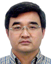 BaoPing Li (李宝平)合作论文数Emergency General Hospital1Jun Wang (王军)合作论文数1




