Bone defect repair remains a significant challenge in orthopedics, particularly for critical-sized bone defects, which often result in nonunion. Traditional treatments have numerous limitations. Recent studies have highlighted the pivotal role of mitochondria as cellular energy and metabolic hubs influencing the function of osteoblasts, osteoclasts, and chondrocytes. Mitochondria regulate energy metabolism, ROS signaling, mitochondrial dynamics, and apoptosis, all of which are essential for maintaining proper bone function. Mitochondrial dysfunction has been identified as a key intrinsic factor contributing to the failure of bone repair. Thus, targeting mitochondria has emerged as a promising therapeutic strategy. This article systematically reviewed the various functional roles of mitochondria in bone repair and evaluated the current progress of mitochondrial-targeted therapeutic strategies. We focused on the mechanisms of action and preclinical advancements related to small molecule compounds, functionalized biomaterials, and advanced cell therapies, offering a theoretical foundation for their potential clinical application. Mitochondrial-targeted therapies show significant promise for enhancing bone repair by improving cellular energy metabolism, restoring redox homeostasis, optimizing mitochondrial quality control, and promoting cell survival. However, this field faces several challenges, including improving targeted delivery efficiency, ensuring long-term safety, and translating these strategies into clinical practice. Future research should prioritize the development of more precise delivery technologies, exploration of multi-target synergistic approaches, and rigorous clinical trials to support the practical application of mitochondrial-targeted therapies for clinical bone regeneration.
Osteosarcoma (OS), recognized as the most common primary malignant bone tumor, presents substantial clinical challenges. The current standard of care, involving extensive surgical resection followed by adjuvant chemotherapy, often leads to critical-size bone defects and is hampered by high risks of local recurrence, metastasis and systemic toxicity. Conventional bone scaffolds are constrained by their purely mechanical role, lacking the inherent bioactivity required for a therapeutic function within the regenerative microenvironment. This significant and unresolved clinical challenge has driven the advancement of sophisticated biomaterial platforms aimed at achieving the dual objectives of effective osteosarcoma elimination and concurrent bone tissue regeneration. This review comprehensively explores the design principles of these scaffolds, detailing their use as a structural base and the integration of key components for antineoplastic strategies and osteogenesis. Furthermore, it delves into advanced smart material systems, including stimuli-responsive drug release platforms and 3D printing technologies for creating patient-specific implants. The discussion also encompasses the critical in vitro and in vivo evaluation models used to assess the efficacy of these platforms. Finally, the review addresses the current challenges in balancing oncotherapy with regeneration and provides insightful perspectives on the future of this promising field, highlighting the potential of these engineered scaffolds to serve as strategic bridges connecting tumor ablation to functional bone reconstruction.
Neuroinflammation following secondary injury in spinal cord injury (SCI) constitutes a major obstacle to functional recovery. Endogenous danger signals, particularly mitochondrial DNA (mtDNA) released post-injury, can activate the cGAS-STING pathway, thereby modulating the activation state of microglia and macrophages toward a pro-inflammatory phenotype. This process unfolds within a highly interconnected network involving Toll-like receptor (TLR) signaling, reactive oxygen species (ROS), NLRP3 inflammasome assembly, and cytokine cascades. Through crosstalk with the NLRP3 inflammasome, the cGAS-STING pathway can enhance cellular susceptibility to pyroptosis. The ultimate execution of pyroptotic cell death remains contingent upon additional downstream events, including the activation of Caspase-1 and the cleavage of Gasdermin D. The state of microglia and macrophages is dynamically regulated by a convergence of multiple signaling pathways. Furthermore, the cGAS-STING pathway engages in complex bidirectional interactions with autophagy, underscoring its multifaceted regulatory functions. This review explores emerging intervention strategies targeting this pathway, including small-molecule inhibitors/agonists, genetic interventions, and smart nanomaterials, aimed at achieving precise immunomodulation within the neuroimmune microenvironment. However, the clinical translation of this field still faces significant challenges, primarily concerning issues of spatiotemporal-specific modulation, drug delivery efficiency, and interspecies differences. Future research should focus on elucidating these intricate mechanisms and developing advanced drug delivery systems to facilitate the translation of this therapeutic target from a conceptual framework into clinically effective treatment modalities.
Oblique Lumbar Interbody Fusion (OLIF) is widely used minimally invasive techniques for lumbar degenerative diseases. While neurological complications are recognized, they predominantly affect the ipsilateral lumbar plexus. Contralateral L4 nerve root compression is an exceptionally rare and often overlooked complication, with limited understanding of its biomechanical mechanisms and sparse radiological evidence linking symptoms to specific causes. A 61-year-old female underwent L3/4 and L4/5 OLIF via a left retroperitoneal approach for degenerative spondylolisthesis and spinal stenosis. Postoperatively, she developed right lower extremity pain and quadriceps weakness. Imaging revealed a malpositioned interbody cage and a fractured L4 osteophyte fragment compressing the contralateral L4 nerve root. Revision surgery via a transforaminal lumbar interbody fusion (TLIF) approach successfully removed the fragment and decompressed the nerve, leading to complete symptom resolution at 5-month follow-up. This case provides direct radiological and intraoperative confirmation of contralateral L4 nerve root compression due to cage malposition and osteophyte fracture-a rarely documented mechanism. Contributing factors likely included asymmetric cage placement and potential patient positioning inaccuracies. The report underscores the importance of precise surgical techniques, including rigorous fluoroscopic verification, optimal patient positioning, and consideration of intraoperative navigation to prevent such complications. Prompt postoperative imaging and timely intervention are crucial for managing unexpected neurological deficits. This case demonstrates that timely revision surgery successfully resolved the contralateral neurological deficits, resulting in complete functional recovery at 5month follow-up. Enhanced awareness and adherence to preventive strategies can significantly reduce the incidence of this serious, yet preventable, complication in lateral access lumbar surgery.
Atlantoaxial instability (AAI) necessitates surgical stabilization to prevent neurological compromise. This finite element study compares the biomechanical performance of four atlantoaxial fixation constructs under physiological loading: standalone atlantoaxial cage (AAC), C1-C2 pedicle screws (C1-C2 PS), C1 pedicle screws with transarticular screws (C1 PS + TA), and a combined 360° construct (C1-C2 PS + AAC). A validated C0-C7 FE model with simulated AAI was subjected to flexion, extension, lateral bending, and axial rotation. Results demonstrated that the 360° construct provided the highest stability, with the lowest range of motion in all directions. It also significantly reduced peak von Mises stress on screws (by up to 91.7%) and cages (by up to 57.2%) compared to standalone constructs, while minimizing vertebral stress (over 90% reduction vs. C1-C2 PS). Adjacent disc stress remained largely unaffected by fixation type, with consistent stress concentrations at the C2-C3 level. The C1-C2 PS + AAC configuration offers superior biomechanical stability, enhanced load sharing, and reduced implant and bone stress, supporting its potential as an optimal strategy for complex AAI fixation.
Osteosarcoma, a very aggressive bone cancer, poses considerable therapeutic hurdles because it is resistant to conventional therapies. The objective of this work was to develop a novel genistein-based therapeutic formulation by encapsulating the compound into exosomes derived from mesenchymal stem cells (MSCs) and to evaluate its in vitro efficacy against osteosarcoma. The physicochemical characteristics of the genistein-loaded exosomes (Exo-Gen) were analysed by transmission electron microscopy (TEM) and nanoparticle tracking analysis (NTA) Osteosarcoma cell line MG63 were treated with Exo-Gen, and their effects on cell viability, cellular uptake and migration were evaluated using in vitro CCK-8 assay, flow cytometry and Transwell assay. The Western blot assay was used to evaluate the protein expression of the PPAR gamma pathway proteins (PPAR gamma, PTEN, P21, Cyclin B1, Bcl-2 and Survivin). RT-qPCR has been employed to determine the mRNA expression levels of PPAR gamma and PTEN. Compared to the free genistein (Free-Gen), the produced Exo-Gen demonstrated improved cellular uptake efficiency and cytotoxicity in the MG63 cell line, with half-maximal inhibitory concentrations (IC50) of 10.18 mu g/mL for Free-Gen and 6.54 mu g/mL for Exo-Gen. Exo-Gen substantially boosted PPAR gamma expression in osteosarcoma cells, relative to Free-Gen. Genistein is a non-toxic PPAR gamma activator which effectively inhibits the osteosarcoma cell growth when administered through exosomes. In addition, in vivo studies utilizing a tumour-bearing mice model corroborated the enhanced anticancer efficacy and biocompatibility of Exo-Gen. This approach significantly improves the distribution and effectiveness of genistein, rendering it as an ideal platform for future cancer therapy.
The inflammatory cascade triggered by spinal cord injury (SCI) presents a major barrier to tissue repair. This review summarizes recent advances in this field. It outlines the selection criteria for different scaffold materials and stem cells. It also describes key strategies for the coordinated regulation of the inflammatory microenvironment, including structural support and immunomodulation. In addition, the review discusses cell-free therapies and the functionalization of stem cell-derived materials. Intelligent responsive systems are also highlighted. Furthermore, this article evaluates the validation of these approaches in preclinical models. It also examines the challenges associated with their clinical translation. Finally, emerging trends, including dynamic adaptation, functional integration, and precise regeneration, are discussed. This review aims to provide a theoretical foundation and to guide future research on combination therapies for SCI.
Meningiomas constitute the most prevalent primary intracranial tumors, accounting for approximately 39
Mitochondrial dysfunction is a critical factor in secondary injury following spinal cord injury (SCI). Mitophagy is an essential mechanism for mitochondrial quality control. Proper and timely activation of mitophagy clears damaged mitochondria, reduces oxidative stress and cell death, and provides neuroprotection. However, excessive activation can cause energy depletion and worsen injury. The effects of mitophagy depend on the specificity of its spatial and temporal activation as well as the cellular microenvironment. This review summarizes novel therapeutic strategies targeting mitophagy, including pharmacological modulators, gene-based interventions, biomaterials, and cell therapies. These approaches precisely regulate mitophagy via distinct molecular pathways. Challenges remain in precise regulation, clarification of cell-specific mechanisms, and real-time monitoring in vivo. Future research should aim to develop precise spatiotemporal regulatory tools, identify relevant biomarkers, and integrate mitophagy-targeted therapies with existing methods, providing new insights into SCI treatment.
Objective This study aimed to determine the biomechanical stability of C0-C2 vertebrae fixed by atlantooccipital joint-occipital condyle-slope screw fixation (AO-CO-C screws) using finite element analysis. Methods Using computed tomography images, a nonlinear intact three-dimensional C0-2 finite element model (FEM) was developed and validated. Six FEMs were reconstructed: intact model, unstable model, atlantoaxial fusion with C1 lateral mass screw and C2 pedicle screw fixation finite element model (Group A), occipital plate screw with C1 lateral mass screw and C2 pedicle screw fixation finite element model (Group B), transarticular atlantoaxial screw with C2 pedicle screw fixation finite element model (Group C), occipital-cervical screw with counter-odontoid peg-C2 pedicle screw fixation finite element model (Group D). Range of motion and maximum von Mises stresses were compared under flexion, extension, lateral bending, and axial rotation. Results Group D showed the greatest decrease in ROM(range of motion) with flexion, which was higher than that of the other techniques. The maximal von Mises stress on Group A and Group B showed the occipital condyle screw began contact with the occipital condyle bone, whereas Group C and Group D. Group C had maximal von Mises stress on the atlanto-occipital joint region; the maximal von Mises stress on Group A was located in the area where the occipital condyle screw began contact with the occipital condyle bone, whereas Group C and Group D had maximal von Mises stress on the atlanto-occipital joint region, and the maximal von Mises stress on Group B was located in the area of the bottom of the C1 lateral mass screw. Conclusion In this study, AO-CO-C screws fixation is the most stable technique. If surgeons have to use other fixation methods, they should be aware that additional fixation or postoperative immobilization may be required to achieve ROM restriction. Careful observation at the maximum stress site on the screw, including screw loosening, screw-bone interface disruption, or screw fracture, is necessary during follow-up imaging examinations(X-ray and CT scans)after occipitocervical posterior fixation.
Objective Several studies highlighted gaps in understanding laminoplasty outcomes among Cervical Spondylotic Myelopathy (CSM) patients with kyphosis, and those with Ossification of the Posterior Longitudinal Ligament (OPLL) with kyphotic deformity (KD). This study aimed to determine the effect of the underlying disease entity on clinical outcomes and to identify the optimal predictive cutoff angle for postoperative KD. Materials and methods We retrospectively screened 204 patients and included 170 with CSM or OPLL who underwent open-door laminoplasty at our hospital between February 2018 and June 2022. Preoperative and postoperative C2-C7 Cobb, C2-C7 Sagittal Vertebral Axis (C2-C7 SVA), and T1slope (T1s) were measured on lateral radiographs. Clinical outcomes were assessed using the visual analogue scale (VAS) and modified Japanese Orthopedic Association (mJOA) scores. One-way ANOVA was performed using SPSS version 26 to compare preoperative and postoperative angular changes among all four subgroups: CSM with kyphosis (KC), CSM with lordosis (LC), OPLL with kyphosis (KO), and OPLL with lordosis (LO). Receiver operating characteristic (ROC) analysis was performed to identify the optimal cut-off angles in the KC and KO subgroups. Results The mJOA scores improved significantly in all subgroups (p < 0.05). However, postoperative VAS scores showed no significant differences among subgroups. The KC subgroup showed significantly smaller improvements in both VAS and mJOA scores, with a markedly lower JOA recovery rate (15.76 ± 41.85%, p < 0.01) compared with the LC (57.54 ± 26.03%), LO (57.98 ± 35.35%), and KO (51.91 ± 34.5%) subgroups. The KC subgroup demonstrated the greatest kyphotic progression (> 5°) and highest mean C2-C7 SVA (10.72 ± 2.8 mm) change. Preoperative C2-C7 Cobb showed a strong positive correlation with JOA recovery rate in both KO (r = 0.748, p = 0.001) and KC (r = 0.700, p = 0.001) subgroups. Furthermore, ROC analysis identified cutoff values of < -4º in the KO subgroup and < -2º in the KC subgroup as predictors of poorer postoperative outcome. Conclusion Preoperative cervical kyphosis might be associated with poorer postoperative outcomes after laminoplasty. In contrast, patients with OPLL and preoperative kyphosis less than − 4º may still achieve favorable outcomes at the final follow-up.
Technique: While the patient is under local anesthesia, we perform the L5-S1 level percutaneous endoscopic lumbar discectomy. This involves breaking the ligamentum flavum directly through the interlaminar approach to remove the protruding lumbar disc, which relieves nerve compression and quickly eases lower back and leg pain. The operative summary is as follows: After administering sufficient local anesthesia, the surgeon inserts a long, rigid needle into the Kambin triangle area until it reaches the posterior edge of the vertebral body or intervertebral disc. The surgeon then replaces this hard needle with a 0.8-mm guide wire to maintain the puncture site and guide the working channel's insertion. Before installing a fluorescence endoscope, we must insert the working channel into the epidural space. We use nucleus pulposus forceps to remove large clots and fat deposits from the working channel, as well as high-pressure bipolar probes to control bleeding and clean the field of vision of muscles, ligaments, and adipose tissue. To cover the S1 nerve root, we turned the inclined plane inward. This prevented damage to the nerve, allowed us to view the entire protruding intervertebral disc, and facilitated the removal of the protruding tissue. We use biting forceps to open the ligamentum flavum, which reveals the nerve root's pulsation and ensures sufficient decompression. After this, we halt the bleeding and suture the incision. Results: Three hours after the surgery, they discharged the patient. A one-year follow-up assessment revealed that the patient did not present any recurring symptoms. Conclusions: This technology provides specific advantages in treating L5/S1 shoulder-type intervertebral disc herniation. (c) 2025 Asian Surgical Association and Taiwan Society of Coloproctology. Publishing services by Elsevier B.V. This is an open access article under the CC BY-NC-ND license (http://creativecommons.org/licenses/ by-nc-nd/4.0/).
The management of multilevel cervical spondylosis with concomitant foraminal stenosis and instability remains challenging. Anterior-only, posterior-only, and circumferential procedures each present disadvantages regarding decompression adequacy, implant burden, and complication risk. We developed an integrated posterior system combining facet joint fusion and semi-open-door laminoplasty to enable decompression and stabilization through a single approach. Thirty cervical CT scans from healthy adults (C2-C7) were reconstructed in Mimics. Key morphometric parameters relevant to device design were assessed, including lateral mass height and facet dimensions, minimum lamina height, lamina safety length, interfacet gap height, facet inclination, and spinous process screw height. A concept construct comprising a lateral mass plate, interfacet fusion cage, and laminoplasty plate was dimensioned from these data and virtually implanted to evaluate anatomical compatibility. Bilateral and sex-related differences were analyzed (two-tailed α = 0.05) with prespecified assumptions for parametric testing and multiple-comparison control. Key morphometric parameters (e.g., lateral mass height: 11.5–13.3 mm; lamina safety length: 25.3–28.6 mm) supported the dimensional design of the system components. Statistical analysis showed no significant differences related to sex or side (all p > 0.05). Crucially, Mimics-guided virtual implantation demonstrated successful positioning of all components without cortical breach, validating the anatomical compatibility of the construct. CT-based morphometry and Mimics-guided virtual implantation demonstrate that the proposed facet-fusion-integrated posterior cervical semi–open-door system is anatomically feasible and dimensionally compatible with subaxial cervical anatomy. However, as this study is limited to imaging data and virtual simulation without biomechanical or clinical validation, the system should currently be regarded as a proof-of-concept design rather than a clinically established technique.
Background: Osteosarcoma (OS) is a highly aggressive primary bone malignancy with a prominent propensity for metastasis. The identification of the key molecular drivers for OS progression is paramount to developing effective therapies. Although kinesin family member 18A (KIF18A) has previously been suggested to play a role as a potential oncogene in the development and metastatic progression of several types of cancer, little is known about its exact functional role in OS. Methods: OS datasets were retrieved from the GSE126209 database and the TARGET dataset, with a focus on expression data of kinesin family genes. Differential expression analysis of these genes was conducted using R, comparing tumor tissues to paired adjacent non-tumor tissues, as well as between metastatic and non-metastatic cases. In order to illuminate the functional mechanism, pathway enrichment analysis was executed through Gene Set Enrichment Analysis (GSEA), and the tumor immune microenvironment composition was analyzed comprehensively using the CIBERSORT algorithm. Functional experiments were conducted to investigate the effects of OS KIF18A on cell behaviors. In vivo experiments were used to identify the function of KIF18A on tumor growth. In addition, drug sensitivity profiling and analysis of the lncRNA-mediated regulatory network were implemented to seek possible therapeutic relevance. Results: Analysis of the kinesin family gene expression in the GSE126209 OS dataset revealed that KIF18A is markedly upregulated in tumor tissues compared to normal counterparts. Further analysis of the TARGET database indicated that elevated KIF18A expression is associated with metastatic OS, a finding that was validated using clinical samples from OS patients. Our functional assay indicated that KIF18A increased proliferation, invasion, and migration activity of OS cells in vitro and inhibited apoptosis. In line with this, the knockdown of KIF18A remarkably suppressed tumor growth in OS xenograft models in vivo. Pathway enrichment analysis revealed dysregulation of several key signaling pathways associated with KIF18A expression, providing mechanistic insights into its oncogenic role. Immune profiling indicated that high KIF18A expression was linked to an immunosuppressive tumor microenvironment. Furthermore, drug sensitivity analysis indicated that lower KIF18A expression was associated with a higher sensitivity to lapatinib. Additionally, a set of lncRNAs associated with KIF18A expression was identified, implicating potential regulatory networks involved in OS progression. Conclusion: This study reveals that KIF18A is upregulated in OS, particularly in metastatic cases, and is linked to poor clinical outcomes. Functional experiments confirm that KIF18A promotes proliferation, migration, and invasion of OS cells while suppressing apoptosis. In vivo experiments reveal that KIF18A knockdown strongly inhibits tumor growth. KIF18A expression correlates with dysregulation of key oncogenic pathways, an immunosuppressive microenvironment, and potential immunotherapy resistance. These results highlight KIF18A’s role as a pivotal oncogene in OS progression and suggest its promise as both a prognostic biomarker and a therapeutic target.
Type II odontoid fractures show high incidence and a notable risk of nonunion. Whether regional variation in trabecular density at the odontoid base contributes to this vulnerability remains unclear. We retrospectively analyzed cervical CT scans from 136 adults. Standardized oval regions of interest (ROIs) were placed within cancellous bone while avoiding cortex and sclerosis. Mean Hounsfield unit (HU) values were compared across three predefined regions—the odontoid tip, odontoid base, and C2 vertebral body—with age- and sex-stratified analyses using analysis of variance and multiple-comparison adjustment. Two blinded observers performed the measurements, and reproducibility was quantified with intraclass correlation coefficients (ICCs). HU values were highest at the odontoid tip, lowest at the base, and intermediate at the C2 body. On average, HU declined by 51
BackgroundRNA-binding proteins (RBPs), a class of molecules that play a crucial role in regulating gene expression, have attracted considerable attention in cancer biology research. RBPs influence osteosarcoma progression by modulating RNA metabolism and participating in cellular proliferation, differentiation, apoptosis, and interactions within the tumor microenvironment. Understanding the current status and future trends of RBPs is crucial for the advancement of osteosarcoma research.MethodsRelevant literature was sourced from the Web of Science, PubMed, and Scopus databases covering the period from January 1, 1994, to December 31, 2024. Using professional analytical tools such as R bibliometrix, VOSviewer, CiteSpace, and SCImago, we conducted a multidimensional visual analysis of publication trends, contributions from countries and institutions, influential authors, significant publications, and keyword distribution.ResultsResearch on RBPs in osteosarcoma began in 1994, with a notable increase in published studies since 2016. The leading countries for research output were China and the United States, primarily from three major U.S. institutions: the University of Illinois, Harvard University, and UT MD Anderson Cancer Center. Significant contributors to this field included Kannanganattu V. Prasanth, Jean-Yves Masson, Yang Wang. The most cited article was a review titled The potential role of RNA N6-methyladenosine in Cancer progression by Professor Shaoqing Ju from China (2020). Prominent journals within this domain included Cancer Research (USA), Oncogene (England), Cancer Cell International (England), and the Journal of Bone and Mineral Research (USA).ConclusionThis study highlights the critical role of RBPs in osteosarcoma. We conducted a systematic literature review using bibliometric methods to outline the research landscape, identify hotspots and emerging trends, and provide valuable references for future studies. Future research should focus on enhancing international collaboration, exploring molecular mechanisms, and connecting these insights to clinical applications—especially in targeted drug development—to improve treatment outcomes for osteosarcoma patients.
OBJECTIVE:To identify biomarkers predictive of chemotherapy sensitivity and prognosis in patients with osteosarcoma. METHODS:This retrospective case-control study included 237 primary osteosarcoma patients (aged 13-30 years) treated at Ningbo No.6 Hospital between 2000 and 2019. All patients received neoadjuvant chemotherapy (methotrexate/doxorubicin/cisplatin) followed by tumor resection. Chemotherapy response was assessed using the Response Evaluation Criteria in Solid Tumors (RECIST, version 1.1). Based on RECIST criteria, patients were categorized into a high-sensitivity group (n=120; complete or partial response) or a low-sensitivity group (n=117; stable or progressive disease). Long-term prognosis was defined as good (postoperative survival ≥36) or poor (<36 months). Demographic, clinical, and laboratory variables, including inflammatory markers, tumor biomarkers, and p53/Ki-67 expression, were compared. Independent predictors were identified by multivariate logistic regression for chemotherapy sensitivity and Cox proportional hazards regression for long-term prognosis. RESULTS:In the high-sensitivity group, post-treatment levels of inflammatory markers - including C-reactive protein (CRP), interleukin-6 (IL-6), tumor necrosis factor-alpha (TNF-α), platelet-to-lymphocyte ratio (PLR) - as well as isocitrate dehydrogenase 1 (IDH1) mutation frequency, lactate dehydrogenase (LDH), and Ki-67 expression were significantly lower compared to the low-sensitivity group (all P<0.05). Multivariate analyses confirmed that lower levels of CRP (Odds Ratio [OR]=0.766; 95% CI: 0.614-0.956), IL-6 (OR=0.889; 95% CI: 0.822-0.961), TNF-α (OR=0.908; 95% CI: 0.833-0.989), PLR (OR=0.975; 95% CI: 0.959-0.992), IDH1 mutation status (OR=0.005; 95% CI: 0.001-0.359), LDH (OR=0.965; 95% CI: 0.937-0.993), and Ki-67 (OR=0.922; 95% CI: 0.861-0.988) were independent predictors of higher chemotherapy sensitivity (all P<0.05). Additionally, these factors, along with lower Ki-67 expression, were independently associated with better long-term prognosis by Cox regression analysis. CONCLUSION:Lower levels of select inflammatory markers, IDH1 mutation status, LDH, and Ki-67 expression were independently associated with increased chemotherapy sensitivity and improved prognosis in osteosarcoma. These biomarkers may aid in risk stratification and therapeutic decision-making for affected patients.
Purpose Surgical resection of sacral chordomas often results in extensive soft tissue defects with high rates of surgical site infection and wound complications. This study aims to present a graduated, individualized treatment strategy for these complex wounds by illustrating two distinct and successful reconstructive approaches. Method We describe two cases of massive defects following total sacrectomy for chordoma. Case 1, a 65-year-old female with a deep three-dimensional cavity, was managed with a modified lumbar artery perforator flap after initial conventional vacuum sealing drainage (VSD) failed to resolve the dead space. Case 2, a 45-year-old male with a large surface defect, was treated conservatively using a modified VSD approach synergistically combined with silver ion dressings. Results In Case 1, the perforator flap provided stable, vascularized coverage, leading to complete and durable wound healing. In Case 2, the enhanced VSD and silver dressing regimen successfully controlled potential infection and promoted robust granulation, resulting in complete wound closure by secondary intention. At follow-up, both patients had well-healed wounds with no evidence of recurrence or major complications. Conclusion This report highlights that a single reconstructive method is not universally applicable for post-sacrectomy defects. A graduated and individualized approach, ranging from advanced conservative management with modified VSD and antimicrobial dressings to complex perforator flap reconstruction, is essential. The selection of the appropriate strategy, guided by a multidisciplinary team, is critical for minimizing morbidity and achieving successful outcomes.
Cartilage tissue engineering holds great promise for efficient cartilage regeneration. However, early inflammatory reactions to seed cells and/or scaffolds impede this process. Consequently, managing inflammation is of paramount importance. Moreover, due to the body's restricted chondrogenic capacity, inducing cartilage regeneration becomes imperative. Thus, a controlled platform is essential to establish an anti-inflammatory microenvironment before initiating the cartilage regeneration process. In this study, we utilized fifth-generation polyamidoamine dendrimers (G5) as a vehicle for drugs to create composite nanoparticles known as G5-Dic/Sr. These nanoparticles were generated by surface modification with diclofenac (Dic), known for its potent anti-inflammatory effects, and encapsulating strontium (Sr), which effectively induces chondrogenesis, within the core. Our findings indicated that the G5-Dic/Sr nanoparticle exhibited selective Dic release during the initial 9 days and gradual Sr release from days 3 to 15. Subsequently, these nanoparticles were incorporated into a gelatin methacryloyl (GelMA) hydrogel, resulting in GelMA@G5-Dic/Sr. In vitro assessments demonstrated GelMA@G5-Dic/Sr's biocompatibility with bone marrow stem cells (BMSCs). The enclosed nanoparticles effectively mitigated inflammation in lipopolysaccharide-induced RAW264.7 macrophages and significantly augmented chondrogenesis in BMSCs cocultures. Implanting BMSCs-loaded GelMA@G5-Dic/Sr hydrogels in immunocompetent rabbits for 2 and 6 weeks revealed diminished inflammation and enhanced cartilage formation compared to GelMA, GelMA@G5, GelMA@G5-Dic, and GelMA@G5/Sr hydrogels. Collectively, this study introduces an innovative strategy to advance cartilage regeneration by temporally modulating inflammation and chondrogenesis in immunocompetent animals. Through the development of a platform addressing the temporal modulation of inflammation and the limited chondrogenic capacity, we offer valuable insights to the field of cartilage tissue engineering.
PurposeThe primary objective of this study is to evaluate and compare the clinical and radiographic outcomes of the combined spinous process-splitting approach with a Wiltse (SPSW) approach, the combined conventional approach with a Wiltse (CW) approach, and the conventional open (CO) approach in unilateral transforaminal lumbar interbody fusion (TLIF).MethodsThe clinical outcomes were assessed, and intraoperative data and complications were collected. Numeric Rating Scale (NRS) scores for low back pain and leg pain, the Japanese Orthopaedic Association Back Pain Evaluation Questionnaire (JOABPEQ) scores for evaluating functions of the lumbar spine and health-related quality of life, and the modified MacNab standard for assessing satisfaction were analyzed. Radiographic outcomes included disc space height, segmental lordosis, interbody fusion assessment, and the rate of muscle atrophy of the multifidus and the erector spinae muscles.ResultsAmong the three groups, the SPSW group exhibited the shortest operation and drain retention time, lowest intraoperative blood loss, and minimal postoperative blood loss. Notably, the SPSW group displayed the highest level of social life function based on the JOABPEQ, and the highest level of patient satisfaction according to the modified MacNab Criteria, along with the lowest rate of muscle atrophy.ConclusionAll the SPSW, CW, and CO approach TLIF procedures achieved overall satisfactory effects of decompression and fusion for lumbar degenerative diseases. The SPSW approach procedure appears to be associated with the smallest surgical trauma and highest satisfaction because of reducing iatrogenic injury of the paraspinal muscles.
 Weiyu Jiang (蒋伟宇)合作论文数22XuDong Hu (胡旭栋)合作论文数20GuanYi Liu (刘观燚)合作论文数15
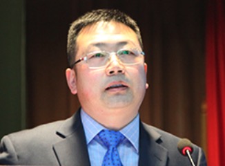 Weihu Ma (马维虎)合作论文数宁波市第六医院骨科15Yun-Lin Chen合作论文数13Chaoyue Ruan合作论文数11
 Rongming Xu (徐荣明)合作论文数Ningbo Mingzhou Hospital11Yang Wang (王扬)合作论文数9ShaoHua Sun (孙韶华)合作论文数6Liujun Zhao合作论文数6Dingli Xu合作论文数5Shuyi Zhou (周树一)合作论文数5Haojie Li (李豪杰)合作论文数5Fan Shu (舒帆)合作论文数5YueHong Li (李跃红)合作论文数5JianJun Sun (孙建军)合作论文数5Wei Chen (陈卫)合作论文数5Yongjie Gu (顾勇杰)合作论文数5Songou Zhang合作论文数4HuaJie Lin (林华杰)合作论文数4HongYong Zhao (赵红勇)合作论文数4
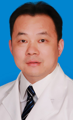 Yong Hu (胡勇)合作论文数Ningbo University Health Science Center;Ningbo No.6 Hospital4Hao Zhang (张浩)合作论文数4Rong Yang (杨荣)合作论文数4kairi shi (施凯日)合作论文数3Kai YANG (杨凯)合作论文数3
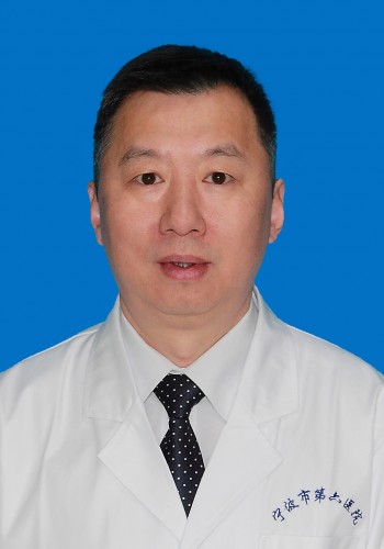 Hong Chen (陈宏)合作论文数Ningbo University School of Medicine3Li Guoqing合作论文数3Dong Wei-Xin合作论文数3Danguo Chen合作论文数2Xiao-Zhou Wang合作论文数2yujie peng (彭宇杰)合作论文数2HuaGuo Zhao (赵华国)合作论文数2YanZhao Zhu (朱彦昭)合作论文数2Jun Tong (童军)合作论文数2Liang Yu (于亮)合作论文数2Biao Liang (梁彪)合作论文数2
 Shareen Shafique合作论文数1Linguo Wan合作论文数1Mujie Liu合作论文数1Yue Zhou合作论文数1Yuheng Wang合作论文数1Wenjie Lu合作论文数1Yuping Quan合作论文数1Jinglong Zhao (赵京龙)合作论文数1Linlin Xie合作论文数1




