During exercise, the muscles' energy demand increases with increasing work load. In humans, the relationship between work load and oxygen uptake is linear until the maximal oxygen uptake (VO2max) is reached and higher exercise intensity requires additional anaerobic energy supply. VO2max is thought to reflect the maximum oxygen transport capacity of the cardiovascular system. We show that at room temperature normal mice could triple running speed at 25% inclination after reaching VO2max in spite of very modest increase of anaerobic muscle metabolism. In mice with cardiac dysfunction due to cardiac disruption of the Serca2 gene (S2KO), VO2max was reduced from week4 to week6 after gene disruption in parallel with progression of cardiac dysfunction. However, S2KO mice maintained maximal running speed at the same the level as the controls. Thus, paradoxically, running economy was better in S2KO than in controls. In S2KO, blood lactate was almost double of that of controls and respiratory exchange ratio was near 1, indicating greater reliance on anaerobic metabolism. However, heat production was lower in S2KO than in controls as reflected by tail temperature. Activity of BAT measured by fluoro-deoxyglucose using PET was reduced by 60±7% during running in controls and by 82±3% in running S2KO mice. In mice, the oxidative metabolism in non-muscle tissue, mainly in BAT, is reduced during exercise to provide more oxygen to the working muscles. This redistribution of oxygen delivery leaves the total VO2 unchanged over a wide range of exercise intensities. When cardiac output and VO2max are abnormally low, exercise intensity can be maintained since muscles can utilize the oxygen normally used by non-muscle tissue such as BAT. We conclude that oxygen consumption of skeletal muscle and BAT is regulated in a reciprocal way.
Human kidney predominant protein, NCU-G1, is a highly conserved protein with an unknown biological function. Initially described as a nuclear protein, it was later shown to be a bona fide lysosomal integral membrane protein. To gain insight into the physiological function of NCU-G1, mice with no detectable expression of this gene were created using a gene-trap strategy, and Ncu-g1gt/gt mice were successfully characterized. Lysosomal disorders are mainly caused by lack of or malfunctioning of proteins in the endosomal-lysosomal pathway. The clinical symptoms vary, but often include liver dysfunction. Persistent liver damage activates fibrogenesis and, if unremedied, eventually leads to liver fibrosis/cirrhosis and death. We demonstrate that the disruption of Ncu-g1 results in spontaneous liver fibrosis in mice as the predominant phenotype. Evidence for an increased rate of hepatic cell death, oxidative stress and active fibrogenesis were detected in Ncu-g1gt/gt liver. In addition to collagen deposition, microscopic examination of liver sections revealed accumulation of autofluorescent lipofuscin and iron in Ncu-g1gt/gt Kupffer cells. Because only a few transgenic mouse models have been identified with chronic liver injury and spontaneous liver fibrosis development, we propose that the Ncu-g1gt/gt mouse could be a valuable new tool in the development of novel treatments for the attenuation of fibrosis due to chronic liver damage.
Proper contraction and relaxation of the heart depends on highly orchestrated and rapid release and reuptake of Ca2+ from the sarcoplasmic reticulum (SR). Normal SR Ca2+ reuptake in the rodent heart is accomplished predominately by the cardiac Ca-ATPase, SERCA2a, which in the failing heart exhibits diminished activity and expression. We have utilized a mouse model of inducible cardiac Ca2+ dysregulation, the Serca2fl/fl mouse, to investigate the relationship between diminished SR Ca2+ flux and heart dysfunction. Conditional deletion of Serca2 initiates the progressive loss of SERCA2 protein, allowing us to examine in fine detail the relationship between titrated loss of SR Ca2+ flux and heart dysfunction. By four weeks post-knockout, SERCA2 protein levels are below 5% of baseline and isolated 4-week KO cardiomyocytes have greatly diminished contractility and Ca2+ handling. The in vivo phenotype at this time, however, is unexpectedly mild, and although all KO mice succumb to congestive heart failure, they do so from 7-10 weeks after knockout. This disconnect between the mild in vivo phenotype and severe ex vivo phenotype is intriguing and prompts the hypothesis that a systemic signaling pathway, such as adrenergic signaling, is responsible for sustaining function in KO animals. We found that in isolated Serca2 KO hearts, ß-adrenergic stimulation elicits a robust inotropic and lusitropic response despite the near-complete lack of SERCA2 protein. This finding will be discussed as it is at odds with prevailing views of Ca2+ regulation of heart function.
The cardiac SERCA2 Ca2+ pump is critical for maintaining normal Ca2+ handling in the heart. Reduced SERCA2a content and blunted Ca2+ reuptake are frequently observed in failing hearts and evidence implicates poor cardiac Ca2+ handling in the progression of heart failure. To gain insight into mechanism we investigated a novel genetic mouse model of inducible severe and progressive SERCA2 deficiency (inducible Serca2 knockout, SERCA2 KO). These mice eventually die from overt heart failure 7-10 weeks after knockout but as yet there have been no reports on intrinsic mechanical performance at the isolated whole heart organ level. Thus we studied whole-organ ex vivo function of hearts isolated from SERCA2 KO mice at one and four weeks post-knockout in adult animals. We found that isolated KO heart function was only modestly impaired one week post-knockout, when SERCA2a protein was 32% of normal. At four weeks post-knockout, function was severely impaired with near non-detectable levels of SERCA2. During perfusion with 10 mM caffeine, LV developed pressures were similar between 4-week KO and control hearts, and end-diastolic pressures were lower in KO. When hearts were subjected to ischemia-reperfusion injury, recovery was not different between control and KO hearts at either one or four weeks post-knockout. Our findings indicate that ex vivo function of isolated SERCA2 KO hearts is severely impaired long before symptoms appear in vivo, suggesting that physiologically relevant heart function in vivo can be sustained for weeks in the absence of robust SR Ca2+ flux.
In human heart failure (HF), reduced cardiac function has, at least partly, been ascribed to altered calcium homeostasis in cardiomyocytes. The effects of the calcium sensitizer levosimendan on diastolic dysfunction caused by reduced removal of calcium from cytosol in early diastole are not well known. In this study, we investigated the effect of long-term levosimendan treatment in a murine model of HF where the sarco(endo)plasmatic reticulum ATPase (Serca) gene is specifically disrupted in the cardiomyocytes, leading to reduced removal of cytosolic calcium. After induction of Serca2 gene disruption, these mice develop marked diastolic dysfunction as well as impaired contractility. SERCA2 knockout (SERCA2KO) mice were treated with levosimendan or vehicle from the time of KO induction. At the 7-wk end point, cardiac function was assessed by echocardiography and pressure measurements. Vehicle-treated SERCA2KO mice showed significantly diminished left-ventricular (LV) contractility, as shown by decreased ejection fraction, stroke volume, and cardiac output. LV pressure measurements revealed a marked increase in the time constant (τ) of isovolumetric pressure decay, showing impaired relaxation. Levosimendan treatment significantly improved all three systolic parameters. Moreover, a significant reduction in τ toward normalization indicated improved relaxation. Gene-expression analysis, however, revealed an increase in genes related to production of the ECM in animals treated with levosimendan. In conclusion, long-term levosimendan treatment improves both contractility and relaxation in a heart-failure model with marked diastolic dysfunction due to reduced calcium transients. However, altered gene expression related to fibrosis was observed.
Highly orchestrated contraction and relaxation of the myocardium depends on rapid release and reuptake of Ca2+ from the sarcoplasmic reticulum (SR). SERCA2a, a cardiac SR Ca2+-ATPase, is a major contributor to re-sequestration of Ca2+ and shows reduced expression with age and in heart failure. Conditional deletion of the Serca2 gene from the hearts of fl/fl mice results in progressive diastolic dysfunction, heart failure, and death over a period of two months. To gain insight into the interplay between Ca2+-mishandling and the evolution of cardiac pump failure we studied mice at 1 and 4 weeks following cardiac specific cre-mediated deletion of the Serca2 gene from fl/fl mice. At each time point, isolated hearts were subjected to paced stimulation across a broad frequency range, followed by ischemia/reperfusion injury. At both 1 and 4 weeks, SERCA2a loss resulted in profound contractile deficits and diastolic impairment relative to normal hearts. However, there was no difference in the relative performance of normal and KO hearts following ischemia/reperfusion injury, suggesting that depleting SR Ca2+ is not protective from ischemia. Curiously, 1 week after Serca2 deletion, when 15-20% of baseline SERCA2a protein remains, diastolic performance in response to stepped increases in pacing frequency was impaired to a similar degree as in 4-week KO hearts containing <5% original protein levels. We are further investigating the consequences of SERCA2a loss shortly after gene deletion, at times less than one week after knockout, to construct a detailed dose-response relationship between titrated changes in SR Ca2+ handling and contractile function.
Cardiomyocyte contraction and relaxation are controlled by Ca2+ handling, which can be regulated to meet demand. Indeed, major reduction in sarcoplasmic reticulum (SR) function in mice with Serca2 knockout (KO) is compensated by enhanced plasmalemmal Ca2+ fluxes. Here we investigate whether altered Ca2+ fluxes are facilitated by reorganization of cardiomyocyte ultrastructure. Hearts were fixed for electron microscopy and enzymatically dissociated for confocal microscopy and electrophysiology. SR relative surface area and volume densities were reduced by 63% and 76%, indicating marked loss and collapse of the free SR in KO. Although overall cardiomyocyte dimensions were unaltered, total surface area was increased. This resulted from increased T-tubule density, as revealed by confocal images. Fourier analysis indicated a maintained organization of transverse T-tubules but an increased presence of longitudinal T-tubules. This demonstrates a remarkable plasticity of the tubular system in the adult myocardium. Immunocytochemical data showed that the newly grown longitudinal T-tubules contained Na+/Ca2+-exchanger proximal to ryanodine receptors in the SR but did not contain Ca2+-channels. Ca2+ measurements demonstrated a switch from SR-driven to Ca2+ influx-driven Ca2+ transients in KO. Still, SR Ca2+ release constituted 20% of the Ca2+ transient in KO. Mathematical modeling suggested that Ca2+ influx via Na+/Ca2+-exchange in longitudinal T-tubules triggers release from apposing ryanodine receptors in KO, partially compensating for reduced SERCA by allowing for local Ca2+ release near the myofilaments. T-tubule proliferation occurs without loss of the original ordered transverse orientation and thus constitutes the basis for compensation of the declining SR function without structural disarrangement.
Impaired Ca2+ handling by the sarcoplasmic reticulum (SR) and Ca2+-dependent arrhythmias are hallmark features of human heart failure. We investigated the control of L-type Ca2+ current (ICa, L) when SR function is reduced and the consequences for arrhythmogenesis. Experiments were performed on cardiomyocytes isolated from conditional SERCA2 KO mice (KO) which had developed heart failure 7 weeks following gene disruption. SERCA2flox/flox (FF) mice served as controls. SR Ca2+ content was reduced to 4% (P<0.05) of FF values in KO cardiomyocytes, and SR Ca2+ release did not occur on a beat-to-beat basis.
Heart failure is associated with reduced sarco(endo)plasmic reticulum (SR) calcium ATPase (SERCA) function. Mice with conditional cardiomyocyte specific knockout of the Serca2 gene (KO) revealed that major reductions in SERCA and SR function are compensated by enhanced Ca2+ cycling across the cell membrane. Thus, cardiac function was maintained at near-normal values, at least in the short term. We hypothesize that this shift from SR to sarcolemmal Ca2+ fluxes is reflected in alterations in SR and sarcolemmal morphology. Seven weeks following gene disruption, some hearts were fixed for electron microscopy by perfusion with 3.5% glutaraldehyde and others for enzymatic isolation of cells for confocal microscopy and patch clamping. Serca2 flox-flox (FF) mice served as controls. Electron micrographs revealed drastically altered SR architecture and density. SR volume was reduced from 1.74±0.16 % of total cell volume in controls to 0.84±0.13 % in KO. The SR area to cell volume ratio was reduced from 0.68±0.06 to 0.32±0.05 µm2/µm3, and 55% of the free SR appeared collapsed, with a virtually occluded lumen. Although overall cardiomyocyte dimensions were unaltered in KO compared to FF, total surface area was increased (cell capacitance = 182 pF vs 147 pF, P<0.05). This resulted from increased t-tubule density, as revealed by confocal images of Di-8-Anepps stained myocytes. Specifically, Fourier analysis indicated a maintained organization of t-tubules which transversely spanned the cell, but an increased presence of tubules in the longitudinal direction. Immunocytochemical data showed that the newly grown longitudinal t-tubules contained Na+/Ca2+-exchanger which co-localized with ryanodine receptors in the SR, but did not contain CaV1.2 channels. Our observations indicate that the SR is greatly reduced and collapses following Serca2 KO. Further, proliferation of longitudinal t-tubules facilitates trans-sarcolemmal Ca2+ flux to compensate for declining SR function.
Sarcoplasmic reticulum Ca2+ ATPases (SERCAs) play a major role in muscle contractility by pumping Ca2+ from the cytosol into the sarcoplasmic reticulum(SR) Ca2+ store, allowing muscle relaxation and refilling of the SRwith releasable Ca2+. Decreased SERCA function has been shown to result in impaired muscle function and disease in human and animal models. In this study, we present a new mouse model with targeted disruption of the Serca2 gene in skeletal muscle (skKO) to investigate the functional consequences of reduced SERCA2 expression in skeletal muscle. SkKO mice were viable and basic muscle structure was intact. SERCA2 abundance was reduced in multiple muscles, and by as much as 95% in soleus muscle, having the highest content of slow-twitch fibres (40%). The Ca2+ uptake rate was significantly reduced in SR vesicles in total homogenates. We did not find any compensatory increase in SERCA1 or SERCA3 abundance, or altered expression of several other Ca2+-handling proteins. Ultrastructural analysis revealed generally well-preserved muscle morphology, but a reduced volume of the longitudinal SR. In contracting soleus muscle in vitro preparations, skKO muscles were able to fully relax, but with a significantly slowed relaxation time compared to controls. Surprisingly, the maximal force and contraction rate were preserved, suggesting that skKO slow-twitch fibres may be able to contribute to the total muscle force despite loss of SERCA2 protein. Thus it is possible that SERCA-independent mechanisms can contribute to muscle contractile function.
BACKGROUND: Alteration of Ca2+ homeostasis is known to be an important mechanism underlying the progression to heart failure. However, a conditional, cardiac specific SERCA2 knockout (SERCA2 KO) mo...
We describe a simulation study of Ca2+ dynamics in mice with cardiomyocyte-specific conditional excision of the sarco(endo)plasmic reticulum calcium ATPase (SERCA) gene, using an experimental data-driven biophysically-based modeling framework. Previously, we reported a moderately impaired heart function measured in mice at 4 weeks after SERCA2 gene deletion (knockout (KO)), along with a >95% reduction in the level of SERCA2 protein. We also reported enhanced Ca2+ flux through the L-type Ca2+ channels and the Na+/Ca2+ exchanger in ventricular myocytes isolated from these mice, compared to the control Serca2(flox/flox) mice (flox-flox (FF)). In the current study, a mathematical model-based analysis was applied to enable further quantitative investigation into changes in the Ca2+ handling mechanisms in these KO cardiomyocytes. Model parameterization based on a wide range of experimental measurements showed a 67% reduction in SERCA activity and an over threefold increase in the activity of the Na+/Ca2+ exchanger. The FF and KO models were then validated against experimentally measured [Ca2+](i) transients and experimentally estimated sarco(endo)plasmic reticulum (SR) function. Simulation results were in quantitative agreement with experimental measurements, confirming that sustained [Ca2+](i) transients could be maintained in the KO cardiomyocytes despite severely impaired SERCA function. In silico analysis shows that diastolic [Ca2+](i) rises sharply with progressive reductions in SERCA activity at physiologically relevant pacing frequencies. Furthermore, an analysis of the roles of the compensatory mechanisms revealed that the major combined effect of the compensatory mechanisms is to lower diastolic [Ca2+](i). Finally, by using a comprehensive sensitivity analysis of the role of all cellular calcium handling mechanisms, we show that the combination of upregulation of the Na+/Ca2+ exchanger and increased L-type Ca2+ current is the most effective means to maintain diastolic and systolic calcium levels after loss of SERCA function.
Several lines of evidence indicate that the sarco(endo)plasmic reticulum ATPase type 2 (SERCA2) is essential for maintaining myocardial calcium handling and cardiac pump function. Hence, a reduction in SERCA2 abundance is expected to reduce work performance and maximal oxygen uptake (VO2max) and to limit the response to exercise training. To test this hypothesis, we compared VO2max and exercise capacity in mice with cardiac disruption of Serca2 (SERCA2 KO) with control mice (SERCA2 FF). We also determined whether the effects on VO2max and exercise capacity could be modified by high-intensity aerobic exercise training. Treadmill running at 85-90% of VO2max started 2 wk after Serca2 gene disruption and continued for 4 wk. VO2max and maximal running speed were measured weekly in a metabolic chamber. Cardiac function was assessed by echocardiography during light anesthesia. In sedentary SERCA2 KO mice, the aerobic capacity was reduced by 50% and running speed by 28%, whereas trained SERCA2 KO mice were able to maintain maximal running speed despite a 36% decrease in VO2max. In SERCA2 FF mice, both VO2max and maximal running speed increased by training, while no changes occurred in the sedentary group. Left ventricle dimensions remained unchanged by training in both genotypes. In contrast, training induced right ventricle hypertrophy in SERCA2 KO mice. In conclusion, the SERCA2 protein is essential for sustaining cardiac pump function and exercise capacity. Nevertheless, SERCA2 KO mice were able to maintain maximal running speed in response to exercise training despite a large decrease in VO2max.
Background: Quantitative real-time RT-PCR (RT-qPCR) is a highly sensitive method for mRNA quantification, but requires invariant expression of the chosen reference gene(s). In pathological myocardium, there is limited information on suitable reference genes other than the commonly used Gapdh mRNA and 18S ribosomal RNA. Our aim was to evaluate and identify suitable reference genes in human failing myocardium, in rat and mouse post-myocardial infarction (post-MI) heart failure and across developmental stages in fetal and neonatal rat myocardium.Results: The abundance of Arbp, Rpl32, Rpl4, Tbp, Polr2a, Hprt1, Pgk1, Ppia and Gapdh mRNA and 18S ribosomal RNA in myocardial samples was quantified by RT-qPCR. The expression variability of these transcripts was evaluated by the geNorm and Normfinder algorithms and by a variance component analysis method. Biological variability was a greater contributor to sample variability than either repeated reverse transcription or PCR reactions.Conclusions: The most stable reference genes were Rpl32, Gapdh and Polr2a in mouse post-infarction heart failure, Polr2a, Rpl32 and Tbp in rat post-infarction heart failure and Rpl32 and Pgk1 in human heart failure ( ischemic disease and cardiomyopathy). The overall most stable reference genes across all three species was Rpl32 and Polr2a. In rat myocardium, all reference genes tested showed substantial variation with developmental stage, with Rpl4 as was most stable among the tested genes.
In the heart, function of the sarco(endo)plasmic Ca(2+)-ATPase (SERCA2) is closely linked to contractility, cardiac function, and aerobic fitness. SERCA2 function can be increased by high-intensity interval training, whereas reduced SERCA2 abundance is associated with impaired cardiac function. The working hypothesis was, therefore, that exercise training before cardiomyocyte-specific disruption of the Serca2 gene would delay the onset of cardiac dysfunction in mice. Before Serca2 gene disruption by tamoxifen, untreated SERCA2 knockout mice (Serca2(flox/flox) Tg-αMHC-MerCreMer; S2KO), and SERCA2 FF control mice (Serca2(flox/flox), S2FF) were exercise trained by high-intensity interval treadmill running for 6 wk. Both genotypes responded to training, with comparable increases in maximal oxygen uptake (Vo(2max); 17%), left ventricle weight (15%), and maximal running speed (40%). After exercise training, cardiac-specific Serca2 gene disruption was induced in both exercise trained and sedentary S2KO mice. In trained S2KO, cardiac function decreased less rapidly than in sedentary S2KO. Vo(2max) remained higher in trained S2KO the first 15 days after gene disruption. Six weeks after Serca2 disruption, cardiac output was higher in trained compared with sedentary S2KO mice. An exercise-training program attenuates the decline in cardiac performance induced by acute cardiac Serca2 gene disruption, indicating that mechanisms other than SERCA2 contribute to the favorable effect of exercise training.
AIMSTo describe the overall role of reduced sarcoplasmic reticulum Ca(2+) ATPase (SERCA2) for Ca(2+) wave development.METHODS AND RESULTSSERCA2 knockout [Serca2(flox/flox) Tg(alphaMHC-MerCreMer); KO] mice allowing inducible cardiomyocyte-specific disruption of the Serca2 gene in adult mice were compared with Serca(flox/flox) (FF) control mice. Six days after Serca2 gene disruption, SERCA2 protein abundance was reduced by 53% in KO compared with FF, whereas SERCA2 activity in field-stimulated, Fluo-5F AM-loaded cells was reduced by 42%. Baseline Ca(2+) content of the sarcoplasmic reticulum (SR) and Ca(2+) transient amplitude and rate constant of decay measured in whole-cell voltage-clamped cells were decreased in KO to 75, 81, and 69% of FF values. Ca(2+) waves developed in only 31% of KO cardiomyocytes compared with 57% of FF when external Ca(2+) was raised (10 mM), although SR Ca(2+) content needed for waves to develop was 79% of FF values. In addition, waves propagated at a 15% lower velocity in KO cells. Ventricular extrasystoles (VES) occurred with lower frequency in SERCA2 KO mice (KO: 3 +/- 1 VES/h vs. FF: 8 +/- 1 VES/h) (P < 0.05 for all results).CONCLUSIONReduced SERCA2 abundance resulted in decreased amplitude and decay rate of Ca(2+) transients, reduced SR Ca(2+) content, and decreased propensity for Ca(2+) wave development.
In ischemic congestive heart failure (CHF), anemia is associated with poor prognosis. Whether anemia develops in nonischemic CHF is uncertain. The hematopoietic inhibitors TNF-alpha and nitric oxide (NO) are activated in ischemic CHF. We examined whether mice with ischemic or nonischemic CHF develop anemia and whether TNF-alpha and NO are involved. We studied mice (n = 7-9 per group) with CHF either due to myocardial infarction (MI) or to overexpression of the Ca(2+)-binding protein calsequestrin (CSQ) or to induced cardiac disruption of the sarcoplasmic reticulum Ca(2+)-ATPase 2 gene (SERCA2 KO). Hematopoiesis was analyzed by colony formation of CD34(+) bone marrow cells. Hemoglobin concentration was 14.0 +/- 0.4 g/dl (mean +/- SD) in controls, while it was decreased to 10.1 +/- 0.4, 9.7 +/- 0.4, and 9.6 +/- 0.3 g/dl in MI, CSQ, and SERCA2 KO, respectively (P < 0.05). Colony numbers per 100,000 CD34(+) cells in the three CHF groups were reduced to 33 +/- 3 (MI), 34 +/- 3 (CSQ), and 39 +/- 3 (SERCA2 KO) compared with 68 +/- 4 in controls (P < 0.05). Plasma TNF-alpha nearly doubled in MI, and addition of anti-TNF-alpha antibody normalized colony formation. Inhibition of colony formation was completely abolished with blockade of endothelial NO synthase in CSQ and SERCA2 KO, but not in MI. In conclusion, the mechanism of anemia in CHF depends on the etiology of cardiac disease; whereas TNF-alpha impairs hematopoiesis in CHF following MI, NO inhibits blood cell formation in nonischemic murine CHF.
 Geir Christensen合作论文数University of Oslo45Ole M. Sejersted合作论文数30Ivar Sjaastad合作论文数24
 William (Bill) Louch合作论文数Institute for Experimental Medical Research, Division of Cardiovascular and Pulmonary Diseases, Oslo University Hospital17Karina Hougen合作论文数11Halvor K. Mørk合作论文数9
 Jan Magnus Aronsen合作论文数Institute of Basic Medical Sciences, Faculty of Medicine, University of Oslo9Ulla H. Enger合作论文数8Heidi Kiil Blomhoff合作论文数8
 Mathis Korseberg Stokke合作论文数7
 Kjetil Taskén合作论文数Univ Oslo, Biotechnol Ctr Oslo, N-0317 Oslo, Norway6Alexandra Vanessa Finsen合作论文数6Odd S. Gabrielsen合作论文数6Geir Florholmen合作论文数6Øyvind Ellingsen合作论文数5
 Jeffery D. Molkentin合作论文数Heart Institute, Cincinnati Children's Hospital;Department of Pediatrics, University of Cincinnati;Division of Molecular Cardiovascular Biology, Cincinnati Children's Hospital Medical Center;Howard Hughes Medical Institute5
 Madelene Ericsson合作论文数5
 Kenneth Chien (钱肯)合作论文数Department of Cell and Molecular Biology, Karolinska Institutet5
 Fredrik Swift合作论文数Institute for Experimental Medical Research;University of Oslo;Ullevaal Hospital;Institute for Experimental Medical Research, University of Oslo5
 Bjørn Steen Skålhegg合作论文数Institute of Basic Medical Sciences, Faculty of Medicine, University of Oslo;Department of Nutrition, University of Oslo5Lisbeth H Winer合作论文数4Erlend B. Smeland合作论文数4
 Nygård Ståle合作论文数Bioinformat Core Facil, Oslo Univ Hosp4
 Ju Chen合作论文数Ju Chen Lab, Department of Medicine, School of Medicine, University of California, San Diego4Sjåland Cecilie合作论文数4Heinis Frazer I合作论文数4Joseph M. Metzger合作论文数4Vidar Hansson合作论文数4
 Torp Sverre H合作论文数3Tore Jahnsen合作论文数3Cathrine Husberg合作论文数3
 Arild C Rustan合作论文数3Ke Draves合作论文数2H B Berntzen合作论文数2A Odegaard合作论文数2Ja Birkeland合作论文数2Bk Erikstein合作论文数2Ul Henriksen合作论文数2I Dale合作论文数2Cl Law合作论文数2
 Giulio Agnetti合作论文数Department of Cell Biology, School of Medicine, Johns Hopkins University2
 Andrew Trafford合作论文数Faculty of Biology, Medicine and Health, The University of Manchester2
 Brage H Amundsen合作论文数2Christoffersen J合作论文数2
 Theis Tønnessen合作论文数Dept Cardiothorac Surg, Oslo Univ Hosp Ulleval2Brendeford E M合作论文数2Yibin Wang (王义斌)合作论文数David Geffen School of Medicine, University of California, Los Angeles2
 Jennifer Van Eyk合作论文数Cedars-Sinai Precision Biomarker Laboratories;Cedars-Sinai Advanced Clinical Biosystems Research Institute2
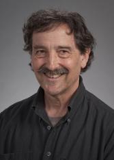 Edward A. Clark合作论文数Department of Immunology, University of Washington;Abacus Bioscience Inc2




