Objective To investigate the effective mechanism of Jiawei Maxing Shigan Tang(JMST, Modified Decoction of Ephedra,Apricot Kernel,Gypsum and Licorice)in treatment of acute exacerbated chronic obstructive pulmonary disease(AECOPD).Methods Wistar rats(n =40)were randomly divided into blank control group,model group,JMST group and clarithromycin group.The blank control group was given normal feeding, and model group, JMST group and clarithromycin group were given airway instillation of lipopolysaccharide(LPS)and smudging for establishing AECOPD model.The model group was given normal saline,JMST group,JMST(8.7 g/kg)and clarithromycin group,clarithromycin suspension(50 mg/kg)continuously for 30 d.The lung function was measured on the 31std.The lung tissue was collected to making pathological sections for observing goblet cells after AB -PAS staining.The expressions of mucoprotein 5AC(MUC5AC)and neutrophil elastase(NE)were detected by using immunohistochemistry technique.Results Compared with blank control group, the ratio of 0.3-second forced expiratory volume to forced vital capacity(FEV0.3/FVC)decreased significantly, and number of goblet cells and expressions of MUC 5AC and NE increased significantly in model group,JMST group and clarithromycin group.Compared with model group, FEV0.3/FVC increased significantly,and number of goblet cells and expressions of MUC 5AC and NE decreased significantly in JMST group.The comparison in FEV0.3/FVC,number of goblet cells and expressions of MUC 5AC and NE had no statistical difference between JMST group and clarithromycin group.Conclusion Jiawei Maxing Shigan Tang can significantly promote FEV0.3/FVC, inhibit proliferation of goblet cells, down-regulate expressions of MUC5AC and NE and relieve airway mucus hypersecretion in AECOPD rats, which may be its one of therapeutic mechanisms in treatment of AECOPD.
目的 观察电针对高血脂合并脑缺血大鼠前脑侧脑室室管膜下区星形胶质细胞活化的影响.方法 采用SD成年雄性大鼠49只,随机分为正常组,模型组、假手术组、电针1组和电针2组.除正常组,其余各组大鼠高脂饲料喂养42 d,电针1组电针丰隆,每天1次,治疗7 d.第50天,模型组和电针组大鼠采用50%FeCl3诱导建立高血脂合并脑血栓模型,术后电针组电针丰隆和百会,每天1次,持续14 d.用神经功能评分方法评定行为学变化,用生物化学法检测血清中总胆固醇(TC)、甘油三酯(TG)、高密度脂蛋白(HDL)和低密度脂蛋白(LDL)水平,采用HE染色观察形态学改变,免疫组化方法检测损伤侧前脑侧脑室室管膜下区胶质纤维酸性蛋白(GFAP)的表达.结果 模型组大鼠高血脂合并脑缺血术后TC、TG、LDL升高,HDL降低,差异具有统计学意义(P<0.05),电针组TC、LDL水平降低(P<0.05).模型组大鼠神经功能评分增高,HE染色可见皱缩神经元,整体组织形态结构显脑缺血症,侧脑室室管膜下区GFAP面密度明显升高(P<0.01),电针治疗可改善神经功能评分,缺血侧细胞核逐渐恢复正常,治疗后面密度持续增加至术后14 d,术后7 d显著,电针1组高于电针2组(P<0.05).结论 高血脂阶段电针丰隆,脑缺血后电针丰隆、百会可通过调节血脂代谢,促进星形胶质细胞活化以及受损神经系统修复.
Objective:To explore the effects of emodin on lung inflammation and inflammatory cytokine levels in BALF of mice with OVA-induced asthma.Methods:The method of OVA sensitization and challenge was used to establish bronchial asthma mice models.20 female C57BL/6 mice were randomly divided into normal group,model group,emodin group and positive controlled group.Each medication group was given relevant medicine for gavage.The overall situations as well as morphological changes from all groups were observed.The levels of cytokines in BALF of mice were detected.Results:Mice from the model group showed shortness of breath,wheezing,sneezing,scratching ear and nose,fecal incontinence etc.Compared with the controlled group,their pulmonary histological results showed a large number of inflammatory cells infiltrated around the bronchi and vessels,uncompletedalveolar structure associated with bronchial epithelial damage and even fibrosis change.What' s more,their levels of IL-4,IL-5,IL-17A in BALF significantly increased (P < 0.01),compared with the controlled group.After treatment with emodin,the above symptoms,signs and pathologic changes from asthmatic mice were alleviated.Their inflammatory levels were also reduced.Conclusion:Emodin might alleviate pulmonary inflammation of bronchial asthma mice modelprobably through inhibiting the levels of IL-4,IL-5,IL-17A.
OBJECTIVE:To observe the effect of electroacupuncture (EA) treatment on the expression of E-prostanoid 2 (EP2) in the spinal cord using a rat model of neuropathic pain, and to examine the effect of different acupuncture prescriptions. METHODS:Rats were randomly divided into six groups: a normal group, model group, sham group, EA 1 group (distal-proximal acupoints), EA 2 group (proximal acupoints), and EA 3 group (distal acupoints). Rats in the EA groups were treated for 20 min, once daily for 7 days. The mechanical withdrawal threshold was evaluated at 1 day preoperatively and at 3, 5, and 7 days postoperatively. At 7 days postoperatively, hematoxylin-eosin (HE) staining was used to investigate spinal morphological changes. Spinal EP2 messenger ribonucleic acid (mRNA) was detected using reverse-transcription polymerase chain reaction. RESULTS:After the EA treatment, the mechanical withdrawal threshold was up-regulated compared with the model group (P < 0.05). The expression of EP2 mRNA was up-regulated in the spinal cord on day 7 after surgery (P < 0.05). EA stimulation tended to down-regulate EP2 mRNA expression compared with the model group (P > 0.05). The EA 1 and EA 2 groups showed a larger treatment effect than the EA 3 group. The HE results showed that EA can improve spinal histomorphology induced by neuropathic pain. CONCLUSION:EA had a strong analgesic and anti-inflammatory effect. The therapeutic effect of EA on the down-regulation of spinal EP2 mRNA expression may be greater when either local acupoints or a distal-proximal combination of acupoints are used than when distal points are used.
OBJECTIVE:To investigate the effect of electroacupuncture (EA) treatment on the expression of cyclooxygenase (COX) 2 and microglia in spinal cord by using rat model of neuropathic pain, and to probe into the relationship between COX 2 and microglia.METHODS:The rats were randomly divided into 6 groups, including normal control group, model group, sham group, EA 1 group (distant acupoints + local acupoints), EA 2 group (local acupoints), and EA 3 group (distant acupoints). Thermal withdrawal latencies were evaluated at 1 day preoperatively and 3, 5 and 7 days postoperatively. At 7 days postoperatively, the spinal COX 2 mRNA was detected by reverse-transcription polymerase chain reaction. Double immunofluorescent staining technology was applied to screen and verify the relationship between altered COX 2 and microglia.RESULTS:Compared with the model group, thermal withdrawal latencies increased after EA treatment (P<0.01). The expressions of COX 2 mRNA were up-regulated in spinal cord of rat on day 7 after surgery (P<0.05). Compared with the model group, EA stimulation (EA 1 and EA 2 groups) reversed the up-regulation of COX 2 mRNA expression (P<0.05). EA 1 and EA 2 groups might have better treatment effect compared with the EA 3 group. Fluorescent images displayed COX 2 and microglia expressed at common areas.CONCLUSIONS:EA was effective in analgesic and anti-inflammatory. EA has decreased the expression of spinal COX 2 mRNA in the trend of the therapeutic effect of "distant acupoints + local acupoints", and "local acupoints" intervention may be superior to that of "distant acupoints" intervention. Microglia may be related to the formation of COX 2.
Objective To investigate the effects of Sanjie Tongluo Decoction on the expression of matrix metalloproteinase 2 (MMP-2),tssue inhibitor of metalloproteinase-2 (TIMP-2) of nephridial tissue in rats with ischemic nephropathy of advanced stage.Methods Male SD rats were randomly divided into sham operation group,model group (both groups were given normal saline 3 mL/d),drug group [Sanjie Tongluo Decoction 10.85 g/(kg·d)] by random number table method.The unilateral renal artery insufficiency ligation was used to establish animal model.In the 28th day of experiment,the levels of 24 hours urinary protein and creatinine of rats were detected.The rats were put to death and nephridial tissue was collected to take light microscope examination,the changes of renal morphology in all groups were observed.Immunohistochemistry was used to detect the expression of MMP-2 and TIMP-2 of nephridial tissue.Results Under the light microscope,the glomerulus and tubulointerstitium in the model group and drug group had damage.Compared with sham operation group,the scores of glomerulosclerotic index and tubulointerstitial lesion in the model group and drug group were all increased (P < 0.01),which of drug group were lower than those of model group (P < 0.01).Compared with model group,the expression of MMP-2 in drug group was increased (P < 0.01),24 hours urinary protein,creatinine and the expression of TIMP-2 were decreased (P < 0.01),which of both groups were higher than those of sham operation group (P < 0.01).Conclusion Sanjie Tongluo Decoction can prevent the lesions of ischemic nephropathy of advanced stage by up-regulating the expression of MMP2 and down-regulating the expression of TIMP-2.
目的 研究慈姑水提部位对异烟肼和利福平合用(INH/RFP)所致小鼠肝损伤的保护作用及其可能的保护机制.方法 利用异烟肼和利福平(各0.1 g/kg)复制小鼠肝损伤病理模型,同时分别灌胃给予低、中、高剂量慈姑水提部位(0.2、0.4、0.8 g/kg)以及阳性对照药水飞蓟素(0.1 g/kg),连续灌胃30 d后,测定小鼠肝指数,血清中丙氨酸氨基转移酶(ALT)、天门冬氨酸氨基转移酶(AST)、乳酸脱氢酶(LDH)活性,肝组织中丙二醛(MDA)、谷胱甘肽(GSH)含量,并观察肝组织病理学切片.结果 与模型组比较,高剂量慈姑水提部位组的肝指数、血清ALT、AST、LDH活性均显著降低,肝匀浆MDA含量显著下降(P<0.01),GSH含量显著升高(P<0.01),组织病理学显示:与模型组比较,随着慈姑水提部位剂量的升高,肝细胞病变明显减轻.结论 慈姑水提部位对小鼠肝损伤具有一定的保护作用,可能与其抗脂质过氧化有关.
Post-traumatic stress disorder (PTSD) is manifested as a persistent mental and emotional condition after potentially life-threatening events. Different animal models of PTSD have been developed for neuro-pathophysiology and pharmacological evaluations. A single prolonged stress (SPS) induced animal model has demonstrated to result in specific neuro-endocrinological dysregulation, and behavior abnormalities observed in PTSD. However, animal studies of PTSD have mostly been performed at one time point after SPS exposure. To better understand the development of PTSD-like behaviors in the SPS animal model, and to identify an optimal period of study, we examined depressive behavior, anxiety-like behavior, physical activity and body weight in SPS model rats for two weeks. Our results confirmed the SPS-induced PTSD-like behavior and physical activity observed in previous studies, and indicated that the most pronounced symptomatic behavior changes were observed on day 1 and 7 after SPS exposure, which may involve stress-induced acute hormone changes and unclear secondary neurobiological changes, respectively. These results provide a solid basis for further investigation into the neuro-pathophysiology of or neuropharmacology for PTSD using the SPS rat model. However, for chronic (pharmacological) studies longer than 7 days, a prolonged PTSD animal model should be developed, perhaps using enhanced stimulation.
Objective To explore the effects ofDachengqiDecoction on morphological changes, lung and spleen indexes, and total IgE levels of mice with allergic asthma; To discuss it relevant mechanism of action.MethodsThe method of OVA sensitization and provocation was used to establish allergic asthma mice models. 20 C57BL/6 mice were randomly divided into control group, model group,Dachengqi Decoction group and dexamethasone positive group. Each medication group was given relevant medicine for gavage. Lung and spleen indexes in each group were detected. The total IgE levels in serum of mice were detected by ELISA, and HE staining was used to conduct pathological observation.Results Compared with control group, the lung index and level of IgE in model group increased significantly and pulmonary histological results showed abundant inflammatory cells infiltrated in the bronchus; compared with the model group, the lung index as well as the levels of IgE decreased significantly in the Dachengqi Decoction and dexamethasone positive group (P<0.05), and the lung tissue organization form was improved significantly.ConclusionDachengqi Decoction has inhibiting effects on pulmonary inflammation of mice with asthma.
Objective To explore Qingjinhuatan decoction’s mechanism on airway mucus hypersecretion of chro-nic obstructive pulmonary disease rat model. Methods Clean level Wistar rats(n = 40)are randomly divided into blank control group(A),model group(B),Qingjinhuatan decoction group(C)and Clarithromycin group(D). Using dripping LPS in bronchus and smoking to intervene on B,C and D group rat in order to build COPD airway mucus hy-persecretion model. B,C and D group rats are separately raised by normal saline,Qingjinhuatan decoction and Clar-ithromycin for 30 days,at the same time blank control group rat are raised normally. All rats are killed on 31th day, and using RT-PCR method to observe HEmRHA,MUC 5ACmRHA expression on lung tissue,Alcian blue periodic acid Schiff staining method to observe hyperplasia of goblet cells on airway epithelium,Immunohistochemical method to test HE and MUC 5AC expression on pulmonary tissue and airway epithelium. Results Model group rats’HEmRHA,MUC 5ACmRHA expression,goblet cells’hyperplasia on airway epithelium,HE and MUC 5AC expression on pulmonary tis-sue and airway epithelium are significant increase than blank control group;Qingjinhuatan decoction and Clarithromy-cin group rats’MUC 5ACmRHA,goblet cells’hyperplasia on airway epithelium,MUC 5AC expression on pulmonary tissue and airway epithelium is significant less than model group;Qingjinhuatan decoction group rats’HEmRAH,MUC 5ACmRHA,HE expression on pulmonary tissue and airway epithelium is significant less than Clarithromycin group;Qingjinhuatan decoction group rats’goblet cells’hyperplasia on airway epithelium and MUC 5AC expression on pulmo-nary tissue and airway epithelium have on difference with Clarithromycin group. Conclusion Qingjinhuatan decoction can reduce airway mucus hepersecrection of chronic obstructive pulmonary disease by HE / MUC 5AC pathway.
Objective To explore the mechanism ofJiedu Qingfei Mixture for airway mucus hypersecretion of rat models with chronic obstructive pulmonary disease (COPD).Methods Airway instilling lipopolysaccharide combining fuming method was used to establish COPD models. Forty clean level Wistar strain rats were randomly divided into blank control group, model group,Jiedu Qingfei group, and clarithromycin group. Model group, Jiedu Qingfei group, and clarithromycin group were given normal saline,Jiedu Qingfei Mixture, and clarithromycin by gavage respectively, while the blank control group was raised normally for 30 d. All rats were killed on the 31st day for taking lung tissue (6 rats from each group were chosen randomly). Pathological changes of lung tissue and mucous glands hyperplasia were observed by HE staining method. NE and MUC5AC mRNA expression on lung tissue were detected by RT-PCR method. Protein expressions of NE and MUC5AC on pulmonary tissue and airway epithelium were detected by immunohistochemical method.Results Compared with blank control group, mucous glands hyperplasia on airway epithelium, mRNA expression of NE and MUC5AC in lung tissue, and protein expressions of NE and MUCA5C on airway epithelium in the model group significantly increased (P<0.05,P<0.01). Compared with model group, mucous glands hyperplasia on airway epithelium inJiedu Qingfei group significantly decreased (P<0.01), as same as clarithromycin group;Jiedu Qingfei group showed better effects on down-regulating NE and MUC5AC mRNA expression in lung tissue compared with clarithromycin group. MUC5AC protein expression on airway epithelium inJiedu Qingfei group significantly decreased (P<0.05), as same as clarithromycin group.Jiedu Qingfei group and clarithromycin group had no difference on NE protein expression in airway epithelium compared with model group.Conclusion Jiedu Qingfei group Mixture can reduce airway mucus hypersecretion of COPD by down-regulating MUC5AC expression through neutrophil elastase.
目的:观察滋阴基础上的清热化痰与益气活血序贯疗法对IPF小鼠肺组织形态学,血清中IL-6、TGF-β含量的影响.方法:SPF级ICR雄性小鼠105只,体重16~20g,随机分为正常对照组、模型组、西药治疗组以及4个中药治疗组,每组15只,除正常对照组外,其余各组采用气管内注射博莱霉素(5 mg/kg)复制小鼠肺纤维化模型.造模第2天起,正常对照组和模型组用生理盐水灌胃,西药组按0.15 mg/g给予甲基泼尼松.4个中药治疗组分别按照不同阶段不同药物不同时限给药,于21 d处死小鼠.HE染色及武兆发简化Mallory氏胶原染色,观察肺组织形态变化;双抗夹心酶联免疫吸附试验(ELISA)法检测血清中IL-6、TGF-β的含量.结果:HE染色及胶原染色显示,模型组小鼠支气管周围及肺泡间隔有大量炎症细胞浸润并伴有出血性渗出,纤维化程度明显.西药组较模型组虽好转但效果不及中药组明显.中药①②组肺泡炎明显好转,肺泡内未见明显的炎性细胞,肺泡隔、细小支气管周围的纤维化病灶较模型组有明显改善,肺泡结构已基本恢复.模型组小鼠血清中IL-6、TGF-β含量均高于正常组,与模型组比较,中药①②组小鼠血清中IL-6 、TGF-β含量降低显著.结论:从以上各项实验指标观察来看,中药①②组能明显控制与改善肺纤维化,降低炎症细胞因子IL-6、TGF-β的含量,提示清热化痰药适宜在肺纤维化早期使用,不宜贯穿使用于整个病程,益气活血药可在病程中后期使用.
[Abstact] Objective To explore the protective effects of the Baimai ( BM ) therapy on focal cerebral ischemia-reperfusion injury in rats and the influence of BM therapy on Jagged1 expression of hipp-ocampal dentate gyrus. Methods The focal cerebral ischemia-reperfusion injury models were induced by suture method, inserting thread 1. 5 h and reperfusion 7 d and 14 d respectively. The rats were randomly divided into sham-operation group, model group, Baimai therapy group ( BMT group) . The area of cerebral infarction was observed by TTC staining, the morphological changes of cortex cell was observed by HE staining, and immunohistochemistry method was used to detect hippocampal dentate gyrus Jagged1 positive cell number of each group rats. Results (1) the TTC staining result showed that normal brain tissue was bright red, infarction area of the brain was pale, and with the prolongation of reperfusion time, the infarct area was enlarged. The areas of cerebral infarction of BMT group were less than model group, and there was a significant difference compared with model group on 14 days (P<0. 05). (2) HE staining showed that degree of injury of cortical neurons in the cerebral ischemia group was lower than that in the model group. (3) BM therapy can increase hippocampal dentate gyrus Jagged1 expression of cerebral ischemia rats, Jagged1 positive cells was higher than model group and control group, the result had significant difference (P<0. 05), after having operation 7 days and 14 days. Conclusions BM therapy has certain protective effect on focal cerebral ischemia-reperfusion injury, and can promote Jagged1 expression in hipp-ocampal dentate gyrus after rats having focal cerebral ischemia-reperfusion injury, the increase of Jagged1 expression may be one of the molecular mechanisms of the proliferation and differentiation of endogenous neural stem cells after cerebral ischemia injury via the Notch pathway.
Objective To observe the effects of Qingjin Huatan Decoction on EGFR/MAPK signaling pathway of airway mucus hypersecretion rats with chronic obstructive pulmonary disease (COPD). Methods Intratracheal instillation of LPS combined with smudging method was used to establish COPD airway mucus hypersecretion rat models. Experimental rats were randomly divided into blank group, model group, Qingjin Huatan Decoction group and clarithromycin group. The blank group was normally fed, while the other three groups were given NS, Qingjin Huatan Decoction, and clarithromycin respectively for gavage, once a day for 30 days. All rats were killed on the 31st day, and pathological changes of lung tissue and mucous glands hyperplasia were observed by HE staining method. The gene expressions of EGFR and MUC5AC in lung tissue were detected by RT-PCR method. The protein expressions of P-EGFR, P-ERK, P-JNK, P-p38 and MUC5AC in pulmonary tissue and airway epithelium were detected by immunohistochemical method. Results Compared with the blank group, mucous glands hyperplasia on airway epithelium, protein expressions of P-EGFR, P-ERK, P-JNK, P-p38 and MUC5AC on airway epithelium significantly increased in the model group (P<0.01); gene expression of MUC5AC of lung tissue increased (P<0.05). Compared with the model group, mucous glands hyperplasia on airway epithelium, P-p38, P-ERK and MUC5AC protein expression on airway epithelium in Qingjin Huatan Decoction group significantly decreased (P<0.05, P<0.01); the protein expression of P-JNK increased significantly (P<0.01). EGFR and MUC5AC mRNA in lung tissue in Qingjin Huatan Decoction group decreased significantly (P<0.01). Conclusion Qingjin Huatan Decoction can reduce airway mucus hepersecrection of COPD by inhibiting ERK and p38 signal pathway on EGFR downstream.
Objective To study the different prescriptions of eletcro-acupuncture ( distal-proximal acu-points, proximal acupoints and distal acupoints) on pain-related behaviors and expression of microglia marker OX42 and cyclooxygenase 2 ( COX2 ) in spinal dorsal horn in rat model of transplantation of autol-ogous nucleus pulposus, so as to provide experimental basis for the mechanism of the different prescrip-tions. Methods 54 healthy male SD rats were randomly divided into six groups ( each n=9 )-normal group, model group, sham operation group, distal-proximal acupoint group, proximal acupoint group, distal acupoint group. After modeling, the rats in three acupuncture groups were treated for 20 minutes, once daily for consecutive 7 days. Those in distal-proximal acupoint group were needled at bilateral “L5 Jiajixue” ( Ex-B2 ) ,“Dachangshu” ( BL 25 ) ,“Weizhong” ( BL 40 ) and“Kunlun” ( BL 60 );those in proximal acupoint group were needled at bilateral“L5 Jiajixue” ( Ex-B2 ) and“Dachangshu” ( BL 25 ) ;the rats in distal acupoint group rats were needled at bilateral “Weizhong” ( BL 40 ) and “Kunlun” ( BL 60). Thermal withdrawal latencies were evaluated. The expressions of OX42 and COX2 in the left spinal dorsal horn were detected using immunohistochemical methods. Results Thermal withdrawal latencies and the expression of OX42 and COX2 increased in rat model of transplantation of autologous nucleus pul-posus. After treatment, thermal pain threshold was improved (P<0. 05), and the expression of OX42 and COX2 were inhibited ( P<0 . 05 ) . The inhibitory effects of distal-proximal acupoint group and proxi-mal acupoint group were superior to that in distal acupoint group. Conclusion Each acupuncture pre-scription reduced anti-inflammatory effects by inhibiting inflammatory signaling pathways in spinal dorsal horn, with the prescription of distal-proximal acupoints and proximal acupoints superior to distal acu-points.
目的:建立小鼠流感性肺炎模型,明确呼吸道流感病毒感染过程中是否存在特异性的肠道病理损伤,并探讨了其在“肺与大肠相表里”研究中的应用.方法:BALB/c小鼠48只随机分为正常组、病毒组.病毒组给予50 μL、50LD5o病毒液滴鼻建立流感病毒感染的小鼠肺炎模型,感染后第3、5、6天分别取材,HE染色检测肺脏、大肠、小肠、心脏、肝脏、肾脏组织病理结果,血生化检测血清谷草转氨酶(AST)、谷丙转氨酶(ALT)和尿素氮(BUN)水平.结果:流感病毒感染后,病毒组小鼠肺组织出现明显的炎性病变外,随时间延长肺组织充血水肿出血加重;小肠黏膜腺体排列紊乱,绒毛粗大、部分脱落,黏膜固有层炎性细胞浸润:大肠黏膜固有层、肌层大量炎性细胞浸润,腺体结构消失、脱落;而心脏、肝脏、肾脏无明显病理改变;病毒组小鼠血清AST、ALT、BUN较正常组相比,差异无统计学意义.结论:小鼠流感病毒性肺炎模型中存在小肠、大肠特异性免疫病理损伤,是研究中医“肺与大肠相表里”理论及相关临床治法的理想模型.
 Dongyu Ge (葛东宇)合作论文数School of Traditional Chinese Medicine, Beijing University of Chinese Medicine43
 Cuiling Feng (冯淬灵)合作论文数Peking University People's Hospital8
 XuDan Wang (王旭丹)合作论文数5
 Ligang Gu (顾立刚)合作论文数Beijing University of Chinese Medicine5
 Zeji Qiu (邱泽计)合作论文数School of Traditional Chinese Medicine, Beijing University of Chinese Medicine5WenZhong Xu (许文忠)合作论文数4Jun Wu (吴珺)合作论文数4Ping Feng (丰平)合作论文数4Jun Wang (王骏)合作论文数4Ying Chen (陈英)合作论文数4
 Angran Fan (范盎然)合作论文数School of Traditional Chinese Medicine, Beijing University of Chinese Medicine4
 Yu Hao (郝钰)合作论文数Dongfang Hospital Beijing University of Chinese Medicine4Qiaofeng Wan (万巧凤)合作论文数4
 Shuyan Wang (王淑艳)合作论文数School of Traditional Chinese Medicine, Beijing University of Chinese Medicine4
 Xiujun Ren (任秀君)合作论文数School of Acupuncture -Moxibustion and Tuina, Beijing University of Chinese Medicine4
 Ying Wu (吴莹)合作论文数School of Traditional Chinese Medicine.BUCM4Zinan XUAN (玄子男)合作论文数3Li-Li Ji合作论文数3Xiao-yu QI (齐晓宇)合作论文数3LiJun Li (李丽君)合作论文数3Qian Wu (吴谦)合作论文数3
 Lina Li (李丽娜)合作论文数School of Traditional Chinese Medicine, Beijing University of Chinese Medicine;Dongzhimen Hospital, Beijing University of Chinese Medicine;Third Affiliated Hospital, Beijing University of Chinese Medicine3
 Shujing Zhang (张淑静)合作论文数School of Traditional Chinese Medicine, Beijing University of Chinese Medicine3
 Wei Li (李伟)合作论文数Beijing University of Chinese Medicine Third Affiliated Hospital3
 Yinchu Si (司银楚)合作论文数School of Traditional Chinese Medicine, Beijing University of Chinese Medicine3
 Feng Li (李峰)合作论文数School of Traditional Chinese Medicine, Beijing University of Chinese Medicine3
 Ya Tu (图娅)合作论文数School of Acupuncture -Moxibustion and Tuina, Beijing University of Chinese Medicine3
 Shuyu Li (李姝玉)合作论文数School of Traditional Chinese Medicine, Beijing University of Chinese Medicine3Da-ling ZHONG (钟大玲)合作论文数2Gui-mei CHEN (陈桂梅)合作论文数2ZhuoYun Wu (吴卓耘)合作论文数2WanChun Wei (尉万春)合作论文数2MengWei Guo (郭孟玮)合作论文数2LiYun Zhao (赵丽云)合作论文数2Jiajia Wu (吴佳佳)合作论文数School of Rehabilitation Science, Shanghai University of Traditional Chinese Medicine2
 Xudong Tang (唐旭东)合作论文数Xiyuan Hospital of China Academy of Chinese Medical Sciences2
 Yin Lin (林殷)合作论文数School of Traditional Chinese Medicine, Beijing University of Chinese Medicine2
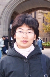 Qian Wang (王谦)合作论文数School of Traditional Chinese Medicine.BUCM2
 Yan Liao (廖艳)合作论文数School of Traditional Chinese Medicine, Beijing University of Chinese Medicine2Jing Liu (刘静)合作论文数2
 Meng Mao (毛萌)合作论文数School of Traditional Chinese Medicine, Beijing University of Chinese Medicine2
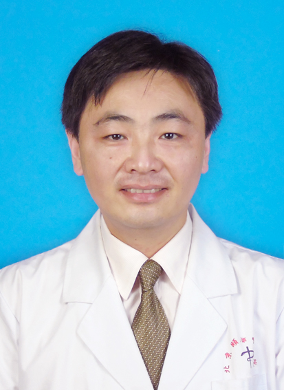 Meng Chen (陈萌)合作论文数School of Traditional Chinese Medicine, Beijing University of Chinese Medicine2
 Lanying Liu (刘兰英)合作论文数Third Affiliated Hospital, Beijing University of Chinese Medicine2
 Guiying Peng (彭桂英)合作论文数School of Life Sciences, Beijing University of Chinese Medicine2YIN Sheng-jun (殷胜骏)合作论文数2Jie Ma (马捷)合作论文数1Zi-rui LIU (刘茈蕊)合作论文数1Liu Zhaolan合作论文数1
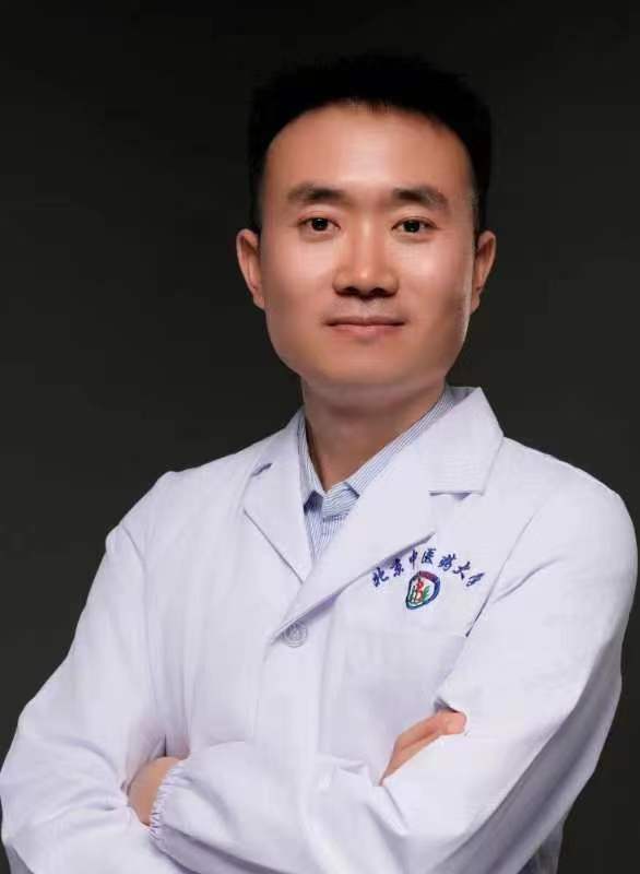 Huili Jiang (姜会梨)合作论文数北京中医药大学1




