IMPORTANCE:Despite the clinical use of three-dimensional transrectal ultrasound (3D-TRUS) in pfCD, no validated model exists to predict early biologic treatment failure. OBJECTIVE:Our objective was to develop and validate such a predictive model using 3D-TRUS-derived parameters. DESIGN:This study was a double-center retrospective study. SETTING:Patients with pfCD who underwent biologic therapy at two medical centers and completed both 3D-TRUS and magnetic resonance imaging (MRI) assessments prior to treatment initiation and at approximately 12 weeks post-treatment (range: 6-18 weeks) were included in the study. PARTICIPANTS:A total of 102 patients were included in the analysis, with 80 patients from Hospital A formed the training/internal validation cohort; 22 from Hospital B comprised the external validation cohort. EXPOSURE:Biologic therapy for pfCD. MAIN OUTCOME(S) AND MEASURE(S):The primary outcome was early treatment failure, defined as stable or aggravated disease (SD/AD) based on MRI criteria. Patients were divided into a failure group and a non-failure group. Predictive factors were identified using LASSO regression followed by multivariate logistic regression, and a nomogram was constructed based on the final model. Model performance was assessed through internal validation via the Bootstrap method (1000 resamples) and external validation. RESULTS:Multivariate analysis identified three independent predictors of early treatment failure (all P < .05): main fistula length ≥ 2 cm, inflammatory mass size > 2 cm, and internal orifice-anal margin distance (per 1-mm increase). The nomogram showed an AUC of 0.955 (95% CI: 0.915-0.995) in training cohort. Hosmer-Lemeshow confirmed good fit, and decision curve analysis (DCA) demonstrated clinical utility. Internal validation maintained an AUC of 0.955 (0.911-0.987). External validation yielded an AUC of 0.875 (0.700-1.000), with DCA supporting clinical applicability. CONCLUSIONS AND RELEVANCE:3D-TRUS features effectively predict early biologic failure in pfCD. The nomogram provides a tool for risk assessment to guide clinical decisions regarding treatment escalation or switch, supporting more individualized patient management.
Ultrasound-guided percutaneous lung needle biopsy(US-PLNB)is a pivotal diagnostic technique for subpleural pulmonary lesions,offering significant advantages such as real-time imaging,absence of ionizing radiation,and high operational flexibility.To standardize its clinical application and enhance both procedural safety and diagnostic accuracy,a multidisciplinary panel of experts was convened by eleven authoritative Chinese academic societies,including Chinese Society of Ultrasound in Medicine.Based on the latest domestic and international evidence-based medicine,this panel developed Chinese expert consensus on ultrasound-guided percutaneous lung needle biopsy(2025 edition).This consensus systematically elaborates on the indications and contraindications,preprocedural assessment and preparation,intraoperative techniques,complication prevention and management,and post-procedural care for US-PLNB.Furthermore,it puts forward 18 specific evidence-based recommendations.The primary aim of this consensus is to provide radiologists and clinicians with standardized operational guidance,promote the standardized application of US-PLNB,and ultimately improve patient care for individuals with pulmonary diseases.
OBJECTIVES:To predict histologic grade of soft tissue sarcoma (STS) with preoperative ultrasound images, aiding in the selection of personalized treatment plans and improving long-term prognosis. METHODS:In total, 238 patients with histologically proven STS were retrospectively enrolled from April 2016 to December 2023 and divided into the training and internal validation cohorts. Seventy patients were prospectively enrolled from 3 centers between January 2024 and December 2024 as the external validation cohort. Radiomics features were extracted from preoperative grayscale ultrasound images. The dynamic nomogram (DynNom) was developed by using multivariable logistic regression analysis. Predictive performance was evaluated with the receiving operating characteristic curve, calibration curve, Hosmer-Lemeshow test, decision curve analysis (DCA), and clinical impact curve (CIC). RESULTS:The DynNom based on clinical-US characteristics (metastasis status, echogenicity, fascia layer, and vascularity) and radiomics features yielded an optimal AUC of 0.915 (95% CI, 0.873-0.947), 0.87 (95% CI, 0.79-0.93), and 0.90 (95% CI, 0.80-0.96) for predicting the STS histologic grade in the training, internal, and external validation cohorts, respectively. The DynNom outperformed the conventional model and radiomics model (P < .05). Calibration curves and Hosmer-Lemeshow tests indicated its satisfactory calibration ability. DCA confirmed that the DynNom outperformed other models in overall net benefit, meanwhile CIC suggested that the DynNom had great clinical applicability in predicting histologic grade. CONCLUSIONS:The dynamic nomogram is a practical tool that could predict the histologic grade of STS, which might help clinicians to screen histologic high-grade STSs as neoadjuvant treatment candidates. ADVANCES IN KNOWLEDGE:The dynamic nomogram had the potential to accurately predict histologic grade in STS patients before surgery. High-risk patients defined by the dynamic nomogram were potential candidates for preoperative radiotherapy and neoadjuvant chemotherapy.
Vision-Language Models (VLMs) have significantly advanced medical visual question answering, yet their performance in ultrasound remains suboptimal. In clinical practice, sonographers explicitly focus on lesion regions to formulate reports, though diagnostic interpretations sometimes vary due to inherent subjectivity. However, existing VLMs are not explicitly structured to interactively zoom into lesions prior to diagnosis; moreover, they typically treat annotations as unbiased ground truths, failing to account for their inherent subjectivity and ambiguity. In this paper, we propose a framework specifically designed to consider the sonographer's cognitive workflow. We first introduce a structured Zoom-then-Diagnose paradigm, which replicates the interactive search process to enable lesion-focused reasoning. Furthermore, within the Group Relative Policy Optimization (GRPO) framework, we introduce an uncertainty-aware reward derived from stochastic group-wise rollouts to estimate prediction consistency as a proxy for model confidence. Together, these two components encourage the model to reinforce accurate predictions on clear cases while remaining cautious under ambiguity. Experiments across liver, breast, and thyroid datasets show that our framework improves lesion localization by 39.3%, demonstrating that our model has learned the ability to actively look closer and diagnose.
OBJECTIVES:To investigate the performance of an artificial intelligence (AI) diagnostic system for thyroid nodule sonography based on deep learning convolutional neural network (CNN). MATERIALS AND METHODS:We retrospectively included 485 thyroid nodules with definite pathology in two tertiary hospitals. The AI diagnostic system was constructed for automatic detection and diagnosis of nodules based on deep learning CNN equipped with image mode and video mode. One gray-scale ultrasound (US) image of each nodule from the two hospitals was selected for diagnosis in image mode (AI modelimg). A US video of each nodule from the second hospital was analyzed in video mode (AI modelvid). Performance of AI modelimg, AI modelvid, and three radiologists with 3-15 years of US experience was evaluated. Sonographic features probably influencing the accuracy of AI modelimg were screened out by binary logistic regression analysis. RESULTS:Although the experienced radiologist achieved highest sensitivity, accuracy and the area under the receiver operating characteristic curve (AUC) compared to AI modelimg and two junior radiologists, there was no significant difference between AUCs of AI modelimg and experienced radiologist (0.770 [0.718-0.816] vs. 0.799 [0.750-0.843] in first hospital dataset, p = 0.253; 0.731 [0.660-0.794] vs. 0.780 [0.712-0.838] in second hospital dataset, p = 0.105). When US videos were used for diagnosis instead of images, significantly higher specificity (0.575 [0.489-0.661] vs. 0.693 [0.613-0.773], p = 0.003), accuracy (0.667 [0.598-0.736] vs. 0.744 [0.681-0.808], p = 0.002) and AUC (0.731 [0.660-0.794] vs. 0.780 [0.713-0.839], p = 0.016) were achieved by AI modelvid. AI modelimg was more likely to make a correct diagnosis in benign nodules with circumscribed margin (OR = 3.46, p = 0.003), hyperechoic or isoechoic echogenicity (OR = 8.83, p < 0.001) and none of echogenic foci or with large comet-tail artifacts (OR = 2.28, p = 0.041). Respectively, AI modelimg acquired higher accuracy in malignant nodules with hypoechoic or very hypoechoic echogenicity (OR = 3.33, p = 0.034) and irregular margin (OR = 4.51, p = 0.003). In TR1 and TR2 (ACR TI-RADS risk level) nodules, accuracy of AI modelimg was 100% (6 of 6) and 90% (27 of 30). CONCLUSION:The AI diagnostic system is feasible and reliable in automatic detection and diagnosis of thyroid nodules and acquires superior performance applied in US videos. Sonographic features of thyroid nodules are a crucial factor influencing the accuracy of the AI model.
CONTEXT:Nanosecond pulsed electric field (nsPEF) ablation has gradually been applied in clinical practice. However, no studies have reported its application in low-risk papillary thyroid microcarcinoma (PTMC). OBJECTIVE:The study aimed to evaluate the efficacy and safety of nsPEF ablation for patients with low-risk PTMC. METHODS:This prospective study (Chinese Clinical Trial Register: ChiCTR-2200064902) included consecutive low-risk patients with PTMC who underwent nsPEF ablation at 6 hospitals in China. Technical feasibility was assessed by recording the technical success of the procedure and therapeutic efficacy at the 1-, 3-, 6-, and 12-month follow-up. Technical success was evaluated using contrast-enhanced ultrasound immediately after ablation, where the target ablation zone exceeded the tumor edge. Therapeutic complications were evaluated. RESULTS:From September 2022 to September 2024, 85 patients (median age, 38 years; interquartile range, 32-47 years; 58 females) with 85 PTMCs were included, all achieving successful nsPEF ablation. At 1-, 3-, 6-, and 12-month follow-up, median tumor volumes were 0.109, 0.054, 0.020, and 0.000 mL, and the associated median volume reduction ratios were -124.0%, -4.8%, 63.5%, and 100%, respectively. At 12 months, 88.0% (66/75) of PTMCs achieved complete disappearance. Fifty-six (65.9%) patients experienced transient thyrotoxicosis. No recurrent tumors, cervical lymph node metastasis, or distant metastasis were observed during follow-up. CONCLUSION:nsPEF ablation under general anesthesia for low-risk PTMC is effective and safe, with the only major complication being transient thyrotoxicosis in more than 50% of patients, which might be caused by the release of thyroid hormones into the bloodstream because of irreversible electroporation. It appears to offer better absorption of the ablated lesions in the short term, potentially increasing its clinical use. Further short- and long-term study is needed.
ObjectiveThis study aimed to develop a machine learning (ML)-based ultrasound (US) radiomics model for prediction of 6-month local treatment response (LTR) ofthermal ablation (TA) for benign thyroid nodules (BTNs).MethodsBetween January 2018 and July 2021, a total of 388 patients who underwent US-guided TA in three centers were included. US radiomics features were extracted from preoperative grayscale US images and data dimensionality reduced using principal component analysis and least absolute shrinkage and selection operator. Then, support vector machine (SVM), logistic regression, a decision tree, K-nearest neighbors, and random forest were applied to selected key US radiomics features for distinguishing a volume reduction ratio (VRR) ≥50% or <50%. Factors affecting 6-month LTR were assessed using multivariate logistic regression to construct a clinical model. Receiver operating characteristic curves were plotted to compare the predictive performance between radiomics-based ML and clinical models.ResultsAt 6 months post-ablation, patients with VRR ≥ 50% were 75.8% (292/372) in the training and internal test cohorts and 59.4% (19/32) in the external test cohort. Finally, 10 US radiomics features were selected for analysis. Solidity was the only independent clinical predictor associated with VRR<50%. Among the five algorithms, the SVM model achieved the optimal predictive efficacy, with an area under the curve (AUC) = 0.81 in the internal test set. The AUC of the SVM-based US radiomics model was significantly higher than that of the clinical model in both internal (0.81 vs. 0.63, P< 0.05) and external test cohorts (0.77 vs. 0.54, P< 0.05).ConclusionThe SVM-based US radiomicsmodel yielded a satisfactory performance for predicting the 6-month LTR, outperforming the clinical model, and facilitating decision-making in favor of TA.
Immune checkpoint blockade for hepatocellular carcinoma (HCC) is frequently limited by the extracellular matrix (ECM). Through transcriptomic profiling, we identify that acquired anti-PD-1 resistance in HCC models is closely associated with prominent Fibronectin 1 (Fn1) upregulation within the tumor microenvironment. To address these interconnected physical and biological barriers, we developed an ultrasound-responsive nanoplatform utilizing oxygen-defect-abundant 2D BiO2-X nanosheets loaded with Fn1-targeted small interfering RNA (BiO2-X/siFn1). Introducing oxygen vacancies into ultrathin BiO2-X alters its band structure, enhancing sono-thermomechanical energy conversion under localized acoustic excitation. This synchronized sono-thermomechanical effect physically remodels the dense collagen matrix, enhancing intratumoral permeation and spatially facilitating immune cell infiltration without relying on extreme hyperthermia. Concurrently, sonothermal-sonomechanical synergistic enhancement of intracellular delivery of siFn1 efficiently downregulates Fn1 expression. This genetic intervention deprives detached tumor cells of integrin-mediated focal adhesion survival signals, resensitizing them to anoikis and effectively suppressing ECM-associated pulmonary metastasis. The BiO2-X/siFn1 nanoplatform significantly increased the recruitment of CD8+ T cells in tumors and restored the therapeutic effect of inhibiting PD-1 in HCC by combining macroscopic physical ECM remodeling with precise molecular blocking of mechanical conduction pathways. This defect-engineered sonosensitization strategy modulates the solid tumor microenvironment and overcomes mechanically induced immunotherapy resistance.
In patients at high risk for hepatocellular carcinoma (HCC), perfluorobutane-enhanced US incorporating Kupffer-phase findings by using modified Liver Imaging Reporting and Data System criteria was effective for diagnosing HCC in liver nodules (≤20 mm), and diagnostic performance was similar to that of MRI.
To investigate the incremental value of super-resolution ultrasound (SRUS) microvascular imaging in optimizing the contrast-enhanced ultrasound (CEUS) liver imaging reporting and data system (LI-RADS) for the hepatocellular carcinoma (HCC) diagnosis. This prospective study enrolled high-risk patients for HCC from January 2024 to April 2025. Each patient underwent B-mode US, color Doppler flow imaging (CDFI), micro-flow imaging (MFI), conventional CEUS, and SRUS microvascular imaging examinations. Based on the two additional vascular features (capsular vessels and chaotic vessels) displayed by SRUS microvascular imaging, three novel criteria were developed for modified CEUS LI-RADS (LR). The LR-4 or LR-M lesions were reclassified to LR-5 using the following three criteria: (I) the presence of capsular vessels; (II) the presence of chaotic vessels; (III) the presence of capsular vessels or chaotic vessels. The diagnostic performance of modified CEUS LI-RADS using three HCC criteria was evaluated and compared with that of the original CEUS LI-RADS. The reference standard was pathologic confirmation or composite criteria. Overall, 593 patients with 593 lesions were enrolled. The visualization rate of the capsular and chaotic vessel features by SRUS is higher than that of CDFI, MFI, and the arterial phase of CEUS (all P < 0.05). The inter-observer agreement was substantial for capsular vessels (kappa = 0.742) and chaotic vessels (kappa = 0.631). Compared with the original CEUS LI-RADS, three novel criteria for modified CEUS LI-RADS demonstrated higher sensitivity, accuracy, and area under the curve (AUC) (all P < 0.05). Particularly, criterion III achieved the best sensitivity (89.80
Accurate preoperative diagnosis of thyroid nodules via fine-needle aspiration (FNA) biopsy remains challenging, particularly in cases with indeterminate cytology. This prospective, noninterventional, blinded, multicenter study establishes ThyroProt, a diagnostic classifier that integrates targeted mass-spectrometry-based quantification of a 3-protein signature with BRAFV600E mutation status, age, and gender. Developed and validated on 837 FNA samples, the classifier is evaluated in a prospective test set of 322 samples, achieving an area under the curve (AUC) of 0.94 with an overall accuracy of 90.7%. For the critical subgroup of Bethesda III/IV nodules, ThyroProt demonstrates an accuracy of 88.0%, with 82.4% sensitivity and 100% specificity. The classifier's robust performance is further evaluated in two independent multicenter cohorts, where it maintains an AUC of 0.87-0.91 and an accuracy of 84.3%-85.7%. This study supports the clinical utility of mass-spectrometry-based targeted proteomics for improving preoperative diagnosis of thyroid nodules, particularly those with indeterminate cytology.
RATIONALE AND OBJECTIVES:Superficial soft tissue masses (STMs) represent a diagnostic dilemma in clinical practice, with ultrasound (US) being the front-line imaging modality available globally. However, the high complexity of STMs in imaging makes subjective evaluation highly dependent on experience, frequently causing inconsistent malignancy assessments. This inconsistency triggers unnecessary benign biopsies and delays treatment for malignant STMs. We developed ST-USNet, a multitask convolutional neural network framework to classify superficial STMs based on manually drawn regions of interest. MATERIALS AND METHODS:This retrospective study included US images of 3168 patients (median age, 58 years; IQR, 46-68 years) with STMs from four institutions between March 2015 and October 2024. The ST-USNet was developed and validated on multi-center data. Its performance was then evaluated on a separate, independent test cohort. The diagnostic performance of ST-USNet was compared with that of radiologists using McNemar tests. RESULTS:The ST-USNet was composed of four sub-models (SM-1, SM-2, SM-a, and SM-b). In the validation cohort, SM-1 was trained to distinguish malignant from benign STMs (AUC: 0.984); SM-2 was to classify the malignant STM subtypes including sarcoma, lymphoma, and metastatic carcinoma (AUC: 0.932, 0.909, 0.922); SM-a was to discriminate between aggressive and indolent lymphoma (AUC: 0.951). SM-b was designed to explore the identification of metastatic carcinoma origin (thyroid, breast, respiratory, digestive, and reproductive systems) as a preliminary analysis; however, due to limited sample sizes, these results should be interpreted as exploratory. ST-USNet achieved high AUCs on the validation cohort and remained effective, albeit with slightly lower performance, on an independent test cohort. In a preliminary reader study (4 radiologists, 85 cases), ST-USNet either significantly outperformed senior radiologists (p = 0.012) or performed comparably to them (p = 0.267, 0.092, 0.332) in all classification tasks and effectively enhanced diagnostic accuracy for both junior and senior radiologists when used as an assistive tool. CONCLUSION:ST-USNet serves as an effective and practical decision support system for superficial STMs classification in clinical oncology, though multi-center prospective validation and continuous model updating are required before clinical deployment.
Nociceptive hypersensitivity and prolonged wound healing due to the interaction of peripheral neuropathy and local immune disorders are key issues that need to be addressed in diabetic foot ulcer (DFU) regenerative repair. Here, we present a low-intensity focused ultrasound (LIFU)-activated piezoelectric gel bandage for DFU wound repair and neuropathic pain relief. Acting as an artificial "skin", this self-powered, functionalized bandage not only temporarily shields tissue from external environment, but also serves as a drug reservoir that can in situ release nitric oxide upon LIFU to promote macrophage polarization, generate piezoelectric current to desensitize TRPV1 nociceptor for on demand neuropathic pain relief and produce reactive oxygen species to eliminate pathogens in male rodents. More importantly, this regimen can promote CGRP neuropeptides release from sensory neurons to invigorate M2-like macrophages-mediated protective cutaneous immunity. This LIFU-activated immune-nociceptor modulation strategy is potentially applicable to other non-healing tissue regeneration.
Microwave ablation (MWA) represents a highly effective and clinically significant therapeutic modality for the treatment of liver metastases. The proliferation of disseminated tumor cells within the peri-necrotic transition zone (TZ) is a critical factor contributing to the postablative recurrence; to date, no effective method has been identified to specifically target and eliminate these cells. Here, based on an experimental liver metastases model, we leverage single-cell RNA sequencing and flow cytometry analysis, which reveals that TZ exhibits a VEGF-mediated immunosuppressive microenvironment, characterized by a significant increase of CD155+ myeloid cells. We further report the development of engineered cell membrane vesicles encapsulating Bevacizumab, which are fused with TIGIT-expressing membranes and platelet membranes (referred to as Bev@TPNVs). The Bev@TPNVs can specifically target the liver and the TZ, inhibit neovascularization, and restore the anti-tumor functionality of CD8+ T cells. Our findings demonstrate that Bev@TPNVs can effectively suppress liver metastasis after MWA. The intrahepatic metastasis burden is reduced by approximately 10-fold compared with the control group, and the survival rate of mice within 70 days reaches 50%. This work has the potential to establish a novel standard treatment paradigm that could revolutionize combined immunotherapy following liver metastasis ablation.
Metabolic dysfunction-associated steatotic liver disease(MASLD)has become one of the leading causes of chronic liver diseases in China and even globally. Early non-invasive diagnosis and grading MASLD,along with timely intervention,are crucial for assessing the disease condition and slowing the disease progression. In recent years,non-invasive techniques for assessing liver steatosis content based on ultrasound have attracted significant attention. The ultrasound derived fat fraction(UDFF)is an emerging quantitative technique for liver fat assessment using ultrasound,which calculates the percentage value of the fat content by analyzing the radiofrequency signals reflected from the liver tissue,thereby quantifying the degree of liver steatosis. UDFF shows significant potential in assessing liver steatosis. However,the current application guidelines based on this technology do not yet fully meet the clinical needs. To further standardize its clinical application,institutions such as the Ultrasound Medicine Branch of the Chinese Medical Association,the National Clinical Research Center for Aging and Medicine,and the Institute of Ultrasound Medicine and Engineering of Fudan University,together with multidisciplinary experts ultrasound,endocrinology,radiology,and hepatology from across China have formulated this "Chinese expert consensus on the use of ultrasound derived fat fraction in the assessment of metabolic dysfunction-associated steatotic liver disease(2025 edition)" based on the lastest clinical application advances of the UDFF technology. This consensus standardizes the clinical scope of application and scenarios of this technology. It optimizes the technical operation process in various aspects,including pre- examination preparation,the operation process,quality control,the number of measurements,result presentation format,and influencing factors as well. Meanwhile,it summarizes current clinical research results,aiming to ensure the standardization of the UDFF technology in clinical applications.
Objective:The aim of this study was to construct a preoperative ultrasound prediction model, and compare its diagnostic performance with the existing sliding sign to predict the severity of abdominal adhesions in order to reduce the occurrence of intraoperative complications and shorten the duration of surgery. Methods:Between June 2020 and June 2022, 100 patients with a history of abdominal surgery were included in this retrospective study. All participants underwent ultrasound examination of five sites on the anterior abdominal wall before surgery. Eighteen sites were excluded because of surgical stomas, etc. Finally, 482 sites were examined by ultrasound, of which 138 (28.6%) were severe adhesions. Based on the intraoperative findings, the patients were divided into two groups: patients with severe adhesions and patients with non-severe adhesions. Finally, the data of 482 abdominal sites examined were randomly divided into a training cohort (70%) and a validation cohort (30%). The least absolute shrinkage and selection operator (LASSO) regression and multivariate binary logistic regression were used to determine the independent influencing factors, thereby constructing a clinical ultrasound prediction model for severe abdominal adhesions. The sensitivity, specificity, positive and negative predictive value (PPV and NPV) and accuracy of the model were calculated. Prediction models were established, and the area under the receiver operating curve (AUC) between models and compared with sliding sign (MA) using the DeLong test to determine the optimal model. At the same time, intraclass correlation coefficient (ICC) was used to evaluate the inter-observer agreement. Results:The LASSO regression showed that wall syndrome, traction sign, deformation of abdominal organs, two-layer peritoneal bright lines, sliding sign (head-foot direction mobility), and examination site were associated with severe adhesions. Multivariate analysis showed that traction sign, two-layer peritoneal bright lines, head-foot direction mobility and examination site (all P < 0.05) were the independent predictors of severe abdominal adhesion. Based on these predictors, the improved prediction model (referred as MB) was established. It showed that the diagnostic performance of MB was better than MA [AUCMB = 0.943 (95% CI: 0.920-0.967) vs AUCMA = 0.827 (95% CI: 0.920-0.967), P < 0.001] in the training cohort. The results were validated [AUCMB = 0.873 (95% CI: 0.818-0.928) vs AUCMA = 0.803 (95% CI: 0.755-0.851), P = 0.005] in the validation cohort. In the training cohort, the MB improved the specificity (22.5%), PPV (22.6%) and the accuracy (13.6%). In the validation cohort, the MB improved the specificity (19.2%), PPV (10.8%) and the accuracy (6.9%). At the same time, the ICC showed that the ultrasonic parameters had high consistency. Conclusions:In the presence of complex adhesions in the abdominal cavity, the new diagnostic model (MB) significantly improved the diagnostic performance compared with the conventional model (MA). The MB improved the AUC, specificity, PPV, and accuracy. Therefore, this model has the potential to reduce severe intraoperative complications.
Non-invasive steatosis grading tools are critical for the assessment of Metabolic dysfunction-associated steatotic liver disease (MASLD). This study evaluated the performance of ultrasound-derived fat fraction (UDFF) against histopathology for the grading of hepatic steatosis. From August 2022 to September 2024, this prospective dual-center study enrolled 418 participants (376 from Center 1, 42 from Center 2) with confirmed or suspected MASLD, all of whom underwent UDFF and liver biopsy. Diagnostic performance was evaluated using Spearman correlation, Bland–Altman plots, and receiver operating characteristic (ROC) curve analyses. Optimal UDFF cut-offs for steatosis grades (S0–S3) were derived from the training cohort and refined to clinically practical integers with reference to validation cohort performance. A dual-threshold strategy defining an indeterminate zone (sensitivity ≥ 90
OBJECTIVES:To verify the ability of super-resolution ultrasound (SRUS) microvascular imaging in assessing Takayasu's arteritis (TAK) activity and predicting prognosis. METHODS:Between November 2023 and July 2024, 70 patients with TAK were consecutively included; disease activity was assessed per the 1990 American College of Rheumatology classification criteria (26 active, 44 inactive). B-mode ultrasound (US), conventional contrast-enhanced ultrasound (CEUS), and SRUS microvascular imaging examinations were performed at the carotid site with maximal wall involvement using the Resona A20 system equipped with a SL10-3U linear transducer. We compared diagnostic performance of individual markers and combined models for disease activity and evaluated deterioration-free survival with Kaplan-Meier analysis. RESULTS:Carotid vasa vasorum was detected by SRUS microvascular imaging in 14 patients (11 active, 3 inactive), while it was not observed in the remaining 56 cases (41 inactive, 15 active). Presence of vasa vasorum correlated strongly with disease activity (p<0.001), demonstrating 42.3% sensitivity, 93.2% specificity, and 74.3% accuracy. The prediction model constructed based on clinical and US characteristics demonstrated high accuracy in assessing TAK activity (area under the curve=0.900). Among 41 patients completing follow-up (17 active, 24 inactive; mean 8.7±2.7 months), patients with inactive TAK maintained stable disease (only 1 relapsed to active phase). Among patients with active TAK, those with vasa vasorum demonstrated significantly poorer outcomes: only 2/7 (28.6%) achieved remission versus 9/10 (90%) without vasa vasorum (p=0.036). CONCLUSIONS:SRUS detection of carotid vasa vasorum serves as a useful indicator for assessing the activity and severity of TAK.
The minimal residual disease (MRD) following tumor resection remains a major challenge for preventing recurrence. Existing treatments usually exhibit poor specificity for scattered tumor cells at the surgical site. Moreover, few strategies successfully combine real-time MRD monitoring with sustained therapeutic intervention, further limiting their efficacy. To address these issues, we developed a PD-L1-targeted and lactate-responsive DNA hydrogel (Gel@FX11-SPNT). A key innovation lies in its dual-functional PD-L1 aptamers: they bind to PD-L1-positive tumor cells to facilitate in situ enrichment and block the PD-L1/PD-1 checkpoint to reactivate immunity. Structurally, the hydrogel network is crosslinked by lactate-responsive aptamers which are conjugated with fluorophore-quencher pairs. When exposed to lactate (a metabolite abundant in MRD microenvironment), the lactate-responsive aptamers undergo conformational changes which not only activates fluorescence for MRD monitoring but also triggers the hydrogel disassembly, allowing release of mitochondria-targeted FX11-SPNT. Under ultrasound irradiation, FX11-SPNT generates reactive oxygen species (ROS) and suppresses aerobic glycolysis, thereby inducing tumor cell apoptosis and immunogenic cell death, which was evidenced by the upregulation of calreticulin (CRT), high mobility group box 1 (HMGB1), and heat shock protein 70 (HSP70). This process promotes dendritic cell maturation and T-cell activation, thus establishing long-term immune memory that effectively eliminates residual tumor cells and inhibits metastasis.
 Lehang Guo (郭乐杭)合作论文数School of Medicine, Tongji University154
 Xiaoyan Xie合作论文数Department of Medical Ultrasonics, The First Affiliated Hospital, Sun Yat-Sen University, China115
 Liping Sun (孙丽萍)合作论文数School of Medicine, Tongji University;Tenth People's Hospital of Tongji University114
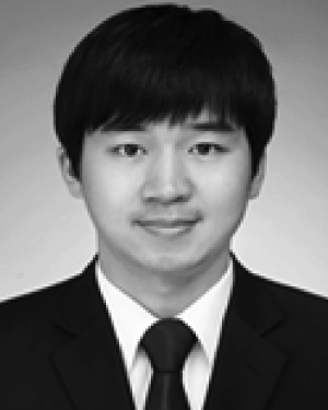 Xiaolong Li (李晓龙)合作论文数110
 Yifeng Zhang (张一峰)合作论文数Shanghai Tenth People's Hospital106
 Chong-Ke Zhao合作论文数85
 Guangjian Liu合作论文数Department of Ultrasound, Sun Yat-Sen University85Zuo-Feng Xu合作论文数84
 Mingde Lv (吕明德)合作论文数The First Affiliated Hospital, Sun Yat-sen University69Jun-Mei Xu (徐军妹)合作论文数66Bo Xiao-Wan合作论文数64Liu Bo-Ji合作论文数60Linna Liu (刘琳娜)合作论文数58Chang Liu合作论文数54
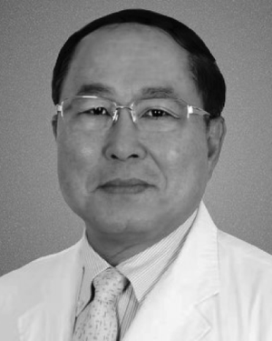 Ming-De Lü合作论文数51
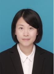 Wenwen Yue (岳雯雯)合作论文数School of Medicine, Tongji University;Tenth People's Hospital of Tongji University45
 Dan-Dan Li合作论文数40Haohao Yin合作论文数39Dan Wang合作论文数Tongji Univ, Shanghai Peoples Hosp 10, Ultrasound Res & Educ Inst, Dept Med Ultrasound,Sch Med, Shanghai 200072, Peoples R China38
 Jinyu Liang (梁瑾瑜)合作论文数The First Affiliated Hospital, Sun Yat-sen University37Qiao Wang合作论文数35Ya-Ping He合作论文数Tongji Univ, Shanghai Peoples Hosp 10, Ultrasound Res & Educ Inst, Dept Med Ultrasound,Sch Med, Shanghai 200072, Peoples R China34
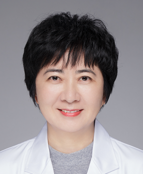 Rong Wu (吴蓉)合作论文数Shanghai General Hospital32Guang Xu合作论文数31Hong Han合作论文数30Wei-Wei Ren合作论文数Tongji Univ, Shanghai Peoples Hosp 10, Ultrasound Res & Educ Inst, Dept Med Ultrasound,Sch Med, Shanghai 200072, Peoples R China30Yanling Zheng (郑艳玲)合作论文数28
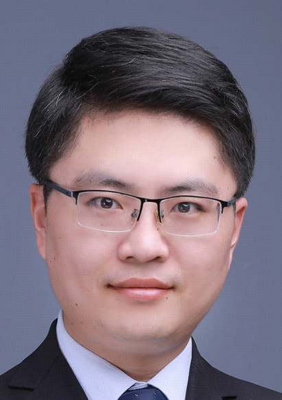 Kun Zhang (张坤)合作论文数School of Medicine, University of Electronic Science and Technology of China28
 Yu Chen (陈雨)合作论文数Shanghai University27Li-Fan Wang合作论文数26
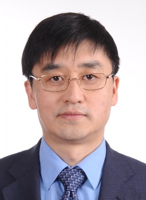 Shen Qu (曲伸)合作论文数School of Medicine, Tongji University25
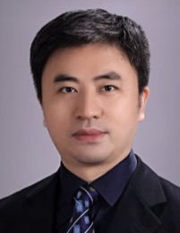 Ming Kuang (匡铭)合作论文数The First Affiliated Hospital, Sun Yat-sen University24
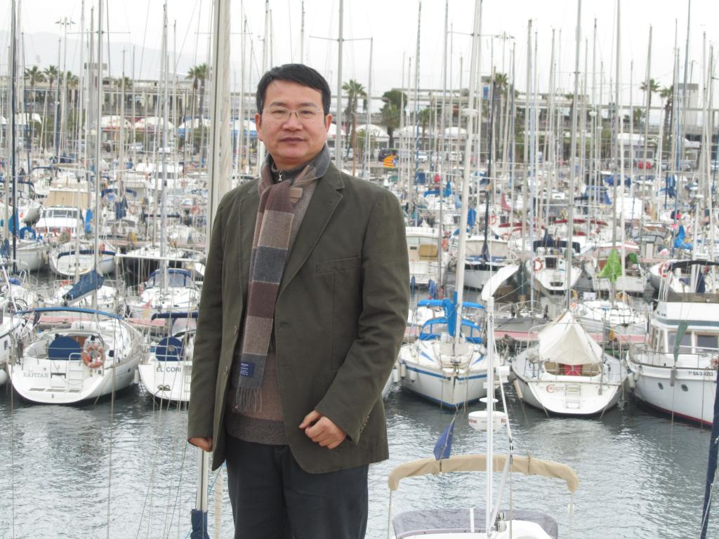 Shuguang Zheng (郑曙光)合作论文数School of Law,Ningbo University,Ningbo ,China24Song-Yuan Yu合作论文数23Bei Huang (黄蓓)合作论文数23Bangguo Zhou合作论文数22
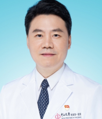 Xiaoyu Yin (殷晓煜)合作论文数The First Affiliated Hospital, Sun Yat-sen University21LiDa Chen (陈立达)合作论文数20Minghua Yao合作论文数20Lu Feng合作论文数19Yi-Kang Sun合作论文数18Bo-Yang Zhou合作论文数18An-Qi Zhu合作论文数18Xu Xiao-Hong合作论文数17
 Hangrong Chen (陈航榕)合作论文数The State Key Lab of High Performance Ceramics and Superfine Microstructure, Shanghai Institute of Ceramics, Chinese Academic of Sciences;University of Chinese Academy of Sciences17
 Xiaohua Xie (谢晓华)合作论文数School of Computer Science and Engineering, Sun Yat-sen University16Yu-Li Zhu合作论文数15Yinying Pu合作论文数Tongji Univ, Shanghai Peoples Hosp 10, Dept Med Ultrasound, Sch Med, 301 Yan Chang Zhong Rd, Shanghai 200072, Peoples R China15Jifeng Yu合作论文数15




