Photoacoustic imaging has emerged as a promising technology in the life sciences, exploiting the relatively weak scattering of sound in biological tissues to overcome the penetration limits inherent in conventional optical imaging. Relying on the photoacoustic effect, this imaging modality enables the identification of a wide range of endogenous molecules by analyzing their unique optical absorption spectra. This review summarizes characteristic molecules commonly employed in photoacoustic imaging and their corresponding biomedical applications across the full spectrum, including key bands such as X-ray, ultraviolet, visible, near-infrared, mid-infrared, terahertz, and microwave. Furthermore, the paper also outlines the main endogenous molecules used in photoacoustic imaging and their successful clinical applications, identifies current challenges in the development of full-spectrum photoacoustic imaging, and offers perspectives on future directions for technological advancement. Continued progress in photoacoustic imaging is expected to broaden its advantages, thereby facilitating advancements in biomedicine.
While fluorescence molecular tomography (FMT) in the second near-infrared window (NIR-II) offers improved penetration and contrast, quantitative deep-tissue recovery remains fundamentally ill-conditioned due to photon diffusion and is constrained by the cost, noise floor, and scalability limitations of short-wave infrared (SWIR) focal-plane arrays. We report a camera-free NIR-II FMT architecture integrating NIR-I spatial-frequency-domain (SFD) structured illumination with NIR-II single-pixel photon-counting detection. Co-registered excitation-reference and fluorescence measurements are fused through a multi-frequency normalized Born formulation to reconstruct the effective fluorescence-yield distribution. Phantom experiments demonstrate millimeter-scale dual-inclusion separability and sub-millimeter axial localization at depths up to approximately 7 mm, with a near-linear fluorescence-yield response. This hardware-efficient framework provides a scalable route toward high-sensitivity NIR-II fluorescence tomography.
Fluorescence molecular tomography (FMT) reconstruction represents an ill-posed inverse problem, and the existing solution methods often serve as estimators that tend to introduce reconstruction errors. In this study, we present a maximally weighted iteration (MWI) based FMT imaging framework designed to mitigate the ill-posedness of the inverse problems by strategically controlling the ill-conditioned weighting matrix through iterative weighted decomposition and a weighted correction term. Furthermore, four types of weighting matrices incorporating L1, L2, Frobenius, and L-inf norms are introduced and quantitatively compared with the conventional Tikhonov regularization method through numerical simulations, phantom and in vivo experiments for both single- and double-target scenarios. These results demonstrate that the MWI-based algorithms, particularly the one using the L2-norm, exhibit superior performance in terms of noise robustness, reconstruction accuracy, and spatial resolution. This study provides an effective strategy to enhance the FMT imaging quality.
Functional near-infrared spectroscopy (fNIRS) enables portable, non-invasive monitoring of cerebral oxygenation, yet its quantitative accuracy in continuous-wave optical topography (CW-fNIRS-OT) is often constrained by conventional methods that rely on an empirical differential path length factor (DPF). The empirical DPF typically assumes homogeneous tissue and ignores the layered head structure as well as inter-subject anatomical variability. In this study, we overcome the DPF-induced quantitative limitations in CW-fNIRS-OT by introducing a time-shift inversion strategy that, in tandem with layer-sensitive time-gated windows, achieves an efficient optical properties inversion while effectively decoupling and compensating for path length-dependent distortions. Leveraging a statistical brain model derived from anatomical atlases, our approach synthesizes subject-specific head geometry from simple anthropometric data, explicitly eliminating MRI dependency. Photon-transport simulations are employed to predefine time gates with preferential sensitivity to superficial and deep layers, while a dynamic time-shift correction compensates for residual inter-subject geometric mismatches. Ultimately, this design permits the precise inversion of layer-specific optical properties via a computationally lightweight single-layer diffusion equation model. Validation through numerical simulations, phantom experiments, and in vivo breath-holding tasks demonstrates that the proposed method significantly outperforms conventional empirical-DPF approaches. It corrects systematic path length errors, achieving a quantitativeness ratio of ∼0.95 vs ∼0.85 for conventional methods, and improves spatial fidelity, contrast-to-noise ratio, and task-state discriminability in SVM classification. Affording personalized, high-accuracy DPF inversion with computational efficiency, this framework provides a clinically viable route to individualized cerebral oxygenation assessment and is poised to catalyze the broader adoption of fNIRS in both neuroscientific inquiry and diagnostic practice.
Foodborne pathogens pose a significant threat to food safety. Therefore, the rapid and accurate detection of these pathogens is of critical importance. Raman spectroscopy, as a rapid, non-invasive, and label-free analytical technique, demonstrates significant potential for their detection. However, in scenarios requiring single-cell level rapid detection, Raman spectroscopy suffers from limited sensitivity. This limitation arises from the short integration times, the inherently weak Raman signal, and the extremely small volume of individual bacterial cells. To address this challenge, this study proposes a spectral restoration approach that combines Mel spectrogram transformation with a generative adversarial network (GAN_Mel). In this approach, the spectrogram transformation process is integrated into the discriminator architecture of the GAN to improve reconstruction efficiency and denoising capability. Experimental results demonstrate that, under a short integration time of 3 s, the proposed method can effectively enhance the spectral signal-to-noise ratio (SNR) to a level comparable to that obtained with a 30-s integration time, achieving a classification accuracy of 92.9% for foodborne bacterial species. This study provides a solid technical foundation for the rapid and accurate single-cell detection of foodborne pathogens using Raman spectroscopy.
Here, we report a wearable functional near-infrared spectroscopy-electroencephalogram (fNIRS-EEG) system to overcome the temporal resolution mismatch between the two modalities, enabling precise investigation of neurovascular coupling dynamics (NVC). By implementing an orthogonal encoding and cyclic decoding scheme, the system achieves fNIRS acquisition at up to 1,000 Hz simultaneously with EEG, while maintaining high signal fidelity across 52 optical and 32 electrical channels. Systematic validation through phantom experiments, physiological tests, and auditory-evoked response measurements confirms the system’s ability to concurrently capture fast neural activity and hemodynamic correlates. Feature-level correlation analysis further reveals strong links between optical neuronal signals and event-related potentials. This technological advance provides a powerful platform for studying rapid neuronal signals, with broad implications for cognitive neuroscience, clinical diagnostics, and brain-computer interfaces.
Smith-Purcell radiation is a fundamental electromagnetic phenomenon that occurs when charged particles are near a periodic structure. Conventionally, the intrinsic loss of the structure reduces its radiation efficiency. Here, we show an exception to this understanding by exploring the interaction of the charged particles with a lossy even-parity phase gradient metasurface. We demonstrate that the intrinsic loss can enable precise control of the SPR spatial distribution while keeping the total radiation energy constant. Remarkably, increasing the loss induces a transition from unidirectional radiation in the lower half-space to a critical state with equal intensities in both half-spaces, and eventually to a regime where radiation in the upper-space dominates. These findings provide an approach to manipulating the SPR effect, with potential applications in free-electron radiation devices and related sources.
Spatial frequency-domain laminar optical tomography (SFD-LOT) enables depth-resolved optical property imaging but remains difficult to extend to dual-band NIR-I/II operation under photon-limited conditions. This limitation arises from the combined constraints of costly short-wave infrared detectors and the ill-conditioned nature of diffuse inverse problems under sparse measurements. Here, we present a dual-band single-pixel SFD-LOT framework that integrates camera-free structured illumination with a lookup-table-assisted algebraic reconstruction technique (LUT–ART). Broadband acquisition at 715, 830, 1050, and 1200 nm is achieved using frequency-multiplexed illumination and SPAD-based single-pixel detection, eliminating the need for InGaAs focal-plane arrays. Surface optical properties estimated by LUT are incorporated as physically grounded background constraints to stabilize volumetric reconstruction, improving conditioning for absorption-focused volumetric reconstruction while providing a framework that can be extended to other contrast mechanisms under appropriate constraints. In this study, the framework is primarily validated through absorption-focused reconstructions. Phantom experiments demonstrate accurate optical-property decoupling (R² > 0.99), stable quantitative response to absorption contrast, millimeter-scale lateral resolution, and reliable depth localization up to 4 mm. In vivo measurements further show improved spectral stability with dual-band acquisition, enabling reduced water/lipid cross-talk and physiologically plausible chromophore and StO₂ distributions. These results establish a practical hardware–algorithm co-designed framework for dual-band diffuse optical tomography under photon-limited conditions.
Foodborne pathogens represent a major global threat to food safety, necessitating rapid and reliable identification technologies. While Raman spectroscopy provides a powerful label free tool for molecular fingerprinting, its performance is often compromised by high biochemical similarity between strains and the bottlenecks of data scarcity in practical scenarios. To address these challenges, we propose the Peak Aware Raman Attention Model (PARAM), a generative deep learning framework designed for high fidelity spectral augmentation. Unlike traditional generative models that often suffer from peak shifts or intensity distortions, PARAM integrates a peak aware attention mechanism. This mechanism explicitly preserves the structural integrity of biochemically significant spectral regions, such as the 1003 cm-1 phenylalanine and 1330 cm-1 nucleic acid modes, through peak guided attention mapping. We evaluated PARAM using four major pathogens under extreme data constraints of only 7 samples per class. Experimental results demonstrate that PARAM generated spectra achieve exceptional consistency with real data, exhibiting 100% peak position matching and a Pearson correlation coefficient of 0.9989. Furthermore, augmenting the training set with PARAM generated data significantly boosted the Random Forest classification accuracy from 87.67% to 97.95%. These findings highlight PARAM as a robust strategy to overcome data scarcity in food safety monitoring.
Rapid and accurate detection of viable leukemia cells is of critical importance for assessment of drug efficacy and prognosis assessment in leukemia. Conventional cell analysis methods generally suffer from long processing time, sample destructiveness, and the cytotoxicity of staining agents, which limit their application in live-cell analysis. To address these issues, a visible and near-infrared(Vis-NIR) snapshot hyperspectral imaging technique enhanced by a dynamic filtering super-resolution demosaicing algorithm is proposed. The technique offers advantages such as high-speed acquisition, label-free operation, and enhanced imaging resolution, making it highly suitable for live-cell subtype classification of leukemia cells. Based on the hyperspectral data acquired by this technique, a three-dimensional convolutional neural network (3D-CNN) is developed to achieve automated leukemia cell classification. Experimental results demonstrate that the proposed method achieves an average classification accuracy of 93.6% across four leukemia cell types, representing a 12.5% improvement compared to the method based on original image. In addition, the acquisition speed reached 780 cells per minute, further demonstrating its suitability for high-throughput applications. This approach provides a robust technical solution for high-throughput, non-destructive detection of leukemia cells, supporting future applications in precision medicine.
Objective and Impact Statement: This study examines prefrontal cortex (PFC) hemodynamic responses in children with idiopathic central precocious puberty (ICPP) versus normals and constructs a noninvasive diagnostic model using functional near-infrared spectroscopy (fNIRS) augmented by machine learning. Introduction: Current ICPP diagnosis relies on invasive and time-consuming gonadotropin-releasing hormone stimulation tests. While fNIRS offers a noninvasive alternative, the neural mechanisms underlying ICPP remain unclear, and reliable automated diagnostic tools distinguishing patients from healthy peers are lacking. Methods: fNIRS data were acquired from 167 participants (82 ICPP and 85 normal) during a mental arithmetic (MA) task. General linear models and statistical tests were employed to analyze group and gender-specific activation patterns. Multidimensional features were extracted from hemodynamic signals, and a conditional denoising diffusion probabilistic model (C-DDPM) was introduced for data augmentation. Results: Analysis revealed gender-specific disparities, with the normal group exhibiting more extensive PFC activation than the ICPP group. In classification, a decision tree model using features from key negatively correlated channels achieved 86.57% accuracy. Notably, integrating C-DDPM-generated synthetic data further improved classifier performance metrics. Conclusion: The study elucidates the mechanisms of PFC activation in both normative and ICPP-affected cohorts during MA tasks and validates the effectiveness of machine learning in distinguishing between normal and ICPP children. This study provides a scientific basis for the development of automated, noninvasive rapid diagnostic tools for ICPP.
Diffuse optical tomography (DOT) is a noninvasive imaging technique with promising biomedical applications; however, its reconstruction is severely ill-posed, leading to low spatial resolution, quantitative accuracy, and pronounced robustness when using conventional algorithms. In this study, an attention-enhanced deep learning post-processing method, termed ART-U-Net-CBAM, is proposed to improve DOT image reconstruction. The method combines the physics-based algebraic reconstruction technique (ART) with a U-Net network integrated with a convolutional block attention module (CBAM), enabling adaptive emphasis on informative spatial and channel features. Trained exclusively on simulated data, the proposed network was evaluated using both numerical simulations and phantom experiments involving circular and elliptical targets. Quantitative results demonstrate that ART-U-Net-CBAM consistently outperforms ART and ART-U-Net in terms of reconstruction accuracy, noise robustness, spatial resolution, and structural similarity. These findings indicate that attention-enhanced deep learning post-processing provides an effective and generalizable strategy for enhancing DOT image quality.
We demonstrate a camera-free NIR-II fluorescence tomography platform using single-pixel spatial frequency domain imaging. NIR-I structured excitation and SPAD-based compressive detection enable millimeter-scale shallow separability and depth-dependent localization in tissue-mimicking phantoms.
Cherenkov imaging provides real-time video of beam incidence upon the patient, for verification of safe and accurate radiotherapy delivery. However, the optical signal is inherently weak and is affected by non-optical radiation leakage and stray x-ray noise from the medical linear accelerator (Linac). This frequently leads to low signal-to-noise ratio (SNR) frames with background clutter, limiting video image clarity and beam visualization. To address this challenge, a wavelet-based deep video denoising method was proposed. The method was validated with three regular square fields and clinical data from two volumetric modulated arc therapy (VMAT) fractions administered to breast cancer patients. Image quality was assessed using the global gamma pass rate ( γ pass ) with 3%/3 mm criteria. The decision-making process of the network was visualized for interpretability. Results show that Cherenkov frames accumulated over five Linac pulses achieved γ pass of 96–97% for all square beams. For clinical VMAT cases, accumulated frames from selected control points reached γ pass exceeding 95%. We believe this to be the first demonstration of a deep video denoising framework sufficiently fast for real-time Cherenkov imaging.
Diffuse optical techniques such as time-domain diffuse optical spectroscopy (TD-DOS) and diffuse correlation spectroscopy (TD-DCS) enable noninvasive assessment of tissue hemodynamics and oxygenation, yet their quantitative accuracy in layered tissues is fundamentally limited by depth-dependent sensitivity and parameter coupling. Here, we propose a time-gated hierarchical TD-DOS/TD-DCS framework, implemented on a fully synchronized multi-wavelength dual-modal platform, that exploits the temporal encoding of photon path length to achieve depth-resolved recovery of optical, hemodynamic, and metabolic parameters, where TD-DOS provides both optical preconditioning for TD-DCS inversion and oxygenation information derived from absorption measurements, complementing blood flow metrics obtained from TD-DCS. Under a unified time-resolved acquisition scheme, early- and late-arriving photons are sequentially analyzed first to estimate superficial optical properties using a single-layer model, followed by stable recovery of deep-layer absorption and blood flow using a constrained two-layer model. This approach improves parameter identifiability without increasing model complexity. The method was validated using bilayer liquid phantoms and in vivo experiments. In phantoms, the proposed strategy reduced deep-layer absorption error from ∼26.5% to ∼9.8% compared to the conventional single-layer fitting, and improved sensitivity to deep-layer flow changes by ∼2.7×. In forearm occlusion measurements, the recovered deep-layer blood flow exhibited significantly greater dynamic contrast than conventional estimates. Breath-holding experiments further demonstrated robust recovery of cerebral blood flow, oxygenation, and metabolic parameters, with a positive association between relative cerebral blood flow (rCBF) and relative cerebral metabolic rate of oxygen (rCMRO2) (R2 = 0.67 ± 0.06). These results support the feasibility of the proposed framework for depth-resolved estimation in layered tissues and provide a unified approach for simultaneous assessment of blood flow, oxygenation, and metabolism in diffuse optical measurements.
Background and objective Diffuse optical tomography (DOT) is a non-invasive imaging technique with promising biomedical applications. However, its reconstruction quality is severely limited by the ill-posed inverse problem, leading to low spatial resolution and quantitative accuracy. This study aims to improve DOT image reconstruction performance by enhancing multi-scale feature extraction and feature reuse through an advanced deep learning framework. Methods A deep neural network-based DOT reconstruction framework integrating dilated convolution and densely connected networks (DenseNet) is proposed. To systematically evaluate the contributions of dense connectivity and dilated convolution, four models involving ResNet, ResNet with dilation convolution (DResNet), DenseNet, and DenseNet with dilation convolution (DDenseNet) are constructed. Numerical simulations and physical phantom experiments are conducted under varying target sizes and absorption contrasts. Reconstruction performance was quantitatively assessed using Mean Absolute Error (MAE), Quantitativeness Ratio (QR), Contrast-to-Noise Ratio (CNR), and Structural Similarity Index Measure (SSIM). Results Both simulation and phantom results demonstrate that the proposed DDenseNet consistently outperforms the other methods. It achieves the lowest Mean Absolute Error, the highest Contrast-to-Noise Ratio, and values of Quantitativeness Ratio and Structural Similarity Index Measure closest to 1.0 across different experimental conditions. Conclusions By combining dense connections with dilated convolution, the proposed DDenseNet effectively enhances multi-scale feature learning and reconstruction accuracy. This framework provides a robust and accurate solution for high-quality DOT imaging and shows strong potential for future clinical applications.
Hemodynamic monitoring of human tissues, particularly cerebral blood flow assessment, is of significant clinical importance. Time-gated diffuse correlation spectroscopy (TG-DCS) enables non-invasive blood flow measurements at varying depths by analyzing temporal intensity fluctuations and time-of-flight (TOF) characteristics of scattered light. While late gated detection offers advantages in measuring the deep tissue blood flow index (BFi), its accuracy is compromised by contamination from superficial tissues and systemic physiological interference. To address these limitations, this study introduces a least mean square (LMS) adaptive filtering approach, which innovatively uses the results of early gated as a reference channel to enhance late gated measurement precision. Furthermore, considering higher dynamic changes in shallow particles compared to deep layers, we designed a novel experimental protocol to validate the recoverability of deep layer diffusion coefficients under significant superficial variations. We developed a TG-DCS measurement system incorporating a software-based autocorrelation algorithm for flexible photon data processing. The system’s performance was evaluated by assessing the influence of shallow layer diffusion coefficient changes on deep layer measurements and comparing deep diffusion coefficients across multiple source-detector separations. Results demonstrate that the optimized late gated approach achieves accurate deep layer particle dynamics detection with a high signal-to-noise ratio and spatial resolution. Finally, in vivo experiments on healthy subjects’ foreheads, including controlled breath-holding protocols, confirmed that the optimized late gated method provides measurements closer to the true physiological values than conventional techniques, even under superficial blood flow variations. The breath-holding experiments further validated the system's ability to accurately track deep tissue hemodynamic changes while minimizing superficial contamination. These comprehensive validations demonstrate the significant advancement of our adaptive filtering innovation and underscore TG-DCS’s potential for reliable brain function monitoring and deep-tissue hemodynamic assessment.
Rapid detection of foodborne pathogenic and spoilage microorganisms is critical for ensuring food safety and quality in liquid matrices. While Raman tweezers spectroscopy (RTS) enables label-free single-cell analysis, its application in high-throughput inline inspection faces a fundamental bottleneck: high flow rates required for efficiency induce severe motion blur and low signal-to-noise ratios (SNR), which blind automated control systems and destabilize optical trapping. To overcome this, we present a Spatiotemporal Video-Enhanced Raman Tweezers (SVERT) system integrating a deceleration-optimized microfluidic chip with a deep learning-based visual feedback loop. We propose a Local-Global Unified Denoising Network (LGU-Net) tailored to recover high-fidelity bacterial structures from low-SNR video streams, achieving a deterministic processing latency of ~0.49 ms. Experimental results demonstrate that SVERT improves the optical trapping success rate from 21.27% ± 2% to 91.47% ± 1.8% compared to raw video input, enabling a four-fold increase in spectral acquisition efficiency. Leveraging the acquired high-quality dataset, we achieved a classification accuracy of 96.74% across four bacterial species of relevance to food safety and quality. Crucially, we validated the system's practical robustness by successfully isolating and tracking trace E. coli in an unpurified commercial beverage. This capability to effectively mitigate natural background interference demonstrates the system's promising potential to be expanded for broader applications in liquid food safety screening.
Infrared imaging is a valuable technology for gas leakage detection due to its high sensitivity, long detection range, and high efficiency. Conventional target detection methods depend on manually extracting image features, which often leads to limited accuracy, low adaptability, and slow detection speeds. Deep learning technology offers a potential solution to these challenges; however, the increasing depth of neural networks imposes significant computational demands, posing challenges to real-time detection. This article presents a compact and energy-efficient gas detection system, implemented with a ZYNQ platform and an infrared camera. We propose a ZYNQ-based convolution accelerator to enhance gas plume detection from images captured by the infrared camera. Operating at a clock frequency of 130 MHz, the accelerator is capable of reaching a peak performance of 37.44 Gop/s, with power consumption of only 4.12 W. The system achieves a processing speed of 0.235 s per image, enabling real-time gas leakage detection.
 Huijuan Zhao (赵慧娟)合作论文数College of Information Technology, Shanghai Ocean University166
 Limin Zhang (张丽敏)合作论文数School of Precision Instruments and Opto-electronics Engineering, Tianjin University113
 Jiao Li (李娇)合作论文数Biomedical Photonic Imaging Laboratory, School of Precision Instrument and Optoelectronics Engineering, Tianjin University92
 Zhongxing Zhou (周仲兴)合作论文数School of Precision Instrument and Optoelectronics Engineering, Tianjin University55
 Dongyuan Liu (刘东远)合作论文数School of Artificial Intelligence, Tianjin University47
 Mengyu Jia (贾梦宇)合作论文数Medical School of Tianjin University;Biomedical Photonic Imaging Laboratory, Tianjin University;School of Precision Instruments and Opto-electronics Engineering, Tianjin University33
 Yukio Yamada合作论文数Department of Human and Engineered Environment Studies, The University of Tokyo/Neurosciences and Biomedical Engineering, University of Electro-Communications29Xi Yi合作论文数27Linhui Wu合作论文数27
 Wenjuan Ma合作论文数26
 Fei Gao (高飞)合作论文数School of Information Science and Technology, ShanghaiTech University25
 Wei Zhang合作论文数School of Electrical and Information Engineering, Tianjin University25
 Zhang Yao合作论文数22Zhuanping Qin合作论文数20
 Yihan Wang (王艺涵)合作论文数School of Life Science and Technology, Xidian University19Yukari Tanikawa合作论文数Institute for Human Science and Biological Engineering, National Institute of Advanced Industrial Science and Technology19
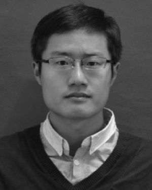 Wang Bingyuan合作论文数18Weiting Chen (陈玮婷)合作论文数18
 Yanqi Zhang合作论文数17
 Daohuai Jiang合作论文数ShanghaiTech Univ, Sch Informat Sci & Technol, Hybrid Imaging Syst Lab, Shanghai 201210, Peoples R China17
 Jingying Jiang (蒋景英)合作论文数School of Medical Science and Engineering, Beihang University16Dan Mai合作论文数15Wan Wenbo合作论文数15
 Tong Lu合作论文数14
 Yiwen Ma (马艺闻)合作论文数School of Precision Instrument and Optoelectronics Engineering, Tianjin University14Xiaoqing Zhou合作论文数13
 Hengrong Lan合作论文数ShanghaiTech Univ, Sch Informat Sci & Technol, Hybrid Imaging Syst Lab, Shanghai, Peoples R China13
 Liu Lingling合作论文数12
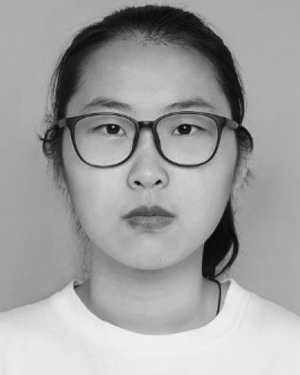 Yuting Shen合作论文数11
 Xin Wang (汪欣)合作论文数河南大学生命科学学院11
 Lixin Zhang (张力新)合作论文数School of Precision Instrument and Optoelectronics Engineering, Tianjin University11
 Li Tongxin合作论文数10
 Pengrui Zhang合作论文数9PingQiao Ruan (阮平巧)合作论文数9
 Qifeng Li (李奇峰)合作论文数School of Precision Instrument and Opto-electronics Engineering, Tianjin University9
 Shinpei Okawa合作论文数Department of Mechanical Engineering and Intelligent Systems, The University of Electro-Communications9
 Tiantian Pan合作论文数8Lin Zhang合作论文数United Imaging Healthcare8
 Yiming Lu合作论文数Department of Bioengineering, University of Washington8Ying Fan合作论文数7Huiyuan He合作论文数7Qingzhen Zhu合作论文数7Andhi Marjono合作论文数Department of Mechanical Engineering and Intelligent Systems, The University of Electro-Communications7TANIKAWA Y合作论文数6WANG Ting-ting合作论文数6Liu Ming合作论文数6
 Lu Bai合作论文数6Panpan Yan合作论文数6Guoyan Yin (尹国艳)合作论文数6




